According to most medical studies, computed tomography CT scans is considered one of the top five medical advancements in the past 50 years. CT imaging has proven its value as a medical diagnostic tool to the extent that the 1979 Nobel Prize in Medicine was awarded to its inventors. In this article, we will delve deeper to understand this type of medical imaging and explore when and why it is used. Let’s get started.
What is CT Scanning?
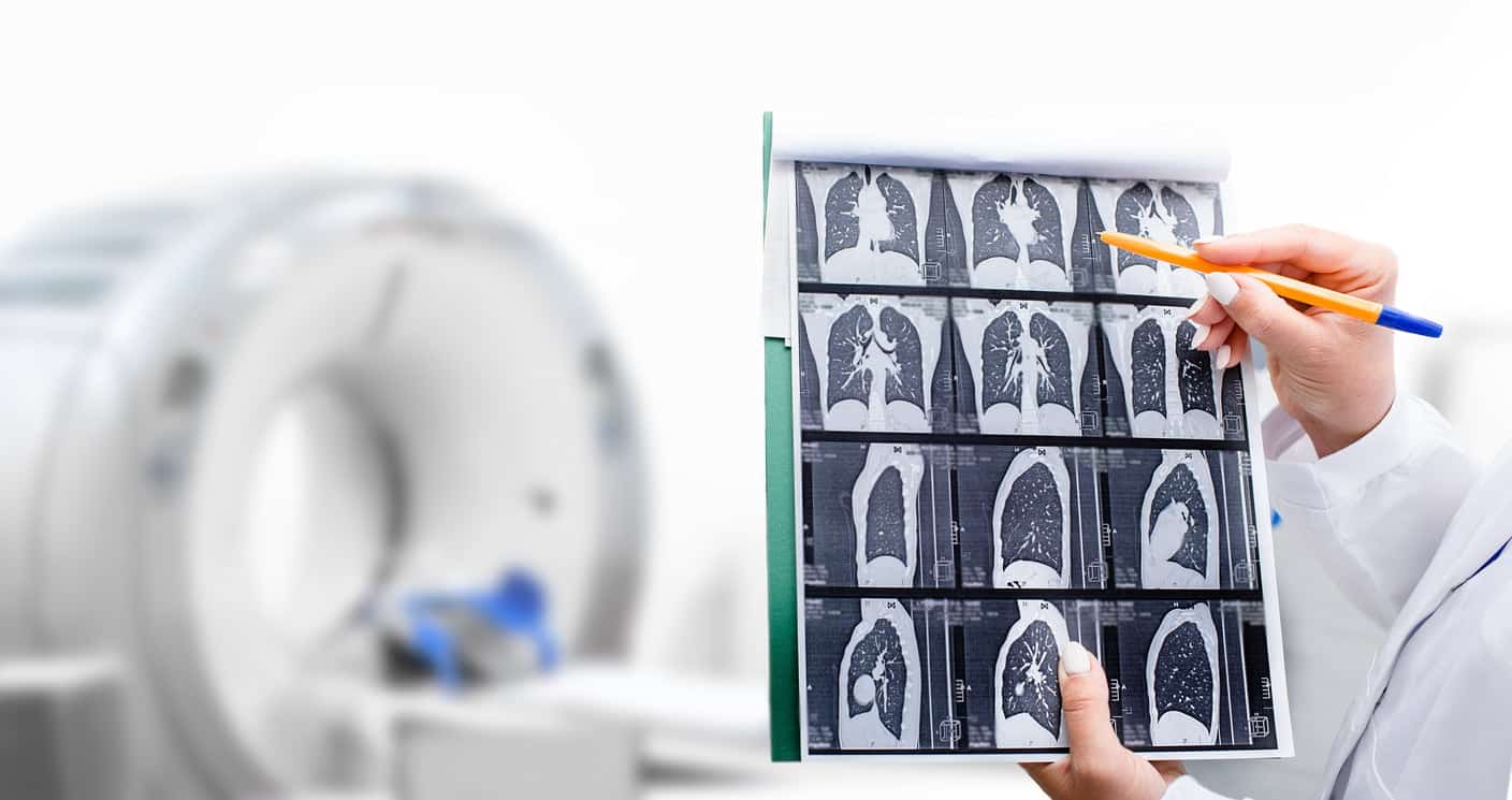
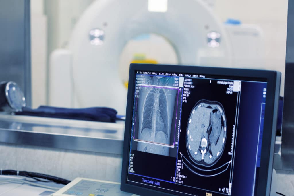
Computed Tomography (CT) or CT scanning is a diagnostic procedure that uses a combination of X-rays and computer technology to produce internal images of the body. This procedure provides accurate images of any part of the body, including bones, muscles, fat, organs, and blood vessels. CT scanning can be used to diagnose tumors, check for internal bleeding, assess injuries or internal damage, and even guide biopsies of tissues or fluids.
CT imaging is more detailed than regular X-rays. In traditional X-rays, an energy beam is directed to the area being examined, capturing differences in the beam as it passes through the skin, bones, muscles, and other tissues. However, X-rays do not provide much detail about internal organs or fine structures.
In CT scans, the X-ray beam rotates in a circular motion around the body, allowing multiple-angle views of the same organ or structure and providing much clearer details. The X-ray data is sent to a computer, displaying images in 2D, and with modern technology, 3D images can also be produced.
Reasons You May Need a CT scan
After understanding what CT imaging is, the critical question arises: why do doctors request CT scans? These scans are used worldwide in hospitals and specialized medical centers as one of the modern diagnostic tools for various reasons. Below, we discuss 10 of the most common reasons why your doctor may request a CT scan.
1. Examining Blood Vessels
CT scans provide sufficient detail for doctors to examine blood vessels and check for blockages or other potential issues. The images produced provide essential information to diagnose vascular diseases without requiring exploratory surgeries or surgical biopsies.
2. Diagnosing Abdominal Issues
Abdominal CT scans may use barium contrast to produce highly detailed images of organs, including the liver, kidneys, gallbladder, spleen, ovaries, and uterus. The addition of iodine based IV contrast enhances the images further, enabling doctors to diagnose a wide range of health issues.
CT scans can detect:
- Abdominal tumors
- Causes of unexplained weight loss
- Potential obstructions in the small or large intestines
- Intestinal infections
- Kidney stones
A specific type of CT scan, known as CT Urography, is used to diagnose the kidneys, bladder, and ureters.
3. Examining Small Bones
Bones in areas like the hands and feet are exceptionally small, making injuries in these areas difficult to detect using X-rays alone. CT scans provide doctors with clear and detailed images, aiding surgical repairs.
4. Examining Tumors
CT imaging often serves as the first step in surgical treatment for tumors. The more information surgeons have about a tumor’s location and size, the better equipped they are to perform procedures like biopsies efficiently. CT scans also help determine the spread of the tumor into surrounding tissues, improving surgical outcomes.
5. Guiding Cancer Treatment
CT scans are frequently used to diagnose cancer and develop treatment plans. During radiation therapy, CT scans are used to pinpoint tumor locations. Additionally, CT imaging helps assess the effectiveness of chemotherapy by showing the extent and speed of cancer progression.
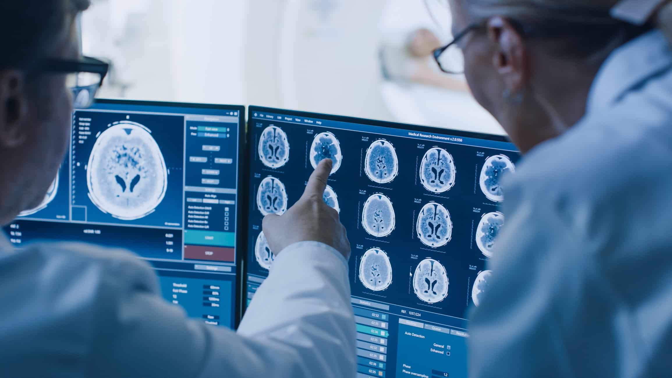
6. Assessing Head and Brain Injuries
Head CT scans provide brain images that help assess:
- Head injuries
- Severe and chronic headaches
- Persistent dizziness
- Bleeding
- Stroke
- Brain tumors
They are also occasionally used to:
- Evaluate soft tissue and bone damage following facial trauma
Plan reconstructive surgeries - Diagnose problems in the temporal bone of the skull that may cause
- hearing issues
- Detect sinus inflammation
- Evaluate aneurysms
7. Diagnosing Soft Tissue Damage
Traditional X-rays reveal very little about soft tissues, whereas CT scans offer a dual advantage by showing both bone and soft tissue conditions. This provides a more comprehensive understanding of injuries to aid in diagnosis and recovery planning.

8. Diagnosing Spine Problems and Chronic Pain
Chronic back pain or spinal injuries are among the most common reasons for CT scans. Doctors may also request CT imaging for the spine to:
- Evaluate spinal fractures
- Assess spinal conditions before and after surgeries
- Identify the cause of spinal pain, such as herniated discs
- Measure bone density to predict fracture risks in patients with osteoporosis
CT imaging is also helpful alongside MRI for patients with spinal canal narrowing, infections, or arthritis.
9. Investigating Post-Accident Injuries
Accidents leading to severe internal injuries often require CT scans. Internal injuries cannot typically be identified with X-rays alone. In emergencies, CT imaging is the first choice for doctors, especially after car accidents or other traumas.
10. Obtaining Images When MRI Isn’t Feasible
While MRI and CT are similar, certain situations make MRI unsuitable, while CT remains a viable option. MRIs can take considerable time to complete. For patients unable to remain still during the procedure, CT scans produce more accurate images in less time.
Note: Individuals with medical implants of any kind cannot undergo MRI and should opt for CT scans instead.
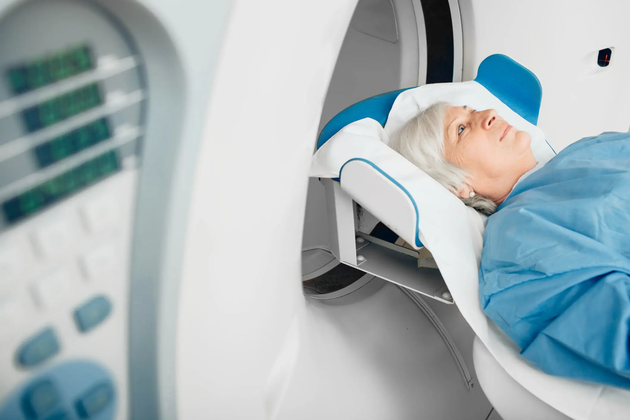
How CT Scans Work
CT devices are large, donut-shaped machines with an X-ray tube and sensors on opposite sides. The center contains a short tunnel where the patient lies on a table that slides in and out. During the scan, the X-ray equipment rotates around the patient, producing vital internal images. The entire procedure takes only a few minutes, making it less stressful for those with claustrophobia.
Preparing for a CT scan depends on whether contrast material is needed. If contrast is required, the patient may need to refrain from eating for a few hours prior to the scan. With IV contrast, patients receive an injection of a dye that may cause warmth, flushing, or a metallic taste in the mouth. Removing all jewelry or metal objects is necessary before the scan. Afterward, patients can continue their daily activities as usual, but if contrast was used, drinking several glasses of water is recommended to help flush out the dye.
Why Is Contrast Sometimes Used in CT scans?
Contrast is a substance taken orally or injected to highlight a specific organ or tissue more clearly. If contrast is needed, fasting for a certain period may be required, and the doctor will provide instructions.
- Notify your doctor if you’ve experienced a reaction to any contrast material before or if you have kidney problems.
- Seafood allergies are not a contraindication to using iodine-based contrast.
- Always inform your doctor of any health conditions.
In conclusion, we hope this article provided a clear understanding of CT imaging, its applications in medical diagnostics, and how it works. At HSI, pioneers in biomedical engineering and healthcare solutions, we offer specialized courses on this type of medical imaging with a focus on training.
Source: Reasons You May Need a CT scan