When it comes to diagnostic imaging techniques, X-ray and MRI are two of the most commonly used tools to help doctors evaluate and diagnose a wide range of medical conditions. While both provide vital diagnostic information, each technology has its own uses and distinct advantages.
Differences Between X-ray and MRI
This guide will help you understand the processes, benefits, limitations, and key differences between X-rays and MRI. With all the necessary information, you’ll feel reassured and well-informed about the diagnostic option most suitable for your health condition.
X-ray
X-rays are an imaging test that uses electromagnetic waves (radiation) to create two-dimensional (2D) images of the body’s tissues and skeletal structures. The radiation used in X-rays is similar to ultraviolet (UV) radiation from the sun but has much higher energy. However, it is used in very low doses that vary depending on the area being imaged.
X-rays rely on electromagnetic radiation to create internal images of the body and are particularly effective at imaging bones and diagnosing fractures, tumors, or infections. X-rays are fast, relatively inexpensive, and well-suited for initial diagnosis, especially in emergency situations. However, they lack the ability to provide detailed views of soft tissues, such as muscles and tendons.
How Do X-rays Work?
The imaging process involves lying on a motorized table (or standing, for chest or mammogram X-rays) between an X-ray source and a detector (such as photographic plates or fluoroscopic screens).
When the X-ray source directs a beam of radiation through your body toward the detector, an image is created based on the shadows cast by different tissues, depending on how much radiation they absorb.
Quick Guide to Understanding Shadows in a Typical X-ray Image:
- Bones: Being very dense, bones absorb most of the X-rays, casting shadows that appear white.
- Fluids, fat, and muscles: These tissues absorb moderate amounts of X-rays, appearing in varying shades of gray.
- Lungs: Filled with air that does not block X-rays, they allow the radiation to pass through and appear black in the image.
Magnetic Resonance Imaging (MRI)
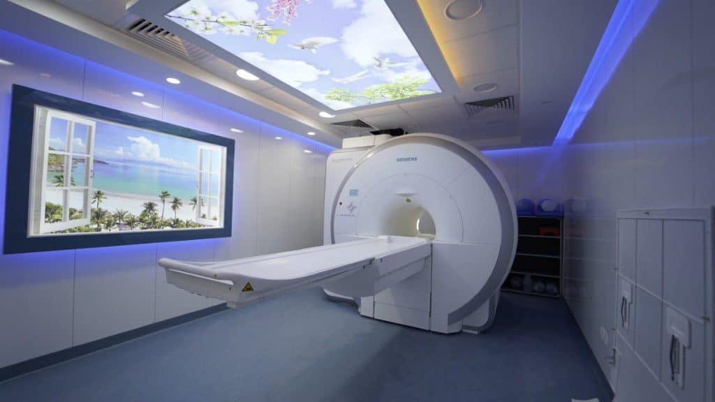
Magnetic Resonance Imaging (MRI) is a painless, non-invasive imaging technique that uses a strong magnetic field and radio waves to create detailed three-dimensional (3D) images of internal structures in the body, including organs, bones, joints, and soft tissues (such as nerves, muscles, and blood vessels).
This technique is ideal for diagnosing disorders of the brain, spinal cord, joints, or internal organs. Unlike X-rays, MRI does not use ionizing radiation, making it a safer option, especially for pregnant women and children. However, MRI is more expensive, takes longer, and may be uncomfortable for patients with claustrophobia.
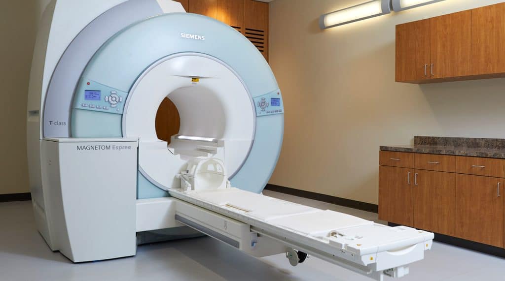
How Does MRI Work?
A traditional MRI machine consists of a large cylindrical scanner that acts as a powerful magnet, with a flat motorized table that moves the patient into the device. Depending on the area being examined, you may enter the machine head-first or feet-first.
MRI works by using the magnetic field to align hydrogen atoms, particularly protons (positively charged particles at the center of atoms). Hydrogen is abundant in both water and fat.
Water makes up about 60% of the human body, flowing freely in the blood and connecting with every cell, tissue, and organ. Fat is distributed throughout the body, around the heart and blood vessels, inside the brain, bones, and nerves, and behind the eyes. This explains MRI’s high sensitivity to conditions involving abnormal fat accumulation (e.g., tumors) or increased fluid presence (e.g., cysts).
Quick Comparison Between X-rays and MRI
X-rays and MRI are both essential diagnostic tools for evaluating various medical conditions. While each technology has unique advantages, the choice of the appropriate method depends on the condition being diagnosed. Below is a quick comparison between the two:
Diagnostic Capability:
- X-rays: Best for imaging bones; highly accurate for detecting fractures, dental issues, and spine problems.
- MRI: Offers a broader range, providing detailed images of bones, soft tissues, and organs. Used to diagnose tumors, neurological disorders, and joint or muscle injuries.
Availability:
- X-rays: Widely available and affordable.
- MRI: Less common and typically reserved for cases requiring detailed imaging.
Risks:
- X-rays: Uses ionizing radiation (which may increase cancer risk with high doses). Doses used are typically very low, but caution is advised during pregnancy.
- MRI: Generally safe since it does not use radiation. However, the magnetic field poses risks for individuals with metal implants. Contrast agents may cause mild side effects, and the procedure can be uncomfortable for those with claustrophobia or limited mobility.
Cost:
- X-rays: Relatively low-cost.
- MRI: More expensive compared to X-rays.
Speed:
- X-rays: Very fast, taking about 5 to 15 minutes.
- MRI: Slower, taking 15 to 90 minutes depending on the area being examined.

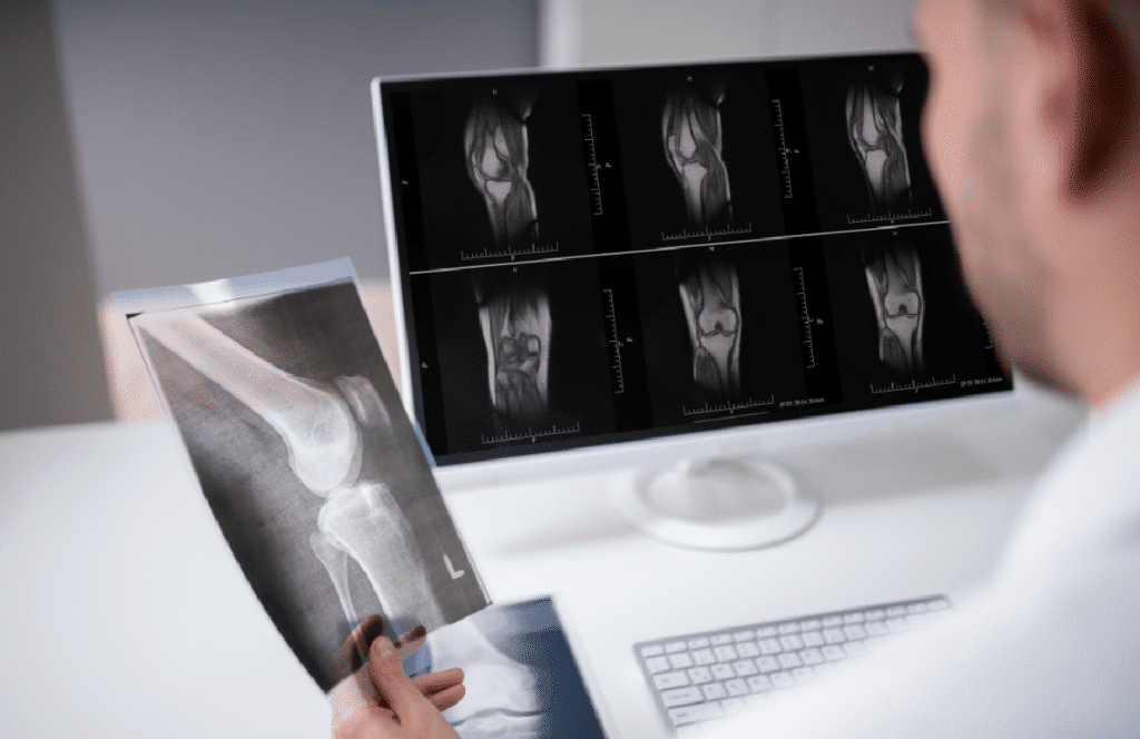
What Can MRI Show That X-rays Cannot?
Magnetic Resonance Imaging (MRI) is more accurate in diagnosing musculoskeletal pain caused by soft tissue injuries that X-rays cannot detect. These conditions include:
- Ligament and tendon injuries
- Degenerative Disc Diseases (DDD)
- Muscle tears or strains
- Nerve compression and damage
- Cartilage injuries (such as meniscal tears in the knee)
Joint disorders (such as osteoarthritis or rheumatoid arthritis)
In cases where X-rays are used as an initial imaging tool to rule out suspected conditions, an MRI or CT scan may be recommended as a complementary test to provide an accurate diagnosis and comprehensive assessment of the problem.
Uses of MRI
MRI can be used to detect abnormalities, infections, degeneration, inflammation, and diseases in soft, dense, and fluid-filled tissues in various parts of the body, including:
- Heart
- Liver
- Adrenal glands
- Kidneys
- Hand, shoulder, elbow, and wrist
- Foot and ankle
- All parts of the spine, including cervical (neck), lumbar
- (back), thoracic (mid-back), and sacral regions
- Brain and head
There are also special types of MRI, such as:
- Magnetic Resonance Cholangiopancreatography (MRCP): Used to detect stones, infections, and diseases in the pancreas, gallbladder, and bile and pancreatic ducts.
- Magnetic Resonance Angiography (MRA): Used to assess the health of blood vessels.
What Can X-rays Detect?
X-rays are used to detect the following conditions:
- Bone injuries such as fractures and dislocations
- Bone tumors, whether cancerous or non-cancerous (additional tests may be required for a definitive diagnosis)
- Osteoporosis (loss of bone density)
- Scoliosis (abnormal curvature of the spine)
- Dental issues, such as cavities, tooth decay, and abscesses
- Lung diseases, including pneumonia and pulmonary nodules (tumors)
- Calcifications (solid calcium deposits) in soft tissues, which may indicate specific medical conditions
- Foreign objects lodged inside the body
Factors Influencing the Choice of Diagnostic Tool
When determining the appropriate choice between X-rays and MRI, the following factors should be considered:
Nature of the Medical Condition
- In cases like bone fractures, X-rays are often sufficient.
- When there are unexplained joint pains or neurological symptoms, MRI may be necessary to provide detailed images of soft tissues.
Patient Condition
- Patients with implanted devices, such as pacemakers or metal implants, may not be suitable candidates for MRI due to the risks associated with the magnetic field.
- Age, medical history, and previous imaging studies play an important role in selecting the appropriate tool.
Comprehensive Diagnosis
In some cases, a comprehensive diagnosis may require both tests to provide a clear and thorough picture.
Conclusion
Both X-rays and MRI are valuable diagnostic tools, each serving a specific role in identifying various medical conditions. The choice of the most suitable tool depends on the nature of the condition, the patient’s history, and diagnostic requirements. Therefore, it is essential to consult with a healthcare provider to determine the best option for your case, ensuring an accurate diagnosis and effective treatment.
Source: MRI vs X-ray: The Difference Between MRI and X-ray and Which to Choose