The Future of MRI Technology | Trends and Innovation

The development of MRI technology has relied on numerous contributions from scientists over the 20th and 21st centuries. Physicists such as Sir Peter Mansfield, Edward Purcell, and Felix Bloch played a prominent role in advancing this technology, alongside chemists like Paul Lauterbur and Eric Audeblad. Today, this technology continues to evolve, becoming a fundamental tool in medicine, especially in the fields of disease prevention and early detection of serious conditions such as cancer.

In addition to diagnosing many other medical conditions, MRI allows doctors to differentiate between healthy tissues and cancerous cells, greatly improving diagnostic accuracy and guiding appropriate treatments. In this article, we will explore the history of MRI technology, its applications, its significance in the medical field, its future prospects, and the role of artificial intelligence in its advancement.

A Brief Overview of MRI

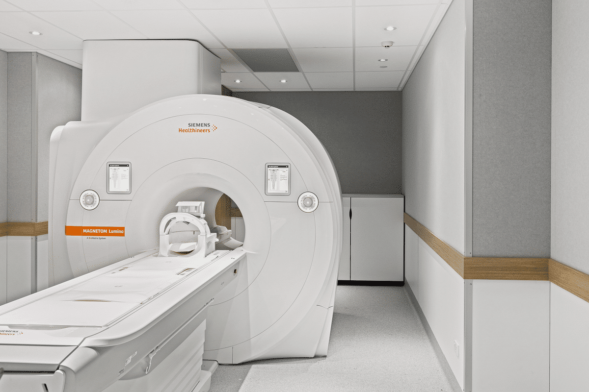
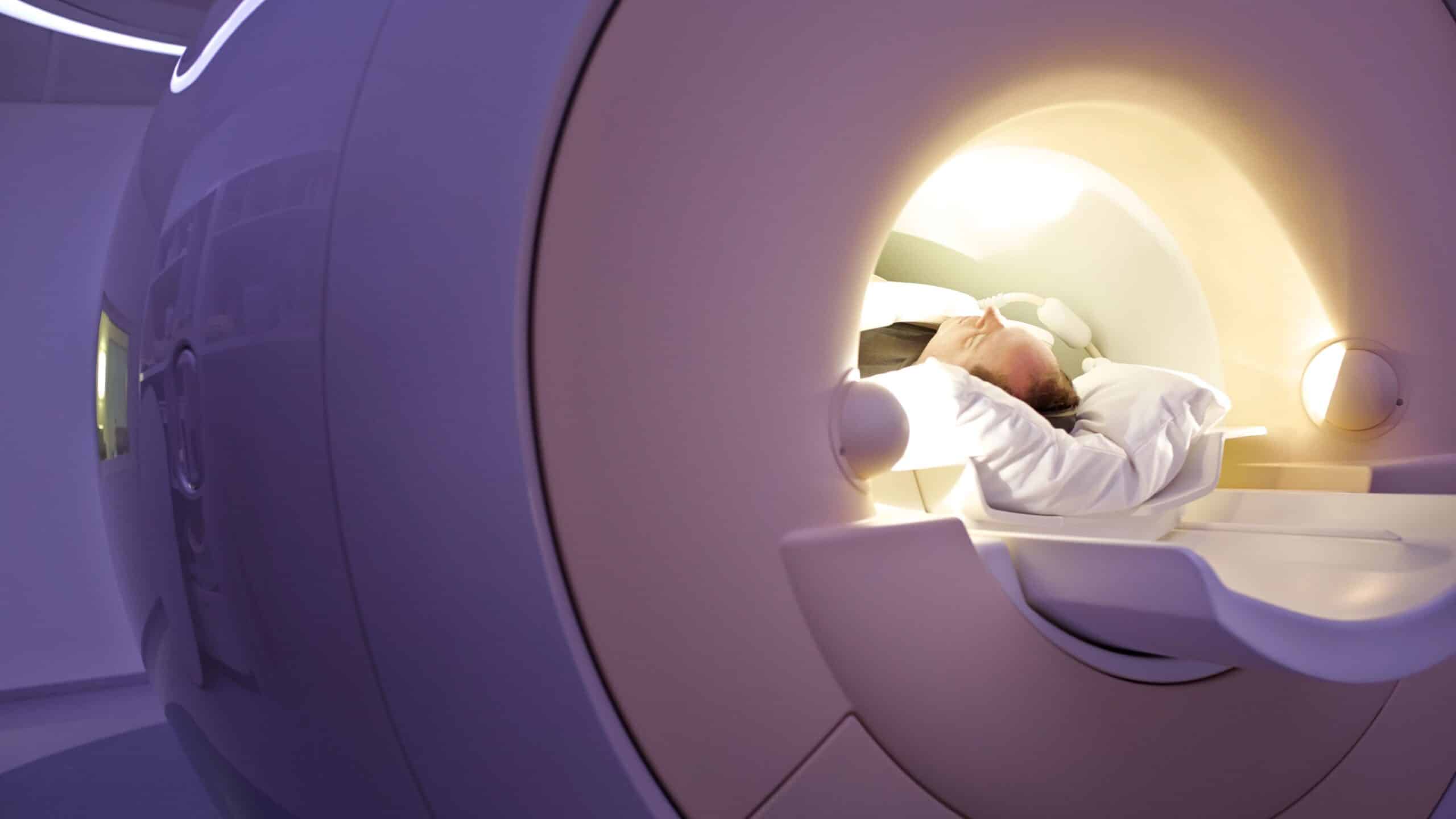
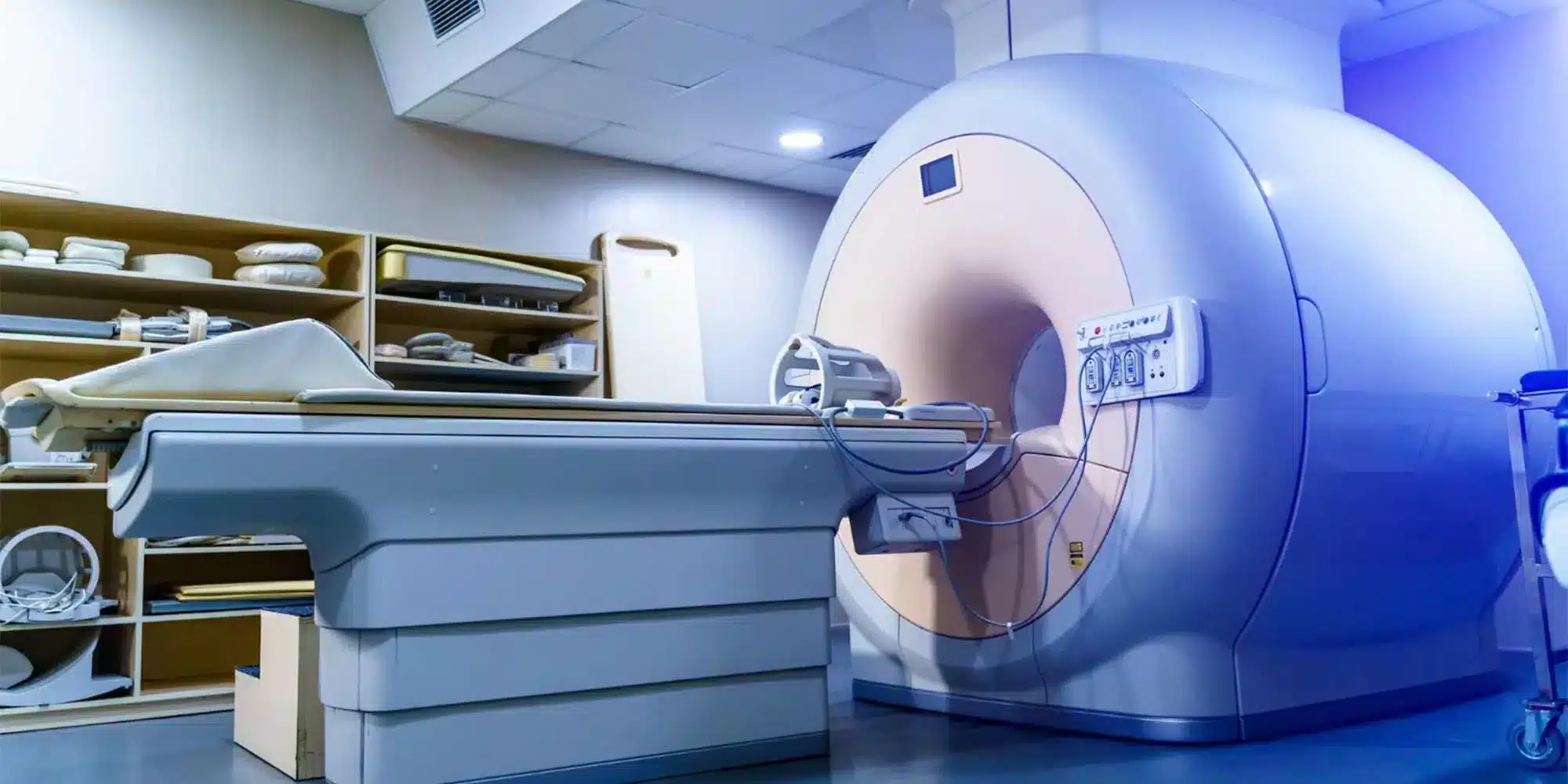
Magnetic Resonance Imaging (MRI) is a non-invasive imaging technique that produces detailed three-dimensional anatomical images, often used for detecting diseases, diagnosing them, and monitoring a patient’s treatment journey. It relies on an advanced technique that excites and detects changes in the orientation of the proton spins found in water, which forms the tissues of the body.

The first device used in magnetic resonance imaging

MRI machines use powerful magnets to create a magnetic field that reorients the protons in the body to align with it. When radiofrequency pulses are applied, the protons are stimulated, causing them to fall out of alignment. When the pulses are stopped, the machine measures the energy emitted as the protons realign with the magnetic field. The time it takes for the protons to realign, and the amount of energy emitted depend on the tissue characteristics and the chemical nature of the compounds. These differences help doctors distinguish between different tissues and diagnose medical conditions with greater accuracy.

The History of MRI and Its Development Over Time

The history of MRI technology dates back to the study of magnetic resonance and how the nuclei of electrons and atoms respond to magnetism. In the 1930s, physicist I.I. Rabi developed a technique for measuring the magnetic properties and movement of atoms, which laid the foundation for what is now known as Nuclear Magnetic Resonance (NMR), which became the basis for medical MRI.

In the 1940s, physicists Felix Bloch and Edward Purcell studied the magnetic resonance properties of materials, paving the way for the use of water content in the body to develop MRI images. In 1952, Purcell and Bloch were awarded the Nobel Prize in Physics for their work.

In 1969, Dr. Raymond Damadian proposed the possibility of using MRI to distinguish cancer cells from healthy cells, and he successfully demonstrated his hypothesis in mice. Damadian discovered that MRI could differentiate between tissues based on differences in relaxation times, leading him to develop a full-body MRI scanner. In 1972, Damadian filed the first patent for this technology, and in 1977, he captured the first MRI image of the human body, a cross-sectional image of his assistant’s chest, marking the beginning of MRI imaging as we know it today.

Applications of MRI Technology

The development of MRI technology has been a significant achievement in the medical field, allowing doctors and scientists to examine the inside of the human body in fine detail using a non-invasive tool. MRI is used to diagnose a wide range of conditions, including:

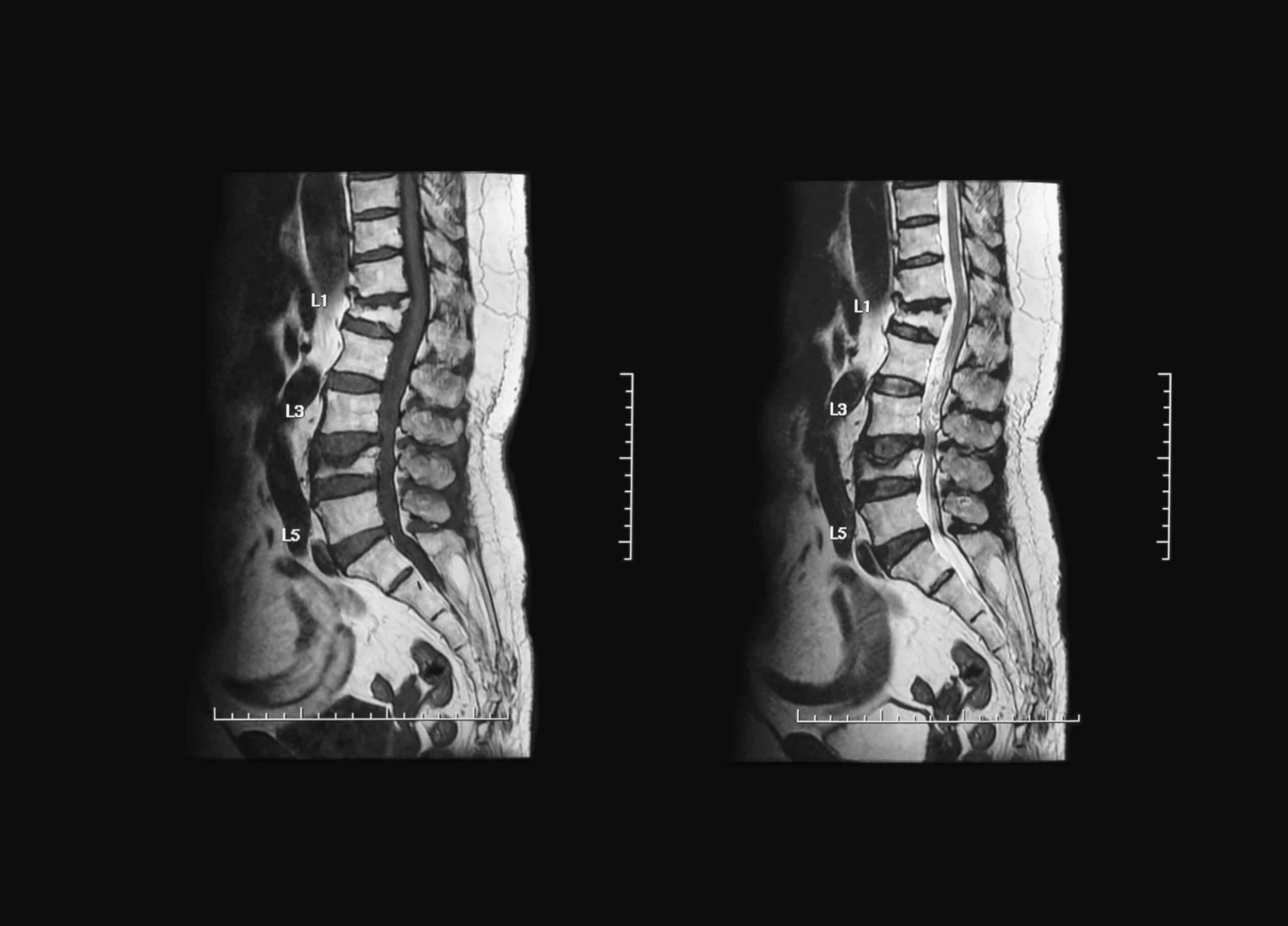
  • Brain and spinal cord abnormalities
  • Tumors, cysts, and lesions in different parts of the body
  • Early screening for breast cancer in women at high risk
  • Injuries or deformities of joints such as the back and knee
  • Certain types of heart problems
  • Liver diseases and other abdominal organ conditions
  • Assessment of pelvic pain in women, such as fibroids and endometriosis
  • Suspected uterine abnormalities in women undergoing infertility evaluation

The Importance of MRI in Medicine

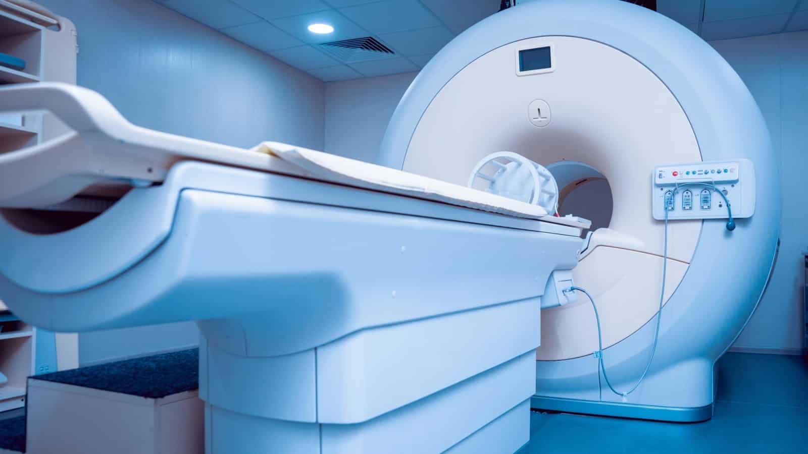
MRI stands out for its ability to capture detailed images of any part of the body from any imaging direction, making it an essential tool for providing high contrast of soft tissues compared to other imaging techniques like CT scans. This ability to distinguish between fat, water, muscles, and soft tissues helps doctors diagnose a variety of medical conditions more effectively.

MRI

Among the key benefits of MRI is its ability to provide detailed three-dimensional images of the targeted area, which enhances doctors’ ability to diagnose diseases accurately and efficiently. It also does not use radiation, as in X-rays or CT scans, making it a safer option for radiation-sensitive individuals such as pregnant women and children.

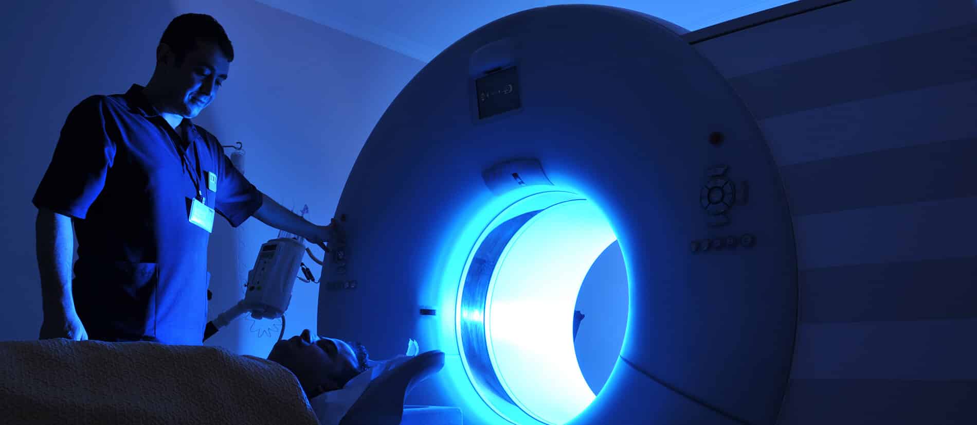
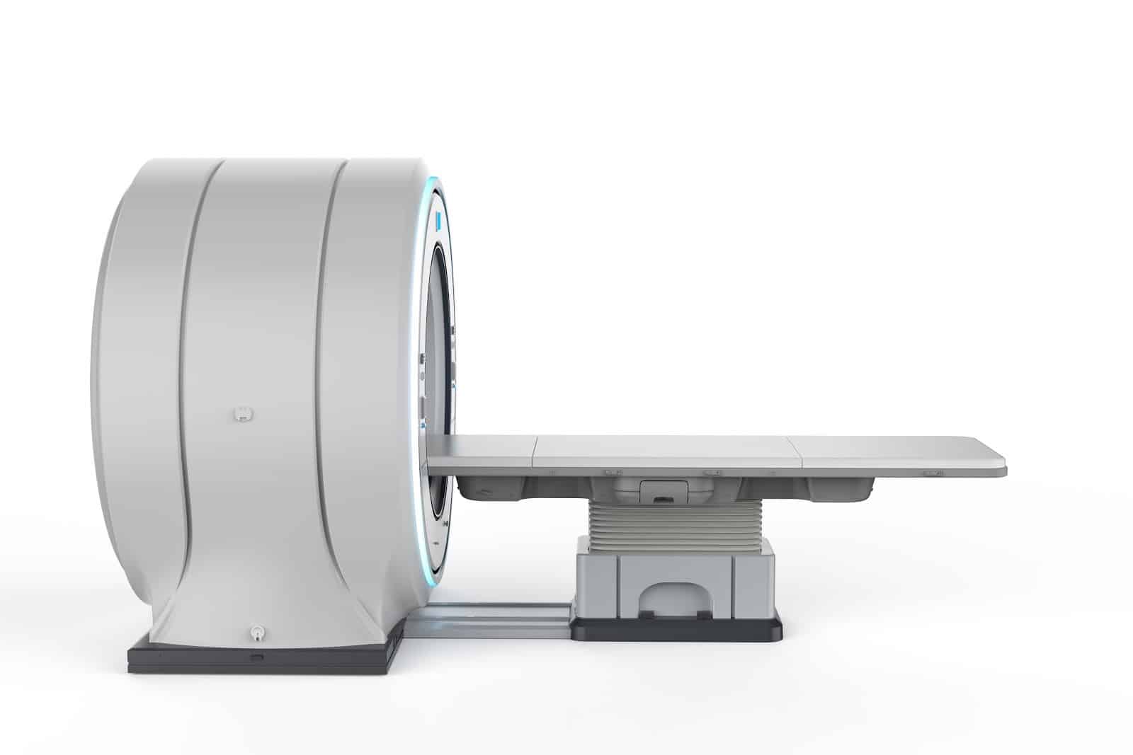
In addition, MRI scans have become faster and more comfortable thanks to recent technological advancements. Scans that typically take between 15 to 30 minutes provide accurate results quickly. Moreover, modern MRI machines with wide openings help reduce patient anxiety, making the scan more comfortable and secure.

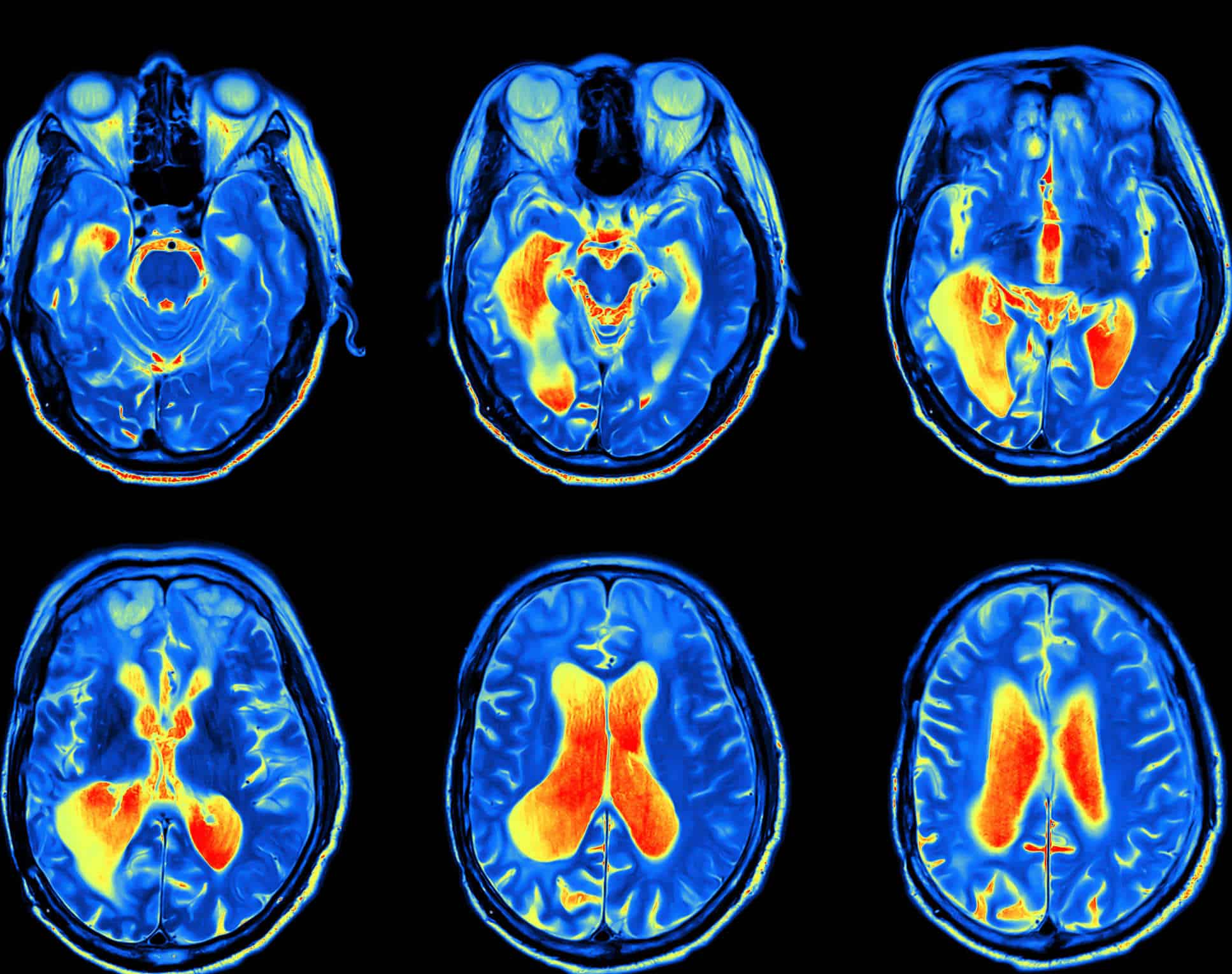
Functional MRI (fMRI) also measures what the tissues are doing rather than just their appearance, helping doctors assess brain activity. By identifying regions involved in vital functions like speaking, movement, sensation, or planning, fMRI can be used to accurately assess the risks of brain surgery.

Uses of Functional MRI

  • Determining the impact of tumors on the brain
  • Assessing the effects of strokes
  • Studying head and brain injuries
  • Monitoring neurodegenerative diseases like Alzheimer’s

Technological Advancements in MRI

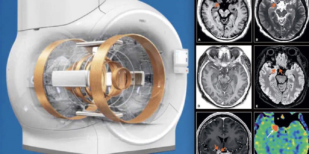
The field of MRI has seen remarkable progress with the emergence of new techniques that improve image accuracy and scan speed. High-field MRI systems such as 7T and 10T have been developed, offering higher image resolution and more detailed visuals than traditional systems with 1.5T or 3T. Additionally, fast imaging techniques have been developed that reduce scan time without compromising image quality. Functional MRI (fMRI) now enables real-time brain activity monitoring. There has also been significant advancement in imaging techniques for white matter in the brain, as well as advanced methods for examining brain tumors and other brain conditions. These developments help improve medical diagnoses and provide more accurate and faster results.

The future of MRI technology

Recent Innovations in MRI Technology

  1. Advancements in both device technologies and image sequencing have led to faster scan times. Techniques such as parallel imaging, simultaneous multi-slice imaging, and compressed sensing have contributed to reducing scan times while improving image quality.
  2. Functional Magnetic Resonance Imaging (fMRI) allows for direct monitoring of brain activity, providing new insights into blood flow and oxygen levels in the brain. This enhances our understanding of brain functions and disorders.
  3. Diffusion Tensor Imaging (DTI) visualizes the movement of water molecules within tissues, offering a clear view of connectivity within the brain’s white matter. This is particularly useful for detecting changes in conditions like multiple sclerosis and brain injuries.
  4. Magnetic Resonance Spectroscopy (MRS) is an advanced technique used to study brain tumors, strokes, neurological disorders, and other diseases affecting the brain.
  5. Advanced Coil Technology has led to improved designs such as array and surface coils, enhancing image quality and sensitivity. This, in turn, reduces scan times and increases diagnostic accuracy.
  6. Intraoperative MRI (iMRI) integrates MRI imaging with surgical procedures, providing real-time, live images during surgery to guide surgeons and ensure precision.
  7. Weight-Bearing MRI is performed when the patient is in a weight-bearing position, such as standing or sitting. This type of MRI is helpful for diagnosing disorders resulting from changes in posture or load bearing, such as spinal and joint diseases.

How Artificial Intelligence Impacts the Future of MRI Imaging

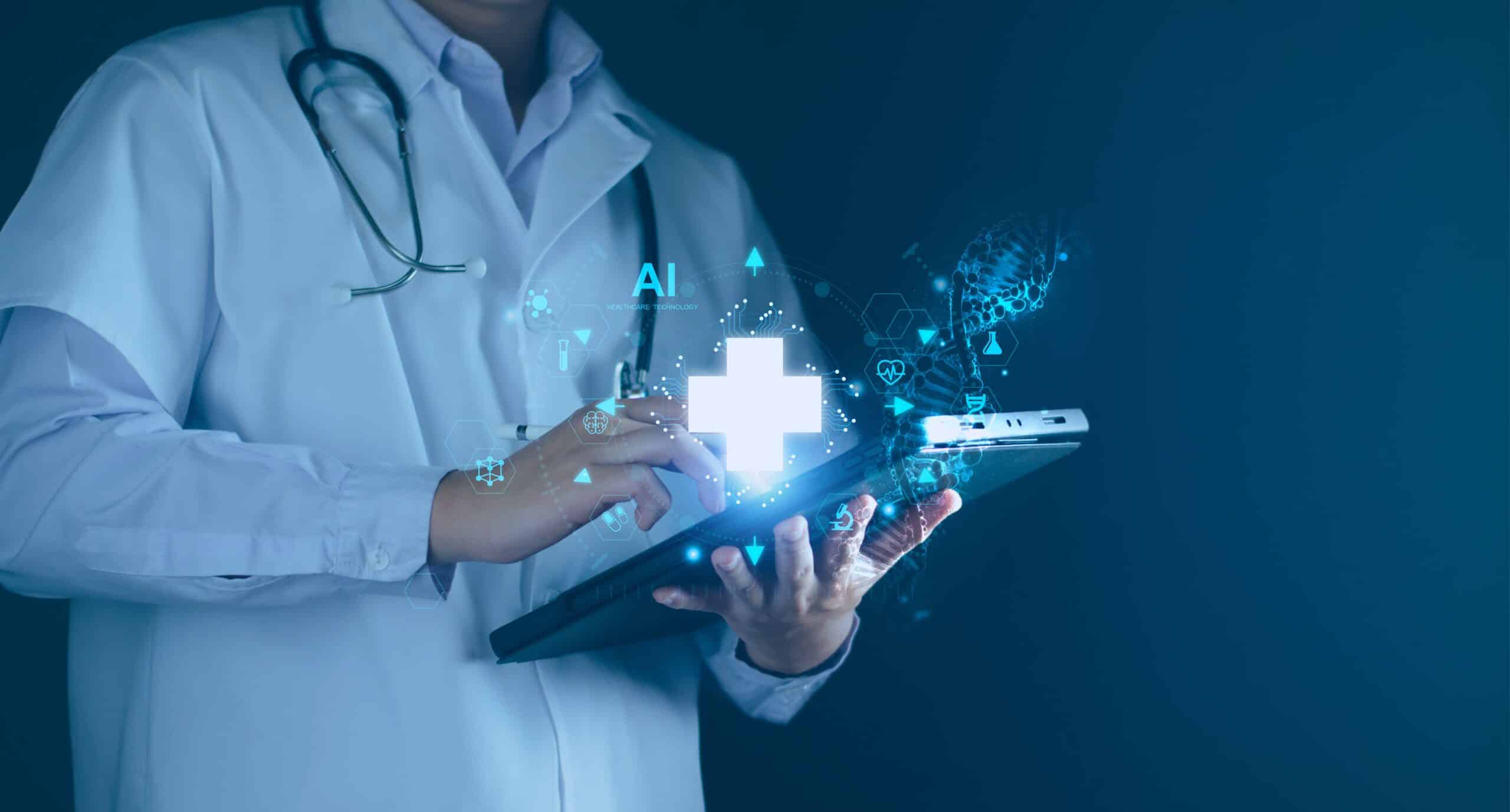
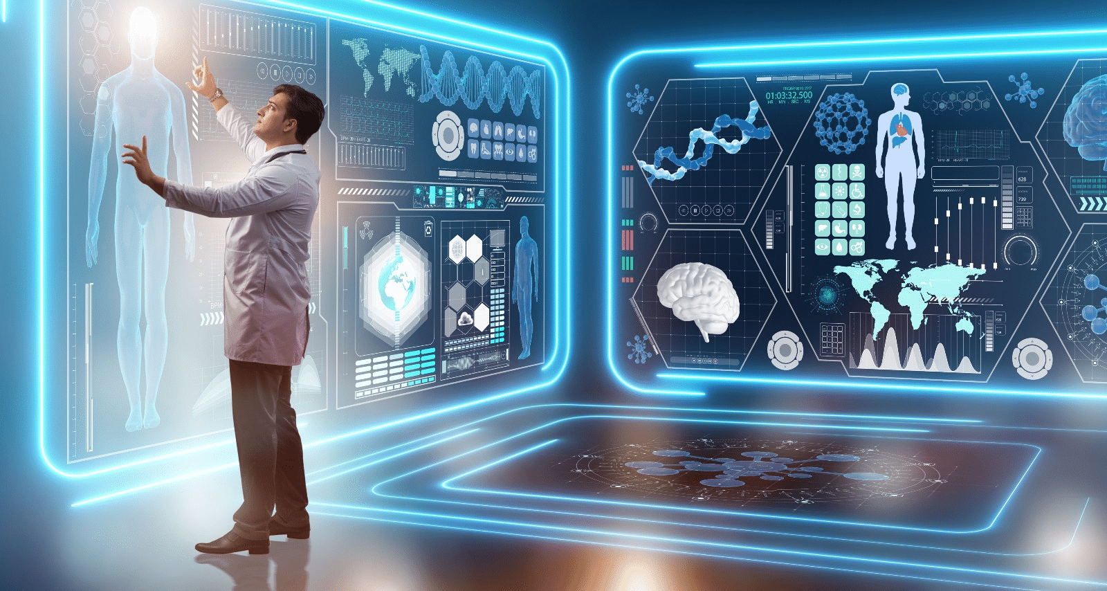
The integration of Artificial Intelligence (AI) into MRI technology represents a significant turning point in medical diagnostics. AI provides substantial improvements in image accuracy and quality, enhancing early and precise diagnosis. Algorithms such as machine learning and deep learning improve scan speed and efficiency, while also automating image analysis and reducing human errors. These advancements open up new possibilities for applying MRI in areas like oncology, neurology, and cardiology, allowing for more personalized and effective medical care.

How Artificial Intelligence Impacts the Future of MRI Imaging

Key Contributions of Artificial Intelligence in MRI

  • Improved Diagnostic Accuracy: AI enhances the precision of MRI image analysis, allowing for more accurate diagnoses.
  • Increased Efficiency: Through the automation of image analysis, AI reduces the time required for interpretation, enabling faster decision-making.
  • Personalized Medicine: AI offers tailored insights into patient health, aiding in the development of customized treatment plans.
  • In Oncology: AI assists in detecting tumors and distinguishing between benign and malignant lesions, improving cancer diagnosis and treatment.
  • In Neurology: AI helps in identifying early signs of neurological diseases such as Alzheimer’s and Parkinson’s, facilitating earlier intervention.
  • In Cardiology: AI is used to analyze heart images and guide treatment decisions regarding heart function and blood flow.
  • Patient Comfort: By reducing scan times and enhancing image quality without compromising accuracy, AI contributes to a more comfortable MRI experience for patients.

Future Directions in MRI Technology

Diagnostic imaging technologies are advancing rapidly, offering more precise, non-invasive methods for early disease detection. Notable developments in this field include artificial intelligence and machine learning, where algorithms are used to analyze medical images with a speed and accuracy that sometimes surpasses that of radiologists. These advancements allow for earlier detection of tumors and other diseases and help in tailoring treatments for individual patients based on their specific needs.

On the other hand, 3D and 4D Imaging provides more detailed views of the body compared to traditional 2D images, facilitating the diagnosis of complex conditions such as bone diseases and heart diseases. Additionally, Molecular Imaging allows physicians to visualize biological processes at the cellular level, aiding in the early detection of cancer.

Portable Point-of-Care Devices are also an emerging technology, enabling physicians to perform real-time scans close to patients, whether in hospitals or remote areas. In conclusion, the future of diagnostic imaging holds great promise for improving diagnostic accuracy, personalizing patient care, reducing costs, and expanding access to healthcare.

In conclusion, with the rapid advancements in the field of Magnetic Resonance Imaging (MRI), it has become essential to keep up with the latest technologies and modern practices to ensure accurate diagnoses and advanced healthcare. Therefore, HSI offers specialized courses in MRI techniques aimed at equipping medical professionals with the practical and scientific knowledge necessary to utilize this technology in the best possible way.

Source: The Future of Magnetic Resonance Imaging (MRI)

Revolutionizing Early Disease Detection: The Role of MRI in Medical Diagnostics

Magnetic Resonance Imaging (MRI) is a non-invasive diagnostic tool that uses a magnetic field and radio waves to produce detailed images of the inside of the human body. It plays a crucial role in the medical field and is considered a revolution in medical diagnostics. The future of MRI technology will see significant advancements in the clarity of the images it produces, and it is expected to develop further alongside the rise of artificial intelligence. In the following sections, we will explore this technology, its role in early disease detection, and how it will continue to evolve in the future.

الرنين المغناطيسي

MRI Technology
MRI works by exposing the body to a specific magnetic field. This magnetic field exploits the physics of protons, which are abundant in the human body due to the high water content in tissues. When the body is subjected to a strong magnetic field, the hydrogen protons in the tissues align with the field. Then, radio pulses are sent through the body, temporarily altering the orientation of these protons.

After a brief period, the hydrogen protons return to their original state and release signals that are captured by the MRI machine. These signals are used to create detailed images of the internal body structures, clearly distinguishing different tissues and organs and enabling precise identification of diseases.

How MRI Can Be Used for Early Disease Detection

MRI is an effective tool for early disease detection due to the high accuracy of its images of body tissues and organs. It can be used in the early stages of tumor development, increasing the chances of successful treatment. Additionally, MRI is valuable for diagnosing various neurological conditions such as strokes and multiple sclerosis. One of its key roles in neurology is identifying changes in brain tissue even before clinical symptoms appear, significantly aiding early detection and effective treatment.

الرنين المغناطيسي

One of the important uses of MRI is in the early detection of diabetic retinopathy, a complication of diabetes. This is done by creating a digital image of the retina and examining it for early signs of the condition.

MRI is also used for early detection of breast cancer, as traditional imaging methods may not detect the disease in its early stages. Furthermore, MRI can help detect possible artery blockages or narrowing that could lead to heart muscle problems. When it comes to bones, MRI can detect early signs of arthritis and changes in surrounding bone tissue.

Innovative Solutions in MRI Technology

While MRI itself is a revolutionary tool, it is continuously evolving to offer innovative solutions for disease detection and treatment. Some of the key innovations include:

Improved Image Accuracy

MRI imaging has been enhanced using mathematical equations, particularly by applying statistical science to improve the visibility of tumors. One of the methods used is calculating the probability mass function for each image in a given time sequence. This process involves analyzing the distribution of different values within the image, helping to identify changes in tissues and internal organs.

Another technique called Kullback-Leibler divergence is used to determine the best images for detecting tumors. This mathematical measure compares two different values, helping to identify the contrast between healthy tissue and a tumor, making the tumor more visible.

Advanced Image Processing Techniques

To improve image processing, advanced algorithms like GLCM (Gray Level Co-occurrence Matrix) and PNN (Probabilistic Neural Networks) are used to detect different brain tumors. These technologies have greatly advanced medical image processing, making it easier and more accurate for doctors to identify various tumor patterns for accurate diagnosis.

Use of Artificial Intelligence

The rapid growth of artificial intelligence tools in recent years will significantly enhance MRI technology. AI assists in accurately analyzing images and handling large data sets (Big Data) to recognize complex patterns, thereby improving doctors’ ability to diagnose diseases more accurately and quickly.

Challenges Facing MRI Technology

Despite the tremendous advancements in MRI technology and its applications, there are some challenges:

  • High Cost: MRI machines are expensive to purchase and maintain, and operational costs increase when advanced techniques are used.
  • Complexity: Some modern MRI techniques can be complex, requiring high skill levels and a deep understanding of how to operate the equipment.
  • Special Needs Patients: It can be difficult for patients with special needs to undergo MRI scans, as they may struggle to stay still during the examination.
  • Patient Anxiety: Some patients may be fearful of being confined in small spaces for extended periods, requiring doctors to take extra time to reassure them.
  • Long Exam Duration: Some MRI scans take 30 to 90 minutes, which can be uncomfortable, especially for elderly patients.

The Role of Contrast Agents in MRI

Contrast agents are chemicals injected into the body before an MRI scan to enhance image clarity and improve the accuracy of details around tissues and internal organs. Contrast agents play a crucial role in MRI:

  • Increasing contrast to highlight differences between tissues, helping to distinguish between healthy and diseased tissue.
  • Certain liquid agents, such as gadolinium, are used to improve visibility of tissues.
  • Gaseous contrast agents are used in specialized cases, such as lung imaging, to interact with oxygen or carbon dioxide for clearer images.
  • They also clarify blood vessels and examine blood flow for conditions like aneurysms.
  • They assist in detecting tumors.
  • They help examine internal organs like the liver and kidneys for any pathological changes.

Innovative Contrast Agents in MRI

Over time, the contrast agents used in MRI have been developed to provide the best possible image quality and diagnostic accuracy. Some of the innovative contrast agents include:

Gadolinium-based agents, which are the most commonly used in central nervous system imaging.
Iron-based agents, typically used for liver and vascular imaging.
Manganese-based agents, which are highly effective at targeting specific organs for precise imaging.
These agents are essential components of MRI techniques, and specialists choose the appropriate agent for each specific case.

With the tremendous advancements in the medical field over recent years, it is crucial for biomedical engineers to continuously enhance their skills and expertise. This is where platforms like HSI come into play, offering courses and consultations in the medical field to help you advance in your academic journey.

on our HSI website, we offer specialized training courses and consultations in the field of biomedical engineering, led by a team of certified experts and specialists to ensure you receive the best scientific content that combines theory and practical application. For inquiries, please get in touch with us, and we will respond with all the information you need to deepen your knowledge in the field of biomedical engineering.

Source: How Diagnostic Imaging is Revolutionizing Early Disease Detection

Soft Skills for Biomedical Professionals

Continuously developing personal skills is of utmost importance for everyone. When it comes to the skills required in the medical and healthcare field, this becomes even more critical. soft skills are a key component of the success of professionals working in the medical sector. While practical skills are essential, personal skills are what make performance holistic and consistently progressive. When we talk about personal skills, it’s not only about individual abilities but also about fostering team skills as a whole. In the following lines, we will explore the essential personal skills that all healthcare professionals should possess and their importance.

Soft Skills

The Role of Personal Skills in the Medical Field

Possessing and continuously developing personal skills in healthcare greatly enhances the quality of both individual and team performance, contributing to comprehensive patient care. With the rapid and immense advancements in the medical field in recent years, it has become crucial to improve skills such as effective communication within the healthcare team, critical thinking, and time management to achieve the best possible outcomes in delivering essential healthcare services to patients. It is far better for a patient to be treated by a medical team that excels in communication and interaction than by another team that lacks these vital skills, even if both have the same level of technical expertise.

Key Soft Skills for Success in the Medical Field

Below are the critical personal skills that every individual in the medical and healthcare field must acquire and develop. These skills will elevate you to an outstanding level of professionalism, earning the trust of patients, and colleagues, and enhancing your self-confidence in tackling challenges with expertise.

1. Effective Communication

Effective communication is one of the most important personal skills in the medical field, as it helps reduce errors and improve collaboration among all members of the medical team. This skill is not limited to the medical team but extends to patients as well. It is crucial to communicate with patients in a simple, easy-to-understand language that matches their level of comprehension and addresses their needs. This helps build patients’ trust in healthcare providers. Every individual in the medical field should be capable of simplifying complex concepts and conveying information effectively to both their team and patients.

2. Critical Thinking and Decision-Making

Critical thinking is crucial across all fields today, particularly in the medical sector. It involves the ability to analyze situations and problems and devise innovative solutions based on available data. In healthcare, professionals often encounter complex issues that require diverse approaches to resolve. Thus, developing this skill is essential for overcoming challenges through non-traditional methods.

3. Coordination and Collaboration Across Specializations

Success in the medical field heavily relies on collaborating and working within a multidisciplinary team. For a productive team environment, good communication and mutual respect among all team members are essential to achieve shared goals. Collaboration fosters an exchange of diverse ideas, facilitates problem-solving, and addresses challenges creatively, ultimately improving the quality of healthcare provided to patients.

4. Time Management and Prioritization

Time management and prioritization are fundamental personal skills for all healthcare professionals. Prioritizing tasks significantly helps balance responsibilities and avoid stress or burnout caused by accumulating duties. Developing planning and time management skills enhances productivity and ensures tasks are completed within set deadlines.

5. Empathy and Support for Patients

This skill closely aligns with communication but focuses specifically on offering adequate psychological support to both medical teams and patients. Demonstrating empathy towards patients and actively listening to their needs can greatly improve the quality of healthcare provided, instilling a sense of trust and comfort in patients. Similarly, supporting fellow healthcare team members improves workplace morale and overall performance.

6. Staying Updated with Technological Advancements

With the rapid advancement in medical technology, keeping up with these developments is indispensable for professionals in biomedical sciences. In recent years, artificial intelligence has revolutionized various fields. Attending conferences and workshops provides opportunities to learn about the latest innovations and share knowledge with experts. Additionally, online educational resources offer a flexible way to self-learn, helping professionals stay up to date in this dynamic sector.

How to Choose Skills Based on Career Path

In the healthcare sector, identifying the personal skills that need development should align with your specialization and career path. For example, if you are an emergency room physician, skills like quick decision-making under pressure and time management are crucial for optimal performance in your role. On the other hand, for nurses, effective communication and empathy are pivotal in enhancing patient care.

In areas requiring advanced technology, such as radiology or laboratory diagnostics, focusing on technological proficiency becomes paramount. Assessing your professional needs and understanding the challenges in your role is essential for selecting the most impactful skills for your career. Remember, developing personal skills is a continuous process requiring dedication and commitment. By improving these skills, you can achieve better performance, ensure patient satisfaction, and contribute effectively to enhancing the quality of healthcare services.

How to Develop Personal Skills

After identifying the key skills to focus on, the next step is developing them. Improving personal skills involves setting clear professional goals and enhancing areas that require growth. This can include attending training courses to refine skills such as communication and time management, engaging in professional discussions, and exchanging experiences with colleagues. Regular performance evaluation helps identify strengths and weaknesses, enabling continuous improvement.

To streamline this process, explore the training and educational services provided by HSI. These courses and workshops are designed to equip you with all the skills needed for tremendous success in your professional journey.

Source: Top 10 Soft Skills for Healthcare Professionals

All copyrights reserved to © HSI