Understanding CE Marking for Medical Devices | A Beginner’s Guide

Selling and marking medical devices in European Union (EU) countries requires them to bear the CE marking. Medical device manufacturers must comply with regulatory requirements to ensure their products meet quality and safety standards. International regulations and standards related to medical devices, such as the European MDR 2017/745 and ISO 13485:2016, can be complex but are essential. Non-compliance with these regulations and standards may result in manufacturers ceasing production altogether. In this article, we provide a guide to understanding the key regulations for obtaining the CE marking.

What is the CE marking?

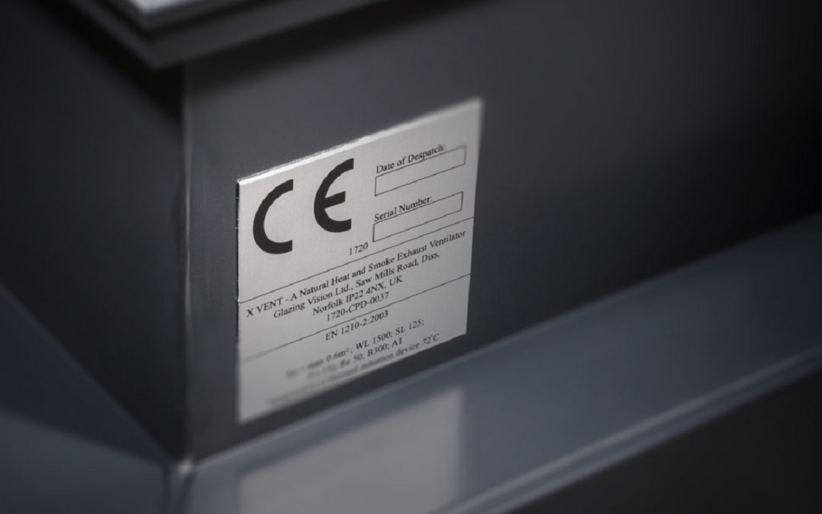
The CE marking stands for Conformité Européenne (European Conformity in French). As the name suggests, it certifies that a product complies with European requirements ensuring safety, health, and environmental protection standards. While this marking is required for many products, for medical devices, the CE marking allows companies to distribute and sell their devices across the 30 countries in the European Economic Area (EEA). This is possible once they comply with the European Regulation 2017/745, also known as the Medical Device Regulation (MDR). This regulation governs the placement of medical devices on the Markin get for human use. Therefore, the presence of the CE letters on medical devices signifies that the product meets all legal requirements for distribution throughout the EEA.

Understanding CE Marking for Medical Devices

Why is the CE marking Important for Medical Devices?

All medical devices that comply with European legislation can obtain the CE marking. The CE marking indicates that a medical device has undergone risk assessment procedures and is a safe and high-quality product for patients.

A product cannot be legally sold or markingeted in EEA countries without this marking—except for devices exclusively used for research purposes, which are an exception. Moreover, compliance with this regulation provides opportunities for many companies worldwide to expand their business.

For example, during the COVID-19 pandemic, numerous infrared thermometers entered the European markinget, regardless of the manufacturer’s country of origin. The CE marking indicated that these devices complied with the requirements and could be legally sold and markingeted in EEA countries.

Here are some key benefits of obtaining the CE marking:

  • Confirms that your device meets the essential legal requirements of the EU.
  • Allows markingeting in all 30 EEA member countries.
  • Some non-EEA countries recognize the CE marking, providing an advantage in entering new markingets.
  • Demonstrates compliance with safety and quality standards.

Standards and Regulations for CE markinging

Understanding the regulations and standards related to medical devices is crucial. Below, we outline the key international standards and regulations necessary for a better understanding of the CE markinging process.

1. Regulation (EU) 2017/745

Regulation (EU) 2017/745, also known as the European Medical Device Regulation (MDR), is the current regulation replacing the previous Medical Device Directive (MDD) and the Active Implantable Medical Devices Directive (AIMD) entirely.

2. Directive 2001/83/EC

Directive 2001/83/EC concerns the placement of medicinal products for human use on the markinget. When medical devices are combined with a medicinal product, such as a drug, manufacturers must determine which component is responsible for the primary function of the combined product.

If the drug enhances the activity of the medical device and cannot be used separately, it becomes an integral part of the device. The combined product is then classified as a medical device and must comply with Regulation (EU) 2017/745.

Understanding CE Marking for Medical Devices

3. Regulation (EC) 276/2004

Regulation (EC) 276/2004 concerns the placement of medicinal products for human and veterinary use on the markinget. For medical devices, it functions similarly to Directive 2001/83/EC.

4. Directive 2004/23/EC

Directive 2004/23/EC establishes quality and safety standards for the donation, procurement, testing, processing, preservation, storage, and distribution of human tissues and cells. Medical devices containing non-viable tissues or cells with a secondary function must comply with MDR.

The general safety and performance requirements in the MDR must be applied to the part of the device containing these elements, regardless of their primary function.

5- ISO 13485:2016

The ISO 13485:2016 standard defines the requirements for a quality management system (QMS) for medical devices. Medical device manufacturers often adhere to this standard, as compliance with it is assumed to align with the QMS requirements in the MDR.

Compliance with this standard ensures adherence to quality management system requirements, including:

  • Quality manual
  • Document and record control
  • Quality management system
  • Human resources
  • Facility structure
  • Contamination control
  • Design, development, and transfer planning
  • Medical device files
  • Supplier evaluation and selection
  • Service activities
  • Sterile medical device requirements
  • Medical device identification and traceability
  • Complaint handling
  • Nonconforming product control
  • Post-markinget surveillance

ISO 13485-2016

6- ISO 14971:2019

ISO 14971:2019 is specifically developed for medical device manufacturers based on principles for applying risk management to medical devices. It serves as a guide for developing and maintaining risk management processes.

Risk management is a requirement in the MDR. However, manufacturers can achieve compliance without necessarily obtaining certification under this standard.

7- FDA 21 CFR Part 820

FDA 21 CFR Part 820 outlines the quality system requirements applicable to medical device manufacturers. Companies aiming to enter the U.S. markinget must have a quality management system (QMS) that complies with FDA 21 CFR Part 820 and obtain FDA approval.

This standard can serve as a guide for MDR quality management system requirements in the European markinget. However, most companies opt to follow ISO 13485:2016, as certification for it can be obtained.

FDA 21 CFR Part 820

Steps to Obtain the CE marking for Medical Devices

The process of obtaining a CE marking can be somewhat complex. To assist you, this guide outlines the general steps to acquire it.

1. Determine the Medical Device Classification

Identify the classification guidelines set by the MDR based on risk level, body placement, and duration of use.

Risk: Devices are categorized into Class I, IIa, IIb, and III. The higher the class, the greater the risk posed to the patient.

Body placement: Devices can be non-invasive (on the body’s surface) or invasive (penetrating the body).

  • Duration of use: Devices are classified as:
  • Transient use: Up to 60 minutes.
    • Short-term use: Up to 30 days.
    • Long-term use: More than 30 days.

2. Appoint a Person Responsible for Regulatory Compliance (PRRC)

Medical device manufacturers must designate at least one individual responsible for regulatory compliance within the company. This person should have expertise in the medical device field.

3. Implement a Quality Management System and Risk Management

The MDR requires manufacturers to have quality management and risk management systems in place. This is why medical device manufacturers choose to comply with ISO 13485:2016, as it is presumed to align with MDR requirements for a quality management system.

MDR

4. Prepare Technical Documentation

Technical documentation, also known as medical device technical files, contains detailed information about the lifecycle of your medical device and is a requirement under the MDR.

5. Implement a Supplier Management System

The MDR requires medical device companies to have a supplier management system in place. Suppliers must be audited to ensure compliance with requirements and standards. Creating a list of approved suppliers based on predefined criteria is useful to ensure that only qualified suppliers provide products and services.

6. Conduct a Clinical Evaluation

Manufacturers must conduct a clinical evaluation to demonstrate compliance with safety and performance requirements. In practice, this means developing a plan to collect and analyze clinical data from relevant scientific literature and clinical investigations involving the specific medical device or an equivalent product.

7. Appoint an Authorized Representative in Europe (if required)

If the medical device manufacturer is not based in the European Economic Area (EEA), they must appoint an authorized representative within an EEA member state.

The authorized representative is responsible for tasks such as:

  • Verifying technical documentation
  • Informing the manufacturer of complaints
  • Registering a physical location for the notified body to receive device samples for inspection

8. Obtain Certification from a Notified Body

A notified body is an independent organization responsible for assessing product compliance before it is placed on the markinget. For medical devices, the notified body audits manufacturers and issues certifications confirming compliance with the MDR.

For higher-risk medical devices, this certification is mandatory, specific to each procedure, and valid for a maximum of five years. After this period, the notified body will audit the manufacturer’s quality management system (QMS) and technical documentation to verify continued compliance with the MDR.

Understanding CE Marking for Medical Devices

9. Prepare a Declaration of Conformity

After obtaining certification from the notified body, manufacturers must prepare a Declaration of Conformity (DoC), taking responsibility for ensuring that the device meets the specific requirements of the MDR.

10. Register Your Device with a Unique Device Identifier (UDI)

To facilitate the traceability of medical devices, each device model must be assigned a Unique Device Identifier (UDI).

The UDI is a unique numeric or alphanumeric code stored in the European Database on Medical Devices (EUDAMED), where essential information about the device can be accessed. The UDI is an additional requirement and does not replace the CE marking or other labeling requirements.

11. Affix the CE marking on the Medical Device

After obtaining approval from national authorities and certification from the notified body, manufacturers can place the CE marking on their medical devices.

The CE marking must be displayed on:

  • The device itself
  • The packaging
  • Any instructions for use

It is essential that the CE marking is visible, legible, and made of a material that cannot be removed or washed off.

For Class II and Class III medical devices, the four-digit number of the notified body must also be printed next to the CE marking.

12. Maintain Post-markinget Surveillance

Before obtaining the CE marking and placing a medical device on the European Economic Area (EEA) markinget, manufacturers must demonstrate that a post-markinget surveillance (PMS) system has already been implemented to address safety and effectiveness concerns.

Medical device companies must collect data on their sold devices through post-markinget surveillance, vigilance activities, and markinget monitoring plans.

This includes feedback related to patient experience with the medical device and the product lifecycle.

Manufacturer Requirements Include:

  • Monitoring complaints, adverse events, and non-conformities
  • Regularly updating safety reports
  • Conducting internal and supplier audits regularly
  • Keeping technical documentation, databases, and records up to date

This surveillance ensures proactive actions to collect and review real-world evidence on device quality and safety. As a result, manufacturers can better handle customer complaints, identify risks, and implement product recalls and implementing product recalls and other market actions.

Medical Devices

Frequently Asked Questions About the CE marking

Here are some common questions about the CE marking for medical devices, which ensures compliance with safety, health, and environmental protection standards.

1- Is the CE marking the same as FDA approval?

Both the European CE marking and the U.S. Food and Drug Administration (FDA) approval aim to evaluate the safety and effectiveness of medical devices. However, they are only valid in their respective markingets.

2- How long is the CE marking valid?

The validity of the CE marking is determined by the notified body and depends on the classification of the medical device. However, it cannot exceed five years. After that, the device must undergo recertification.

For example, a Class IIa device may receive certification valid for only three years. Additionally, annual surveillance audits are conducted between certification renewals.

3- Can a CE marking be placed on a medical device product?

Class I medical devices that are non-sterile and non-measuring can self-declare compliance. However, higher-class devices must be assessed by notified bodies to obtain the CE marking.

4- How long does it take to get CE marking approval?

The approval timeline for the CE marking varies depending on the device’s classification and complexity, as well as whether the manufacturer already has a certified Quality Management System (QMS) under ISO 13485:2016. Generally, obtaining CE marking approval takes between 16 to 18 months from start to finish.

5- How many notified bodies are designated for CE markinging?

According to the NANDO database, as of 2022, there were 34 notified bodies accredited for MDR and 7 for IVDR.

Source: CE markinging for Medical Devices [Step-by-Step Guide]

The Role of AI in the Development of Smart Prosthetics and the Challenges They Face

Over the past few years, artificial intelligence (AI) and smart prosthetics have begun transforming early prosthetic solutions. These technologies are not merely futuristic concepts but practical tools that are already enhancing how individuals embark on their prosthetic journeys.​

Understanding Early Smart Prosthetics and Their Challenges

Before delving into the specifics of this technology, it’s crucial to understand what early smart prosthetics are and their significant importance. These are the initial prosthetic devices provided to patients shortly after surgery, following wound healing and before the residual limb has fully stabilized.​

The Role of AI in the Development of Smart Prosthetics

The Role of Artificial Intelligence in Enhancing Prosthetic Movement

Imagine a prosthetic limb that not only mimics movement but also learns, adapts, and responds as if it were a natural extension of the body. This is what AI technologies offer in the field of prosthetics, revolutionizing their functionality by enabling more intelligent and fluid movements that adapt to the user’s needs. From predictive algorithms to machine learning, AI is fundamentally transforming prosthetics, making them more efficient and their functions more realistic and human-like.​

1- Processing and Adaptation

AI enhances prosthetic movements by processing vast amounts of data collected from sensors embedded in the device. These sensors monitor muscle signals, joint angles, and external conditions such as surface type or temperature. The AI system analyzes this data in real-time, allowing for precise adjustments to ensure smooth movement and immediate responsiveness.

2- Learning and User Adaptation

Machine learning, a branch of AI, drives smart prosthetics capable of adapting to individual user needs. By analyzing repetitive movements, the prosthetic can learn user patterns and understand their movement preferences. Over time, the device becomes more intelligent, anticipating the user’s intentions even before they are executed.

For example, an AI-powered prosthetic arm can distinguish between muscle signals associated with grasping a water cup and those for holding a pen, allowing for seamless transitions between tasks, making the device a natural extension of the body rather than just an assistive tool.

3- Enhancing Balance and Improving Gait

In lower-limb smart prosthetics, AI plays a crucial role in maintaining balance and improving gait patterns. By analyzing how the user distributes their weight and moves across different surfaces, the prosthetic can make precise adjustments to ensure stability.

For instance, walking on a sandy beach requires a different pattern than walking on a flat sidewalk. A smart prosthetic leg can detect these changes and adjust its stiffness, angle, or stride length, accordingly, reducing fatigue and ensuring the user’s safety.

Smart Prosthetics

The Importance of Early Prosthetic Fitting

Fitting a prosthetic limb at an early stage plays a crucial role in the recovery process, as it helps to:​

  • Reduce swelling​
  • Improve blood circulation​
  • Prevent joint stiffness​
  • Allow the patient to begin standing and moving​
  • Preserve muscle strength and balance​

Difficulties in Early Device Fitting

One of the biggest challenges facing early smart prosthetic fittings is the continuous change in the shape of the residual limb. Swelling may increase and decrease, the skin may be sensitive, and the shape of the residual limb is not yet final. Therefore, prosthetic sockets need to be adjusted as healing continues, making it difficult to design a socket that is both snug and comfortable.​

A socket that is too tight may cause pain or injury, while one that is too loose may lead to instability and increase the risk of imbalance. Thus, achieving the right fit requires a delicate balance and continuous care, where smart technology can make a real difference in prosthetic design.​

The technology behind smart prosthetics

​The technology behind smart prosthetics relies on a combination of hardware components and software to function efficiently. Sensors, actuators, and processors work together to collect data, analyze it, and execute precise movements.​

Advanced Algorithms

AI algorithms transform raw sensor data into actionable insights. For instance, a machine learning model can analyze thousands of steps to determine how a user’s gait changes when moving uphill versus downhill. The algorithm uses this knowledge to adjust the prosthetic’s movement automatically, ensuring a more seamless experience.​

Companies developing smart prosthetics should prioritize the accuracy and efficiency of these algorithms. Collaborating with data scientists and software engineers can help refine these systems, ensuring reliable performance under various conditions.​

Artificial foot

Sensor Integration

Sensors are the cornerstone of AI-powered prosthetics, gathering the necessary data to enable the system to learn and interact. These sensors include:​

  • Myoelectric Sensors: Capture electrical signals emitted by muscles.​
  • Gyroscopes: Measure stability and assist in adjusting movement.​
  • Pressure Sensors: Monitor weight distribution to ensure natural walking patterns.​

The data collected by these sensors is transmitted to an integrated processor within the prosthetic limb, where AI algorithms analyze it in real time. This allows the device to respond immediately to any changes in the user’s movement or surrounding environment, providing a smoother and more effective user experience.​

How AI is Transforming Early Prosthetic Design

AI brings speed, precision, and predictive capabilities to the prosthetic design process, facilitating a faster and more accurate match between the prosthetic design and the individual. This is achieved through:​

1- AI-Assisted Socket Design

In traditional socket design, a prosthetist uses measurements and molds to create a well-fitting socket. With smart prosthetics, it’s now possible to digitally scan the limb and input this data into advanced modeling software. This software predicts how the limb will change over time and suggests socket shapes that can adapt accordingly.​

2- Predicting Limb Volume Changes

One of the most challenging aspects of early prosthetic fitting is the continuous change in limb volume, which may shrink or shift weekly. AI can now track these changes using data from sensors and user inputs, even predicting future changes.​

3- Matching Appropriate Components to Each User

AI’s role extends beyond socket design to selecting the appropriate foot or knee for each user. Based on gait patterns, strength, age, and goals, AI can suggest prosthetic combinations that offer the best balance between support and flexibility. This enhances the success of the initial fitting, helps the user move faster with fewer adjustments, reduces trial time, and accelerates the transition to a more stable walking pattern.​

How AI is Transforming Early Prosthetic Design

The Future of Smart Prosthetics

The future of smart prosthetics extends beyond the devices themselves to building integrated systems that enhance user experience by incorporating complementary technologies.​

For example, mobile applications can serve as auxiliary tools, allowing users to monitor their prosthetics’ performance, adjust settings, and access tutorials or troubleshooting guides. These apps can also collect valuable data for companies, providing insights into how users interact with their devices and identifying areas for improvement.​

Additionally, wearable devices like smartwatches or fitness trackers can enhance functionality. By synchronizing with AI-powered prosthetics, these devices can provide real-time feedback on activity levels, body posture, and gait, helping users make informed decisions about their health and mobility.​

Expanding Access by Reducing Costs

While smart prosthetics offer advanced functionality, they often come with high production costs. To make these devices accessible to a broader range of users, companies should adopt cost-effective manufacturing techniques and explore alternative materials.​

Technologies like 3D printing can play a pivotal role in reducing production expenses. By leveraging additive manufacturing, companies can quickly produce complex prosthetic components at a lower cost. When combined with AI, these components provide the same high-level performance as traditionally manufactured parts.​

In conclusion, integrating AI into prosthetic design represents a revolutionary step toward improving users’ quality of life, offering them more natural movements and an enhanced user experience. In this context, HSI Center stands out as a leader in biomedical engineering and healthcare solutions, offering specialized training courses and professional consultations aimed at empowering professionals and organizations to make impactful contributions to the healthcare industry. ​

​The source: How AI and Smart Prosthetics Are Improving Early Fit Prosthetic Solutions.

AI in Drug Discovery: How Machine Learning Accelerates Pharmaceutical Research

The development of new drugs is a complex, resource-intensive process with a high failure rate. In this context, leveraging Artificial Intelligence (AI) and Machine Learning (ML) holds tremendous potential to revolutionize the pharmaceutical industry. These technologies enhance data analysis and predictive modeling, leading to faster and more effective treatments. In this article, we explore the role of AI in drug discovery.

Challenges of Traditional Drug Development

The journey of developing new drugs is typically fraught with challenges. It can take 12 to 15 years, involves a high failure rate, and may cost billions of dollars to bring a single drug to market. Key reasons for these difficulties include:

  • Incomplete understanding of disease complexities.
  • Existing disease models often lack accuracy.
  • Difficulty translating model results into effective treatments.
  • Declining reliability of animal testing as a predictor for human outcomes, along with growing ethical concerns.

Drug Discovery

How AI Addresses These Challenges

Recent advancements—especially in machine learning—have significantly contributed to drug discovery. In the past, scientists spent extensive time on manual tasks like sample analysis and data collection. Now, AI aims to automate and speed up these processes.

Although automation is not a new concept, its integration with AI is a game-changer. AI allows companies to sift through and analyze vast datasets to extract precise insights about drugs. Data management and analysis—pillars of pharmaceutical R&D—are now more efficient, enabling researchers to overcome many recurring obstacles.

A Paradigm Shift in Pharmaceuticals: AI as an Innovation Driver

The pharmaceutical industry is undergoing a fundamental transformation by integrating AI into the complex drug discovery process. As is well-known, developing a new drug requires significant financial investment and years of research. However, integrating AI into pharmaceutical R&D is helping to reduce both the massive costs and extended timelines. Here’s how AI is contributing to this evolution:

1. Accelerating Drug Discovery

AI integration speeds up the transition of drugs from the design stage to clinical trials. Recent analyses show that some AI-powered companies have advanced drug candidates to human trials in less than ten years, with some developed in just two and a half years. A notable example includes a leading medication for respiratory diseases that swiftly reached clinical trials thanks to AI technologies.

2. Digital Compounds and Molecular Visualization
AI’s impact extends into the digital realm, where it analyzes vast molecular libraries and protein structures using tools like 3D databases and large-scale integrations. This accelerates the discovery process from years to just months. Key benefits include:

  • Rapid evaluation of millions of potential molecules.
  • Intelligent search for drug compounds.
  • Designing new molecules using generative models.

AI in Drug Discovery

3. Predictive Analytics and Intelligent Systems

To reduce clinical trial failure rates, AI offers powerful tools such as:

  1. Text analysis of medical records using Natural Language Processing (NLP).
  2. Protein structure prediction and chemical compound optimization.
  3. Analysis of genomic data and biosignals from wearable devices to generate actionable insights.
  4. These tools are also used to understand drug effects post-launch, improving treatment strategies.

4. Identifying Disease Pathways and Therapeutic Candidates

Tackling complex diseases like Alzheimer’s requires precise identification of new therapeutic targets. AI is now employed to analyze genetic data and protein interactions, helping to uncover these targets. It has even facilitated the repurposing of existing drugs—like diabetes medications—to reduce Alzheimer’s risk.

AI Within the Regulatory Framework

AI, especially machine learning, is transforming drug discovery and development. Prior to AI integration, getting FDA approval was a long and costly process. But AI is gradually changing this landscape.

The FDA’s Stance on AI

The U.S. Food and Drug Administration (FDA) has shown a positive attitude toward adopting AI technologies in the pharmaceutical industry. Highlights include:

  • Acknowledgment of AI’s value: The FDA has published position papers outlining its vision for AI in drug development.
  • Surge in submissions: Over 100 AI/ML-related applications were submitted to the FDA in 2021.
  • Risk-based framework: The FDA aims to strike a balance between public safety and innovation potential while safeguarding patient data and drug safety.

AI in Drug Discovery

AI Integration in Pharmaceutical Companies

Incorporating AI into pharmaceutical operations can lead to a major leap in how drugs are discovered and developed. Combining AI with big data analytics has proven to reduce costs and speed up development.

How Can Pharmaceutical Companies Benefit from AI?

  • Cost reduction: Potential to save billions in expenses.
  • Increased efficiency: Enhances R&D productivity through data automation.
  • Advanced technologies: Techniques like Virtual Ligand Screening (VLS) and protein databases help in quicker compound selection.
  • Predictive modeling: Forecasting protein structures and identifying the most suitable therapeutic classes.
  • Regulatory compliance: Ensuring AI usage aligns with FDA guidelines.

Steps for Integrating AI into Pharma

  1. Conduct thorough analysis to identify areas where AI offers the most value.
  2. Develop a protocol for AI implementation aligned with company values and regulations.
  3. Train staff to use AI tools and technologies.
  4. Prepare and test AI models to ensure alignment with business objectives.
  5. Scale usage based on company growth and market needs.

Benefits of AI in Pharmaceuticals

  • Lower operational costs: Reports indicate up to a 50% reduction in hiring costs.
  • Improved task accuracy: Accuracy levels can reach 90% for certain functions.
  • Advances in personalized medicine: Ability to design drugs based on individual genomes and predict future health issues.

Conclusion, in an era of rapid technological advancement, the pharmaceutical industry can no longer overlook the immense capabilities of AI. No longer just a supportive tool, AI has become a strategic partner in accelerating innovation, cutting costs, and improving the success rate of discovering safe and effective treatments. Companies that proactively adopt and intelligently integrate these advanced technologies into their strategies will lead the future and gain a true competitive edge in a market demanding faster responses and more accurate decisions.

Source: AI in Drug Discovery: Accelerating Pharmaceutical Breakthroughs

The Role of AI in the Early Detection of Diseases through Medical Imaging

AI’s Role in Early Disease Detection via Medical Imaging
The use of AI in medical imaging represents a major breakthrough in integrating technology into healthcare. This integration is not just a technological advancement, but a qualitative leap in enhancing diagnostic and treatment plans. AI is leveraged in the early detection of diseases through advanced imaging techniques. In this article, we will discuss this role and how it contributes to improving healthcare.

The Importance of AI in Early Disease Detection

We are not exaggerating when we talk about the importance of AI in healthcare and medical imaging. It goes beyond merely improving image quality and processing speed. AI capabilities in medical imaging have evolved from basic diagnostics to aiding in early disease detection. It’s no longer just about faster and clearer images, but also about saving lives through early intervention and providing personalized treatment options.

Early Detection of Diseases

These advancements have had a significant impact on healthcare, especially in fields like oncology and neurology, where timely and accurate diagnosis is critical. More specifically, these innovations have significantly increased the number of patients undergoing screenings, enhancing not only patient outcomes and safety but also productivity and cost-effectiveness.

AI Capabilities in Medical Imaging

AI technologies have a multifaceted impact on medical imaging. They improve diagnostic accuracy, reduce human error, and enable early disease detection, ultimately raising the standard of patient care. AI can be utilized in various ways to enhance medical imaging.

The full potential of AI can be realized through the effective use of machine learning and deep learning algorithms, which are now playing a pivotal role in advancing imaging technologies.

AI Improvements in Medical Imaging

  1. One of the most notable advancements is image quality enhancement. AI algorithms can analyze massive volumes of imaging data, learning to detect subtle patterns and anomalies that may be invisible to the human eye. The increased clarity and resolution of these images have paved the way for more accurate diagnoses, such as the ability to detect fractures.
  2. Image processing speed is another area where AI makes a significant difference in early disease detection. In the past, analyzing medical images was time-consuming, which affected the timing of diagnoses and treatment.
  3. Thanks to its ability to quickly process and analyze large datasets, AI significantly reduces the time needed to analyze medical images. As a result, AI systems can now interpret and generate complex images much faster than traditional methods, speeding up the diagnostic process.
  4. By integrating deep learning algorithms, AI can identify patterns associated with specific diseases, making it a powerful tool for early detection and diagnosis. Through advanced image analysis, AI can detect abnormalities such as tumors in their early stages, greatly enhancing the patient experience.
  5. AI also plays a major role in personalized medicine and preventive healthcare. By analyzing medical imaging data alongside a patient’s medical history, AI can provide personalized diagnostic insights, paving the way for more tailored treatment plans.

Key Applications of AI in Medical Imaging

Current AI applications in medical imaging are significantly impacting fields such as radiology, oncology, and neurology. By harnessing AI, medical professionals in these areas can improve the accuracy and efficiency of numerous diagnoses and treat medical conditions more successfully.

These three fields demonstrate how AI applications in medical imaging are not only enhancing current diagnostic and treatment methods but also opening new horizons in personalized medicine. From improving diagnostic accuracy in radiology to aiding in the early detection of cancerous cells and neurological diseases, AI is proving its value in the healthcare sector.

Below are the key fields that benefit from AI in the early detection of diseases.

1. Radiology

In the field of radiology, for example, artificial intelligence is primarily used to identify abnormalities in imaging scans. AI algorithms applied to computed tomography (CT) and magnetic resonance imaging (MRI) can quickly detect and segment areas of interest in scans of the lungs, liver, and brain.

2. Cancer Tumor Detection

Particular attention should be given to AI’s role in tumor imaging. It assists in detecting and monitoring cancerous tumors, improving the accuracy of cancer screenings. For instance, deep learning AI techniques have been used to analyze mammograms and accurately distinguish between malignant and benign tumors.

Early Detection of Diseases

3. Neurology

In neurology, AI enhances the analysis of brain images for conditions such as stroke, Alzheimer’s disease, and brain tumors. AI algorithms can detect subtle changes in brain tissue, allowing for early intervention and treatment. A notable example is the use of AI in diffusion-weighted imaging, a type of MRI, to quickly identify stroke-affected areas in the brain.

AI’s Impact on Diagnostic Accuracy

The integration of AI into medical imaging analysis has significantly improved diagnostic accuracy. It reduces human error and enhances early disease detection. This shift toward AI-enhanced diagnostic and imaging methods is reshaping the landscape of medical imaging technology and precision medicine. Below are the key impacts:

1. Improved Diagnosis of Various Medical Conditions

Thanks to its ability to learn from large datasets, AI algorithms have shown remarkable improvement in identifying and diagnosing medical conditions. For example, AI can match—or even surpass—the accuracy of human radiologists in detecting diseases like pneumonia in chest X-rays. This level of precision is especially critical in cases where early detection greatly influences treatment outcomes.

2. Reduction of Human Error

The second major advantage of AI in medical imaging is its consistency, which significantly reduces human error. Unlike human radiologists who may be affected by fatigue or subjective judgment, AI systems provide consistent analyses. AI support can reduce diagnostic errors in mammograms for breast cancer, thereby improving diagnostic accuracy.

Medical Imaging

3. Early Disease Detection

Early disease detection is crucial for effective treatment, and AI is playing a vital role in this area. For example, AI has been instrumental in the early detection of diabetic retinopathy—a condition that can lead to blindness if left untreated. By analyzing retinal images, AI can detect this condition with high accuracy, often before any physical symptoms appear in the patient.

Challenges and Ethical Considerations

Despite the tremendous potential of artificial intelligence in the healthcare sector and medical imaging technologies, it still faces a number of challenges and concerns that slow down its widespread adoption. These challenges include:

1. Data Privacy

The first challenge affecting AI is the concern over data privacy. AI systems require access to large datasets of patient images, which raises significant concerns about confidentiality. Ensuring the security and privacy of this sensitive data is absolutely critical. Healthcare institutions must comply with strict data protection regulations, as safeguarding this information is essential.

2. High Costs

The cost of integrating AI into existing standard imaging technologies presents a major barrier to its adoption. Acquiring AI software, along with the necessary hardware upgrades, can be extremely expensive for many healthcare institutions—especially public ones.

3. Training Medical Staff

For AI to be successfully used in medical imaging, it also requires specialized training for healthcare professionals. Radiologists and technicians must be educated on how to interpret AI results and incorporate them into clinical decision-making processes.

4. Technical Challenges

Technical issues—such as data integration and algorithmic bias—pose significant challenges. Integrating AI into current health IT systems can be complex and demands seamless compatibility. To address this, AI systems must be trained on diverse and comprehensive datasets and undergo rigorous validation processes to ensure accuracy and fairness.

It is essential to ensure the transparency of AI-supported systems in decision-making processes and to clearly define accountability for the outcomes of those decisions. Regulatory frameworks must be established to guarantee the safe and effective use of AI in the medical field. These frameworks should address key issues such as:

  • Validating algorithm reliability.
  • Official certification and approval.
  • Managing potential biases within the systems.

In conclusion, integrating AI into medical imaging is not merely a technical advancement, but a promising paradigm shift that redefines diagnosis and treatment planning. The positive impact of AI integration in imaging systems extends beyond patients, medical practitioners, and healthcare providers—it benefits anyone seeking a more efficient and error-free healthcare system.

Source: The Role of AI in Medical Imaging

The Role of AI in Predictive Maintenance of Medical Equipment

The medical field is undergoing an unprecedented revolution through the integration of artificial intelligence technologies. This innovative advancement is transforming the way doctors diagnose diseases, develop new treatments, perform surgeries, and even maintain medical equipment.

Unexpected equipment failures can lead to significant issues in healthcare institutions. But what if there were a way to predict a malfunction before it occurs? That’s exactly what we’ll explore in this article on the role of AI in predictive maintenance of medical equipment—aimed at reducing the risks of catastrophic or costly problems before they happen.

What is Predictive Maintenance in Medical Equipment?

Healthcare facilities typically rely on scheduled preventive maintenance or immediate repairs when equipment breaks down. However, this approach may not always be effective, often leading to sudden equipment downtime that negatively impacts patient care. This is where predictive maintenance comes in—a proactive approach designed to anticipate and prevent equipment failures.

Predictive Maintenance of Medical Equipment

Predictive maintenance is an efficient solution that relies on automation and AI to foresee equipment failures before they occur. Modern medical equipment is often equipped with sensors that collect real-time data on performance metrics. This data is then fed into AI algorithms that analyze trends and detect potential issues. Through continuous monitoring of equipment conditions, these AI-powered systems can predict possible malfunctions and schedule maintenance interventions in advance.

One of the key techniques used in this type of maintenance is condition-based monitoring, where sensors gather data on equipment health and performance. Machine learning then analyzes this data to detect early warning signs of potential issues, enabling maintenance teams to act proactively.

Predictive maintenance in medical equipment helps:

  • Avoid costly and unplanned downtimes
  • Reduce unnecessary maintenance tasks
  • Extend equipment lifespan by preventing premature failures
  • Improve maintenance scheduling

AI in Medical Equipment Maintenance

The role of AI in predictive maintenance of medical equipment goes beyond simply integrating technology to ensure smooth operations, prolong equipment life, and enhance patient safety. AI remains a driving force in the development of medical technologies. The integration of AI advancements positions predictive maintenance as a critical element in transforming healthcare equipment management and paving the way for a new era of patient-centered care.

By embedding AI algorithms, medical equipment can be programmed to perform tasks independently or with minimal human intervention, setting a new standard in the reliability and efficiency of medical services.

How Artificial Intelligence Works in Predictive Maintenance of Medical Equipment

Artificial intelligence in predictive maintenance relies on a complex process involving predictive analytics, machine learning, and data collection. Therefore, understanding the fundamental AI concepts is essential to grasp how machines learn.

Data Collection and Analysis

The role of AI in predictive maintenance of medical equipment is a modern approach to collecting and analyzing data from medical devices. AI uses sensors installed on medical equipment to continuously collect data, then analyzes trends to detect issues before they become critical.

Predictive Analytics

As part of AI’s role in predictive maintenance of medical equipment, machine learning and data analysis are used to predict the behavior of equipment. This helps reduce risks and enables swift action in healthcare operations.

Predictive Maintenance of Medical Equipment

Supervised Learning

AI models trained on labeled datasets improve the accuracy of predicting equipment failures, with a focus on enhancing AI’s role in medical devices.

Unsupervised Learning

AI is essential for gaining deep insights from medical device data analytics, as it can identify anomalies and unexpected issues without prior labeling.

Reinforcement Learning

This involves training an agent to interact with an environment and learn optimal actions through trial and error. It can be used to enhance maintenance scheduling or resource allocation strategies to minimize unplanned downtime and boost reliability.

The Role of AI in Predictive Maintenance

AI tools are designed to simulate human intelligence in performing specific tasks, allowing these tasks to be completed automatically instead of manually. In the case of predictive maintenance, AI solutions can analyze current operating conditions and search for indicators that a device might fail in the future—even if no apparent operational problems exist. By assessing the current performance against baseline data, AI tools can detect slight efficiency drops that signal the need for maintenance.

The role of AI in predictive maintenance of medical equipment includes the following:

1. Generating Work Instructions

Generative AI can analyze historical maintenance data, equipment specifications, and sensor readings to create customized work instructions for technicians. These instructions are clear, precise, and tailored to the specific needs of each maintenance task, minimizing errors and improving first-time fix rates.

2. Optimizing Work Order Planning

By analyzing historical trends and predicting potential equipment failures, generative AI integration helps schedule maintenance activities more efficiently. This allows preventive maintenance to occur before failures, reducing downtime and associated costs.

3. Supporting Reliability Engineering

AI helps analyze sensor data and maintenance history to identify patterns indicating potential equipment degradation. This enables proactive maintenance actions, ensuring efficient equipment operation and avoiding unexpected failures.

Predictive Maintenance of Medical Equipment

4. Analyzing and Applying Maintenance Standards

AI can be trained on industry standards and regulations related to equipment maintenance. This allows it to review existing maintenance plans and identify areas that may not comply with current standards—improving safety and regulatory compliance.

5. Updating Maintenance Quality

AI can continuously learn and improve from new data. As it analyzes outcomes of completed maintenance tasks, it can identify areas for procedural improvement or where new techniques may be applied. This ongoing feedback loop supports continuous enhancement of the quality and effectiveness of maintenance practices.

Benefits of AI-Enhanced Predictive Maintenance

AI-enhanced predictive maintenance significantly contributes to reducing maintenance costs through proactive measures that address potential failures before they occur. Algorithms analyze sensor and monitoring system data to detect early warning signs, enabling timely intervention to prevent breakdowns and reducing the need for costly emergency repairs.

It helps extend the lifespan of equipment by addressing issues early and avoiding unnecessary stress on the machines. This leads to fewer replacements and minimized downtime.

It improves operational efficiency by optimizing maintenance schedules, allowing maintenance teams to accomplish more with fewer resources, lowering labor costs, and increasing overall technician productivity.

Predictive Maintenance of Medical Equipment

Applications of AI in Predictive Maintenance of Medical Equipment

AI-based predictive maintenance is an advanced step toward improving the quality and efficiency of healthcare by reducing unexpected failures and ensuring the continuous operation of critical medical equipment. In this context, the applications of AI in predictive maintenance of medical devices fall into three main areas: medical imaging equipment, diagnostic devices, and life support systems. Each area includes a range of devices that benefit from AI’s ability to predict failures, enhance performance, and reduce sudden downtime that may negatively impact the quality of healthcare services provided to patients.

MRI (Magnetic Resonance Imaging) Machines

AI-powered predictive maintenance for MRI machines involves continuous monitoring of components to ensure optimal performance and reduce downtime. AI algorithms analyze data patterns to predict potential issues, enabling immediate intervention and avoiding delays in diagnosis.

X-Ray Machines

Predictive analytics, component deviation detection, and failure forecasting are key areas where AI is applied to X-ray machines. By taking a proactive approach, AI helps prevent unplanned interruptions and ensures smooth diagnostic operations.

Blood Analyzers

Real-time monitoring of baseline parameters is a key aspect of AI-driven predictive maintenance for blood analyzers. AI systems forecast potential failures by studying data trends, allowing issues to be addressed before they impact diagnostic accuracy.

ECG (Electrocardiogram) Devices

AI is used in predictive maintenance of ECG machines to monitor device components and signal quality. This proactive approach ensures the continuous availability of accurate cardiac data, aiding in fast and reliable patient diagnosis.

Ventilators

Predicting issues related to airflow, pressure, and sensor performance is a core application of AI in maintaining ventilators. Proactive monitoring ensures the reliability of life support systems, which are critical in emergency care.

Infusion Pumps

AI-powered predictive maintenance for infusion pumps includes monitoring fluid delivery accuracy and pump calibration. AI predicts potential issues by analyzing usage patterns, ensuring the accuracy and continuity of medication dosages.

Source: How AI Is Used in Predictive Maintenance

Wearable Medical Devices: How AI is Shaping the Future of Healthcare

In recent years, the healthcare sector has witnessed groundbreaking technological advancements, with the integration of artificial intelligence (AI) bringing a paradigm shift in patient care. One of the most notable developments is wearable medical devices, which are revolutionizing healthcare. These innovative devices not only transform how we monitor and manage our health but also unlock new possibilities in medical research and personalized medicine. In this article, we will explore these advancements in detail.

What Are Wearable Medical Devices?

Wearable medical devices are small, portable electronic devices worn on the body, capable of collecting, analyzing, and transmitting a wide range of health data. These devices can monitor various vital signs, such as heart rate, blood pressure, and temperature, providing real-time information that can be used for early disease detection or chronic condition management. With technological advancements, these devices have become more sophisticated, offering features such as ECG monitoring, continuous glucose monitoring (CGMs), and even sleep tracking.

Wearable Medical Devices - How AI is Shaping the Future of Healthcare

Benefits of AI-Powered Wearable Medical Devices

The integration of AI in wearable medical devices is transforming healthcare by providing patients and healthcare providers with continuous, data-driven insights. This personalized approach enhances patient care, particularly in managing chronic diseases and promoting preventive health measures. By tailoring interventions to individual needs, AI-powered wearables improve patient outcomes, enhance healthcare services, and empower individuals to take control of their health. Here are some key benefits of incorporating AI into these devices:

1. Enhanced Monitoring and Early Detection

AI-powered wearable devices continuously monitor vital signs and detect irregularities, enabling early detection of health risks such as arrhythmias or sudden changes in blood sugar levels—often before noticeable symptoms appear. This allows timely interventions and has shown promising results in detecting atrial fibrillation, potentially preventing strokes.

2. Personalized Treatment Plans

By analyzing individual health data, AI tailors’ treatment recommendations to meet specific patient needs, optimizing medication dosages, exercise routines, or dietary adjustments for more effective care. For instance, AI can adjust insulin doses for diabetics based on real-time glucose readings or suggest lifestyle modifications for hypertension patients.

3. Better Chronic Disease Management

Continuous monitoring enables more effective management of chronic diseases, particularly where one-size-fits-all treatments often fall short, such as diabetes, hypertension, and cardiovascular conditions. This reduces complications and hospital admissions, leading to more responsive care.

Wearable Medical Devices - Future of Healthcare

4. Empowering Patients

AI-powered wearable medical devices enhance patient engagement by providing real-time health insights, personalized health tips, and reminders, enabling individuals to manage their health more effectively.

5. Improved Patient Outcomes

By facilitating early interventions and continuous data-driven analysis, wearable devices improve long-term healthcare outcomes. Preventing severe complications, reducing hospital admissions, and minimizing emergency visits ultimately enhance patients’ quality of life.

6. A Key Role in Preventive Medicine

AI-powered medical devices play a crucial role in preventive medicine. Through continuous monitoring of vital signs and daily activities, these devices help individuals identify potential health risks and make informed decisions to prevent illnesses. For example, a wearable device tracking physical activity and sleep patterns can provide valuable insights into lifestyle habits that may contribute to obesity or sleep disorders.

7. Enabling Individuals to Take Charge of Their Health

Patients can easily track their progress and share data with their doctors, facilitating informed decision-making in diagnosis and treatment. For example, individuals with chronic conditions can use wearables to monitor symptoms and make necessary lifestyle or medication adjustments, ultimately leading to better health outcomes and quality of life.

How Wearable Medical Devices Support Medical Research

The benefits of wearable medical devices extend beyond patients; they also offer significant advantages to the medical research community. In clinical trials, these devices play a crucial role in collecting accurate and reliable data. Traditional data collection methods often rely on self-reported information or intermittent measurements, which can be prone to errors. In contrast, wearable devices provide continuous and objective data, offering a more comprehensive view of a patient’s health.

By integrating these devices into clinical trials, researchers can gather real-time data on how patients respond to treatments, identify potential side effects, and monitor adherence to prescribed therapies. This wealth of information allows for more precise assessments of the efficacy and safety of new treatments, ultimately accelerating drug development and bringing new medications to market faster.

How Do Wearable Medical Devices Work?

Wearable medical devices are equipped with sensors that continuously collect data on vital signs, physical activity, and other health metrics. This data is then transmitted to a smartphone or another connected device for analysis and interpretation. Some devices provide real-time feedback, while others store the information for healthcare professionals to review later.

Wearable Medical Devices

Types of Wearable Medical Devices

  1. Wearable medical devices come in a variety of forms, catering to different health needs and preferences. Here are some common examples:
  2. Fitness Trackers: Monitor physical activity, heart rate, sleep patterns, and calorie consumption.
  3. Smartwatches: Many smartwatches include health monitoring features, such as heart rate tracking, blood pressure monitoring, and ECG functionality.
  4. Continuous Glucose Monitors (CGMs): Essential for individuals with diabetes, these devices provide real-time blood sugar readings without the need for finger pricks.
  5. Wearable ECG Monitors: Detect irregular heart rhythms and other cardiac issues, enabling early intervention and heart attack prevention.
    Smart Rings: Compact devices offering various health monitoring features, including heart rate tracking, blood oxygen levels, and sleep analysis.
  6. Wearable Biosensors: Advanced devices capable of measuring multiple biomarkers, such as stress levels and skin temperature.
  7. AI-Powered Devices: Enhance data analysis, allowing for precise health monitoring and predictive insights.
  8. Sleep Management Devices: Track sleep patterns and provide actionable insights to improve sleep quality and overall health.
  9. Wearable Hearing Aids: Advanced hearing aids and smart earbuds that offer hearing assistance, health tracking, and seamless connectivity.
  10. Smart Glasses: Enhance vision, provide real-time data, and assist in rehabilitation or accessibility solutions.
  11. Wearable Robots: Aid mobility and rehabilitation, supporting physical therapy and improving movement.
  12. Smart Clothing: Garments embedded with sensors to track physical activity, posture, and health metrics.

The Future of Wearable Medical Devices

The field of wearable medical devices is advancing rapidly, and we can expect to see smaller, more comfortable devices with expanded capabilities. Integration with artificial intelligence (AI) will enable more sophisticated data analysis and personalized recommendations. Moreover, wearable technology will play an increasingly vital role in managing chronic diseases, monitoring mental health, and providing remote patient care.

How AI is Shaping the Future of Healthcare

Challenges Facing Wearable Devices

While wearable medical devices significantly enhance healthcare, they also face several challenges that must be addressed:

  1. Data Privacy and Security: Protecting sensitive health data is crucial to prevent unauthorized access.
  2. Regulatory Compliance: The development and commercialization of these devices must adhere to strict regulatory guidelines to ensure safety and effectiveness.
  3. User Adoption: Ensuring that individuals wear and use these devices correctly is essential for accurate data collection and effective health management.
  4. Battery Life: Limited battery life can be inconvenient and hinder continuous monitoring.
  5. Data Accuracy: These devices rely on sensors to monitor vital signs, but external factors such as body movement, environmental conditions, or sensor placement can impact data accuracy.

Conclusion

Despite these challenges, wearable medical devices have revolutionized healthcare by offering innovative solutions for monitoring, diagnosing, and treating a wide range of health conditions. With advancements in sensor technology, AI integration, and personalized healthcare, these devices are becoming more accurate, user-friendly, and accessible. As they continue to evolve, they hold great promise for improving patient outcomes, enhancing healthcare management, and reducing the burden on traditional healthcare systems.

source: Wearable Medical Devices: The Future of Healthcare

Biomechanics and the Role of Artificial Intelligence in Prosthetic Design

The technological integration in biomechanics and the role played by machine learning and artificial intelligence in prosthetic design have become a boon for individuals with disabilities. Leading manufacturers of rehabilitation aids have utilized the concept of neural networks to simulate various anatomical and biomechanical functions of missing human body parts. The involvement of human interaction with various factors such as electronic circuits, software, robotics, etc., has had a revolutionary impact in the field of rehabilitation, leading to the development of devices like prosthetic legs, human body-controlled prosthetics, and exoskeletons. In this article, we will discuss the role of Biomechanics and artificial intelligence in prosthetic design and rehabilitation.

Prosthetics: Innovation in Medical Compensation

Prosthetics include any artificial device that replaces a missing body part due to injury, disease, or congenital defects. Some companies have developed various types of prosthetics, ranging from purely cosmetic to advanced devices made using titanium and carbon fibers. Regarding the manufacturing of these prosthetics using biomechanics, the process is quite similar to making medical insoles. The end of the amputated limb is digitally scanned, and then a special socket is designed to which the prosthetic is attached. Using electrodes, electrical signals from the muscles are received and sent to an electronic control board inside the prosthetic, which translates these signals into actual movements.

Biomechanics

Understanding Prosthetic and Orthotic Engineering

The field of prosthetics focuses on creating artificial limbs, while orthotic specialists design devices to support or correct musculoskeletal conditions. Traditionally, this field relied on manual measurements, mechanical adjustments, and trial-and-error methods. Although these techniques were effective, they often lacked precision and adaptability to achieve optimal results.

Artificial Intelligence in Prosthetic Design is changing this landscape by integrating machine learning, robotics, and sensing technology. These tools transform static devices into dynamic systems capable of learning and evolving based on individual user needs. The result is a new level of functionality that mimics natural movement and intelligently responds to unique biomechanical challenges.

History of Artificial Intelligence in Prosthetics and Orthotics

The first intelligent prosthetic limb was developed by Charles A. Blatchford & Sons, Ltd in 1993, with an improved version in 1995 called the Intelligent Prosthesis Plus. In 1998, Blatchford developed an adaptive prosthetic limb combining three operating mechanisms—hydraulic, pneumatic, and microprocessor control. A fully microprocessor-controlled knee was developed in 1997 by Ottobock, known as the C-leg. In 2015, Blatchford introduced Linx, the world’s first fully integrated limb with seven sensors and four CPUs throughout the leg, allowing coordination and synchronization of the knee and ankle joints by sensing and analyzing data about the user’s movement and activities to make them more natural.

The first prosthetic hand was launched by Touch Bionics in 2007, featuring individually powered fingers and a thumb with gripping capabilities. The design was later integrated with a rotating thumb known as i-limb ultra and i-limb revolution designs, implemented using Biosim and the My i-limb application. Bebionic was commercially launched in the market in 2010 by RSL Steeper.

Biomechanics in Prosthetic Design

The concept of a brain-computer interface (BCI) implemented by neural prosthetics or mind-controlled prosthetics that can recognize real-time data and tools to achieve nearly natural function is a current requirement. A mind-controlled smart prosthetic arm based on electroencephalography was presented at the IEEE Conference in 2016 but has not yet been commercialized. Researchers are on the path to developing more complex devices that mimic the natural brain by implementing artificial intelligence on onboard computers that read and respond to neural signals transmitted to prosthetics and robotic orthotics that enhance the function of amputated and paralyzed body parts.

Application of Artificial Intelligence in Prosthetic Design

Artificial intelligence (AI) is driving a significant transformation in the engineering of prosthetics and orthotics, converting traditional devices into intelligent, adaptive tools. These innovations not only enhance mobility but also improve the overall quality of life for individuals with physical disabilities. By leveraging the latest advancements in AI for prosthetic design, these devices have become more responsive, intuitive, and integrated into daily routines. As AI progresses, it bridges the gap between human capability and technological support in ways previously deemed impossible.

The integration of AI with biomechanics has significantly improved prosthetic control, enabling amputees to operate prosthetic limbs more effectively. Recently, a brain-controlled prosthetic (a form of myoelectric control) was introduced as the latest advancement in AI-assisted control systems. A joint project between the Pentagon and Johns Hopkins University’s Applied Physics Laboratory (APL) led to the development of a modular prosthetic limb entirely controlled by brain-implanted sensors. This innovation even aims to restore the sense of touch by sending electrical pulses from the limb to the sensory cortex.

Types of AI Applications in Prosthetic Design

The application of biomechanics and Artificial Intelligence in Prosthetic Design can be categorized based on the affected area:

  1. Lower limb prosthetics and orthotics
  2. Upper limb prosthetics and orthotics
  3. Rehabilitation aids such as automated mobility devices

Prosthetic

Role of Biomechanics and AI in Prosthetic Design

  1. Adaptive Prosthetics: AI-powered prosthetics utilize algorithms to instantly adapt to user movements and environmental changes.
  2. Dynamic Adjustment: AI-driven prosthetic legs adjust gait and force distribution dynamically when transitioning from flat surfaces to stairs or uneven terrain.
  3. Customized Design: AI-enhanced technology leverages imaging data and user feedback to design devices that offer optimal fit and improved function.
  4. Generative Design Tools: These tools craft prosthetic sockets that precisely fit individual anatomy, enhancing comfort and reducing pressure points.
  5. Neural Integration: AI-based brain-machine interfaces allow users to control prosthetics via neural signals.
  6. Neural Signal Interpretation: AI interprets brain activity to execute precise movements in prosthetics, providing users with greater autonomy.
  7. Predictive Maintenance: AI technology tracks device usage to predict wear and tear, ensuring timely maintenance and longer lifespan.
  8. Smart Orthotic Braces: Equipped with sensors, these braces alert users to potential issues before failures occur.
  9. Biomechanical Simulation: AI simulates human movement to optimize prosthetic and orthotic designs before production.
  10. Digital Assessment: Digital tools evaluate balance, alignment, and weight distribution, allowing for precise adjustments during design.

Technological Innovations in AI

  1. Generative AI in Design: AI maximizes functionality while minimizing material usage by creating efficient, user-specific designs.
  2. Virtual Modeling: Virtual models simulate device performance, allowing for precise adjustments and reduced development cycles.
  3. Self-Learning Prosthetics: Prosthetics equipped with self-learning systems improve through use and feedback over time.
  4. Advanced Sensor Integration: AI-driven sensors analyze movement patterns and pressure, continuously enhancing device performance.

Application-of-AI-in-Prosthetic-Design

Challenges and Ethical Considerations

  1. Data Privacy: Protecting sensitive user information collected by AI systems.
  2. Accessibility: Ensuring these technologies are available to underserved populations.
  3. Bias in AI Models: Mitigating errors that could affect device functionality.
  4. Regulatory Compliance: Meeting stringent medical standards.

Future Trends

  1. Neuroprosthetics: Improved brain-device communication may restore sensory feedback.
  2. Hybrid Bio-Devices: Combining AI with genetically modified tissues could achieve seamless body integration.
  3. On-Demand Manufacturing: AI and 3D printing can streamline custom prosthetic fabrication, reducing costs and wait times.
  4. Emotionally Intelligent Devices: Future devices may adapt to user emotions, providing additional support during stress or fatigue.

In conclusion, the role of biomechanics and Artificial Intelligence in Prosthetic Design has revolutionized the engineering of prosthetics and orthotics by transforming them into intelligent tools that enhance independence and mobility. Despite significant advancements in recent years, there remains a need for more affordable and accessible prosthetics to ensure availability for individuals with disabilities, regardless of their financial means, without hindering their opportunity to live normally.

Herein lies the role of HSI Center in offering innovative solutions in the field of medical engineering. HSI is committed to advancing the sector through a comprehensive range of specialized courses and high-quality healthcare solutions, contributing to enhancing the quality of life for individuals in need of these technologies.

Source: AI in Biomechanics: From Creating Cosmetic Prosthetics to Making Metahumans

The Role of Mammography in Breast Cancer Detection

Mammography, also known as a mammogram, plays a pivotal role in the early detection of breast cancer. It can reveal changes in the breast years before they can be felt by the patient or the doctor. Current guidelines from the American College of Radiology (ACR) and the National Comprehensive Cancer Network (NCCN) recommend annual mammograms for women starting at age 40. Research has shown that annual mammograms help with early cancer detection, improve treatment plans, and increase survival rates. In this article, we will explore the importance of mammograms in breast cancer screening and diagnosis.

What is Mammography?

Mammography is a specialized medical imaging technique that uses a low-dose X-ray system to view the internal tissues of the breast. Mammograms assist in the early detection and diagnosis of breast diseases in women.

image shows Mammography in Breast Cancer Detection

Mammography can detect abnormal breast tissue, but it cannot diagnose cancer on its own. It shows irregular areas that might require further testing, such as a breast biopsy, which can confirm whether the tissue is cancerous or benign.

Mammogram developments

Three key advancements in mammography include digital mammography, computer-aided detection (CAD), and breast tomosynthesis.

Digital Mammography:

Also known as full-field digital mammography (FFDM), this system replaces traditional film with digital sensors that convert X-rays into breast images. Like digital cameras, it provides clearer images with less radiation exposure. The images can be reviewed by radiologists on computers and stored for long-term access. The patient’s experience during digital mammography is similar to that of conventional mammograms.

Computer-Aided Detection (CAD):

CAD systems analyze mammogram images to identify areas of unusual density, mass, or calcifications that might indicate cancer. These areas are highlighted for the radiologist to review more carefully.

Breast Tomosynthesis:

Also known as 3D mammography or digital breast tomosynthesis (DBT), this technique captures multiple images of the breast from different angles, which are then reconstructed into a 3D image. Similar to a CT scan, the process provides more detailed, layered images.

Mammography in Breast Cancer Detection

Breast tomosynthesis can:

  • Detect small cancers that might be hidden in traditional mammograms
  • Reduce unnecessary biopsies or additional tests
  • Improve the detection of multiple tumors
  • Provide clearer images of dense breast tissue
  • Offer more accurate information about tumor size, shape, and location

Important Note: Mammograms expose patients to a small amount of radiation. However, research indicates that the benefits of early detection far outweigh the risks. The radiation dose is comparable to 18 weeks of natural environmental exposure, and modern mammography machines use minimal radiation to maintain high-quality imaging.

Types of Mammograms

Mammography can be categorized into two types:

Screening Mammogram:

This test is performed on women with no symptoms of breast cancer to detect tumors too small to be felt. It can identify cancers as tiny as a grain of rice. For women over 50, screening mammograms are the most effective method for early breast cancer detection.

Diagnostic Mammogram:

This type is used to investigate symptoms such as lumps, nipple discharge, or abnormalities found in a screening mammogram. Radiologists may take additional images to get a more detailed view of the suspicious area.

an-image-showing-the-results-of-a-mammogram

The Importance of Mammograms in Cancer Detection

Mammograms reduce the risk of death from breast cancer by helping doctors find cancer early, when it is most treatable. They detect various types of breast cancer, including invasive ductal carcinoma and invasive lobular carcinoma. Early detection provides more treatment options and increases the likelihood of successful outcomes. Benefits of Screening Mammograms:

1- Increased Survival Rates:

Studies show that for every 1,000 women screened biennially from age 50 to 74, approximately 8 lives are saved.

2- Improved Early Detection:

Mammograms can find cancers too small to be detected by touch. Detecting cancer early significantly improves the chances of effective treatment.

3- Better Treatment Outcomes:

Cancers detected early are more likely to be smaller and easier to treat, often requiring less aggressive therapies and offering a higher quality of life during and after treatment.

Conclusion:

Mammography plays a crucial role in the early detection of breast cancer by identifying abnormalities before symptoms appear. It remains the most reliable tool for routine breast cancer screening, helping save lives through early detection. If you are interested in learning more about radiology, HSI Center offers specialized training programs in diagnostic imaging, including mammography, led by experienced professionals in the medical engineering field.

Source: The Vital Role of Mammograms in Early Cancer Detection

Five Essential Considerations for Designing a Highly Efficient Operating Room

Designing a Highly Efficient Operating Room properly is a critical factor in enabling hospitals to have efficient operating rooms that can adapt to ongoing innovations in healthcare and better safeguard patient health. Given the significant role that operating rooms play in saving patients’ lives, attention must be paid to the environment where surgeries are performed, regardless of their type.

Considering the current social and economic situation in the healthcare sector, hospitals must optimize their resources more than ever. Efficiency, performance, and innovation must integrate to reduce healthcare spending without compromising patient-centered care.

Operating Room Zoning

Modern hospital operating rooms are committed to creating distinct and differentiated areas within the surgical zone. The primary goal is to prevent potential contamination and infection of patients, as the major source of postoperative wound infections stems from improper handling of shared spaces between medical staff and patients.

designing Efficient Operating Room

Therefore, dividing the structure of the operating room into three different zones reduces and eliminates sources of contamination:

  • Black Zone: This is the initial isolation area upon entering the operating room. Here, the patient is prepared with appropriate surgical attire. It is the area where patients are received, with semi-restricted access, and includes administration and changing rooms.
  • Gray Zone: The clean area where individuals present must wear sterile masks and caps. Patient companions and medical staff are allowed in this area. It contains the pharmacy, storage for surgical instruments, and access areas to operating rooms.
  • White Zone: An area with maximum restrictions, where the operating room and sterile access corridors are located. In this zone, doors must remain closed at all times.

Operating Room Design

In improving hospital and healthcare center facilities, surgical intervention areas, or operating rooms, are among the most critical areas to consider. One of the key steps to enhance them is innovation in operating room design, taking into account essential design considerations, which we will discuss in this article.

When designing surgical rooms, the location and flow of patients, staff, and materials should be considered as the three main elements during the design stages of the operating room. Additionally, isolating it from the main traffic in the hospital, having direct access to recovery units, communication with pharmacy units, radiology, and emergency entrances are important. The surgical area should also have good lighting focused on the surgical table and provide proper air conditioning.

Operating Room Zoning

Consideration should be given to providing a sterile protection area, changing rooms for all medical staff, rooms for administrative staff, a cleaning room, and equipment storage.

It should also include several sub-areas such as:

  • Preoperative registration or reception area.
  • Preparation area.
  • Anesthesia room.
  • Post-anesthesia care units.
  • Facilities for cleaning staff.

Basic Requirements When Designing Operating Rooms

  • Double Circulation Design: Operating areas should be designed with a double circulation system to ensure proper workflow protocols.
  • Separation of the Clean Area: The clean area in the operating room should be defined and separated from other areas.
  • Proper Air System: An appropriate air system is essential, and positive pressure gradients must be maintained.
  • Surgical Handwashing Area: A surgical handwashing area with a sensor-operated sink should be provided.
  • Size of Surgical Rooms: Operating rooms should have a minimum area of 25 square meters.
  • Floor Materials: The floor should be made of semi-conductive materials connected to the ground, with attention to electrical installations and their maintenance.
  • Doors: Doors should be tightly sealed or sliding and operate automatically.
  • Surface Coverings: Walls, ceilings, and floors should be covered with smooth materials, free from cracks or openings.
  • Avoiding Gaps: Gaps such as corners and crevices should be avoided.
  • Emergency Communication System: Rooms should have an optimal communication system for emergencies, including telephones, intercoms, buzzers, emergency lights, and a computer area.
  • Lighting: Lighting should be appropriate, preferably using LED lights.

Operating- Room

Key Considerations When Designing Operating Rooms

Designing operating rooms is a critical task that combines functionality, safety, and comfort. The environment where surgical procedures are performed must be meticulously planned, as its design plays a significant role in the success of surgical operations. Therefore, it is essential to consider several factors to ensure that these spaces meet the needs of healthcare professionals and provide the best possible outcomes for patients.

1- Attention to Operating Room Layout

The design should facilitate the smooth flow of personnel and equipment, minimizing the risk of contamination during procedures. An effective layout typically includes designated areas for surgical instruments, a sterile zone where the surgical team operates, and clear pathways for easy access to essential medical supplies. Additionally, integrating advanced technology, such as surgical lighting and imaging systems, should be seamless to enhance the room’s functionality.

2- Infection Control Measures

Preventing infections is paramount when designing an operating room, as these environments can be breeding grounds for bacteria and viruses. It’s crucial to implement measures to prevent the spread of infections. One advantage of maintaining a lower temperature in the operating room is that microorganisms grow more slowly at reduced temperatures. Key considerations for infection control include:

  • Using non-porous, easy-to-clean materials in the construction of the operating room.
  • Designing walls, floors, and ceilings to minimize dust accumulation and facilitate maintenance.
  • Carefully planning the ventilation system to ensure proper air circulation and filtration, thereby reducing the risk of airborne contaminants.

3- Adequate Lighting

Sufficient lighting is essential for the surgical team to have clear visibility and perform precise procedures. Utilizing a combination of ambient lighting and task lighting can provide the necessary illumination.

4- Facilitating Movement

Ergonomics cannot be overlooked in operating room design. The layout should promote comfort and ease of movement for the surgical team. Factors such as the height of surfaces, accessibility of equipment, and the positioning of monitors can significantly impact the efficiency and effectiveness of work in the operating room.

5- Integrating Technology into Operating Room Design

Advanced surgical equipment and integrated technology can enhance surgical precision and improve communication among the surgical team. Ensuring that all technological components are seamlessly incorporated into the operating room design can streamline workflow.

In conclusion, designing operating rooms is a multifaceted process that requires careful consideration of various factors, including layout, infection control, lighting, ergonomics, technology integration, and collaboration with healthcare professionals. By focusing on these elements, designers can create operating rooms that support the surgical team in delivering high-quality patient care and achieving successful surgical outcomes.

At HSI, we offer specialized courses to guide and assist healthcare professionals, including effective strategies for designing operating rooms that serve patients and enhance overall medical services.

Source: All the Keys to the Correct Design of an Operating Theatre

How to Effectively Design an Intensive Care Unit

There is no doubt that the Intensive Care Unit (ICU) is one of the most crucial departments within medical facilities. However, the spotlight on the importance of ICU design gained momentum with the COVID-19 pandemic, which highlighted the critical need for such units. Modern ICUs are no longer limited to a single unit handling all critical cases. Facilities with specialized departments, such as surgery, pediatrics, neonatology, and cardiology, often have separate ICUs tailored to each specialty. Let’s explore how ICUs have evolved over time to meet patients’ needs.

Designing an Intensive Care Unit

Establishing an effective ICU requires specialized architectural and structural design to meet the demanding needs of intensive care. Studies indicate that efficient ICU design has a direct impact on patient morbidity and mortality rates, reduces medical errors, shortens patient stays, improves outcomes, and helps lower treatment costs.

Considerations for ICU and CCU Design

ICU vs. Critical Care Units

What is the difference between an Intensive Care Unit (ICU) and a Critical Care Unit (CCU), also known as a Coronary Care Unit? Both are designed to provide specialized medical care for critically ill patients.

Patients may be admitted to an ICU for various reasons, including planned admission after surgery, unplanned admission due to an accident, or a severe decline in health.

ICU teams consist of highly skilled professionals, such as ICU nurses, doctors, and specialists, trained to provide critical care to patients suffering from diverse medical, surgical, and trauma-related conditions.

Some hospital ICUs specialize in specific medical conditions or injuries, such as major trauma, severe burns, respiratory failure, organ transplants, complex spinal surgeries, and cardiothoracic procedures.

Levels of Critical Care

There are three levels of adult critical care:

  • Level 1: Requires basic monitoring or minor interventions after discharge from advanced care.
  • Level 2: Focuses on pre-operative optimization or extended post-operative care.
  • Level 3: Involves advanced respiratory support or support for at least two organ systems.

Intensive Care Unit

Key Elements in Designing an Intensive Care Unit

Design guidelines for Intensive Care Units (ICUs) have recently gained significant recognition for their ability to create safer and more efficient healthcare environments for both patients and staff. These designs focus on optimizing the layout and functionality of ICU rooms to enhance patient care while supporting the needs of medical teams.

These designs are not based solely on personal preferences or assumptions; they incorporate research findings and scientific evidence to guide decision-making regarding patient care, staff needs, room design, and critical care. The following guidelines emphasize room layout, organization, staff training and expertise, and overall unit design.

1. Reducing Stress Levels

One of the primary benefits of evidence-based ICU design is its ability to reduce stress levels for patients, families, and medical team members in critical care rooms. The design of patient care rooms plays a vital role in influencing the emotional state and overall well-being of both staff and patients.

Evidence-based designs consider factors such as noise reduction in patient care rooms, privacy for critical care patients, exposure to natural light in staff areas, and access to natural views. These elements help create a calming environment that promotes healing and recovery.

2. Enhancing Lighting

Lighting is a critical element in ICU design, as it impacts various aspects of patient care, room functionality, and staff performance. Evidence-based guidelines emphasize the importance of appropriate lighting conditions in critical care rooms to support clinical tasks and ensure patient comfort.

Designing an Intensive Care Unit

When implementing lighting solutions, staff should consider the room layout. Adequate lighting levels in critical care rooms improve visibility for medical staff while maintaining a warm and comforting atmosphere for patients.

Incorporating natural light in ICU rooms has been proven to reduce depression rates and improve sleep quality for both patients and staff. Effective lighting design is essential for creating a therapeutic environment.

3. Improving Ventilation

Proper ventilation is crucial for maintaining optimal air quality within ICUs, ensuring better patient care and reducing the risk of airborne infections for both patients and staff.

Good air circulation in critical care units minimizes the concentration of pathogens and ensures a consistent supply of fresh air for patients and medical staff. Adequate ventilation also helps regulate temperature within critical care units, creating a comfortable environment conducive to recovery.

4. Soundproofing

External noise is one of the most disruptive factors in healthcare facilities, especially in ICUs where constant monitoring and medical interventions are conducted. This can disturb both patients and staff. However, thoughtful design can minimize noise levels, creating a more peaceful environment for everyone.

ICU rooms design that incorporate sound-absorbing materials, such as acoustic ceiling tiles and soundproof wall panels, help reduce noise levels. This enhances patient comfort and improves communication among healthcare workers.

5. Allocating Sufficient Space for Equipment, Staff, and Patients

A key consideration in critical care unit design is ensuring sufficient space for patients and staff. This includes enough room to accommodate essential medical equipment, supplies, staff, and patients within the ICU layout. The design should facilitate ease of movement and accessibility for both medical teams and patients without feeling crowded or constrained.

Key Elements in Designing an Intensive Care Unit

6. Incorporating Infection Control Measures

Infection control is vital in any healthcare setting, especially in ICUs where critically ill patients and post-surgery recovery patients are more susceptible to infections. Design plays a pivotal role in ensuring effective infection control measures are implemented.

Designing ICUs with infection control guidelines in mind helps minimize the spread of diseases within critical care units, ensuring patient safety.

Considerations for ICU and CCU Design

  1. Key considerations for designing an ICU or a Critical Care Unit (CCU) include creating patient rooms tailored to critical care needs.
  2. Ensuring adequate space for patient beds, medical equipment, and staff movement is essential for effective healthcare delivery.
  3. Proper lighting and ventilation in patient rooms create a safe and comfortable environment, following guidelines for equipment placement and spatial requirements.
  4. Easy access to emergency equipment and supplies is critical for providing intensive care and ensuring patient safety in critical situations. Quick response times are essential for patient care, making accessibility to emergency equipment and supplies a top priority. These resources must be well-organized and easily accessible throughout patient rooms and other facility areas.
  5. Soundproof rooms reduce noise and provide patients with a quiet, restful environment. These rooms should also be equipped with necessary medical equipment.
  6. Effective monitoring and care in healthcare rooms require clear visibility and communication between patients and staff. Design plays a crucial role in ensuring optimal health outcomes.

In conclusion, ICU rooms are among the most critical units in hospitals, offering life-saving care for patients in critical conditions. This requires meticulous design to meet patient needs, support medical teams, and ensure safety. At HSI, we offer specialized courses to assist professionals in designing ICUs that cater to patients’ needs while supporting doctors and nurses in their work. In this article, we have outlined some essential considerations to help achieve the best design outcomes.

Source: Basic Intensive Care Unit Design Guidelines

The Future of AI in Healthcare: What to Expect

The role of artificial intelligence (AI) in healthcare has emerged as a transformative force, offering unprecedented opportunities to enhance patient care and advance the medical field. By 2025, the impact of AI has become evident with numerous applications in medical imaging, diagnostics, personalized medicine, and virtual health assistance. However, the true potential of AI in healthcare has yet to fully unfold, with significant advancements and challenges expected in the coming years. In today’s article, we will explore the current and anticipated role of AI in healthcare and the long-term impact it is likely to have on the medical field.

What is the Role of AI in Healthcare?

Artificial intelligence refers to the ability to simulate human intelligence and perform complex automated tasks using computers and machine learning technologies. AI-powered devices aim to replicate human cognitive abilities but can also surpass them in many areas, particularly when it comes to quickly analyzing vast amounts of big data to identify patterns, variations, and trends. In healthcare, AI is already transforming the patient experience, how doctors practice medicine, and how the pharmaceutical industry operates.

AI in Healthcare

The Role of AI in Healthcare

AI in healthcare encompasses a wide range of technological advancements that enable machines to perform tasks such as learning, problem-solving, and decision-making—tasks that typically require human intelligence. By identifying patterns and connections, AI-powered systems analyze and interpret text, images, audio, and video. These algorithms are essential for machine learning, a branch of AI that allows healthcare systems to learn from data and continually improve across various functions. This includes recommendation systems, natural language processing, and image recognition.

Current Applications of AI in Healthcare

Artificial intelligence is rapidly being integrated by healthcare organizations across several domains, including:

1- Drug Development and Discovery

By analyzing massive datasets to identify promising drug candidates, AI accelerates the process of discovering and developing new medications. AI-powered simulations can save time and costs by modeling drug interactions and predicting drug efficacy.

2- Predictive Analytics and Risk Assessment

Through the evaluation of patient data and environmental variables, AI supports public health by predicting the likelihood of disease outbreaks. Early interventions based on predictive analytics improve patient outcomes by managing health issues and preventing hospital readmissions.

The Future of AI in Healthcare

3- Disease Detection and Diagnosis

AI-driven algorithms can accurately identify diseases such as cancer, fractures, and cardiovascular disorders by analyzing medical images like MRIs, CT scans, and X-rays. Machine learning algorithms enhance early disease detection by recognizing patterns and abnormalities in medical data.

4- Treatment Planning and Personalization

AI assists medical practitioners in creating personalized treatment plans based on patients’ clinical, genetic, and lifestyle information. Tailoring treatment recommendations to unique health profiles improves therapeutic outcomes.

5- Administrative Efficiency

AI-powered applications help healthcare organizations automate administrative tasks such as billing, scheduling, and patient inquiries. Robotic process automation (RPA) ensures more efficient claims processing and medical data management.

6- Remote Monitoring and Telemedicine

Real-time health data collection is made possible by AI-enabled sensors and smart devices, facilitating remote patient monitoring. Telemedicine systems use AI to conduct virtual consultations, enabling patients to access medical care from anywhere in the world.

The Future of AI in Healthcare What to Expect

The Future of Artificial Intelligence in Healthcare

Artificial intelligence (AI) is a broad term encompassing various interconnected processes. Below are some of the most common applications of AI in healthcare:

  • Machine Learning (ML): The process of training algorithms to perform tasks such as information classification and outcome prediction using datasets like medical records.
  • Deep Learning: A subset of machine learning that creates neural networks capable of handling more complex tasks using larger datasets, longer training periods, and multiple layers of machine learning algorithms.
  • Natural Language Processing (NLP): The application of machine learning to human spoken and written language. In healthcare, NLP is used to comprehend published research, reports, notes, and documentation.

AI is also employed in computer systems to automate administrative and clinical processes through Robotic Process Automation (RPA). Many healthcare organizations use RPA to streamline daily operations in their facilities and enhance patient experiences. AI can be applied in three key areas within healthcare:

  1. Patient-Oriented AI
  2. Physician-Oriented AI
  3. Administration and Operations-Oriented AI

Will AI Change the Future of Healthcare Delivery?

AI is reshaping healthcare delivery and diagnostics across various stages of the value chain. AI systems can accurately evaluate medical images to assist healthcare professionals in diagnosing issues. AI-powered technologies expedite the analysis of pathology slides, improving diagnostic precision. Additionally, AI models leverage patient data and medical histories to predict disease outbreaks and develop personalized treatment systems based on lifestyle, clinical history, and genetic variables.

The Future of AI in Healthcare

AI-enabled telemedicine systems offer remote consultations, while wearable technologies and sensors facilitate real-time health monitoring and early intervention. With tailored educational materials and recommendations, AI solutions enhance administrative processes and boost patient engagement.

AI also improves surgical outcomes through robotics designed to stabilize movements and provide real-time feedback to physicians. Furthermore, AI scans vast databases to extract valuable insights for policymaking, drug development, and resource allocation in healthcare.

Opportunities for Transformation with AI

It’s no surprise that AI offers extensive opportunities to improve standard medical procedures, from diagnosing diseases to identifying the most effective treatment paths for patients with critical conditions like cancer. By reducing physical inconsistencies and providing up-to-date information throughout medical procedures, AI-equipped robotic surgery technology enables physicians to perform operations with greater precision.

1. Enhancing Quality

By leveraging massive datasets and advanced algorithms, AI significantly improves diagnostic accuracy, treatment plans, and personalized care, ultimately leading to better patient outcomes and an overall enhancement in healthcare quality.

2. Reducing Costs

AI-driven automation, predictive analytics, and optimized resource allocation streamline processes, minimize waste, and reduce the overall costs of delivering healthcare services.

3. Improving Accessibility

AI can bridge the gap in healthcare access between urban and rural areas. Even in the most remote locations, high-quality treatment becomes feasible through AI-enabled telemedicine and remote monitoring.

What Are the Challenges of AI in Healthcare?

In conclusion, despite predictions about the future of artificial intelligence in healthcare, several challenges persist. These include concerns about data privacy, integration with existing systems, the need for high-quality data, addressing ethical issues, and ensuring that AI tools are unbiased and transparent in their decision-making processes. To address these anticipated challenges, HSI offers training courses in medical AI, empowering healthcare professionals to stay updated with ongoing advancements and effectively leverage AI to serve patients better.

source: Artificial intelligence in medicine: current trends and future possibilities

The Use of Contrast Agents in Medical Imaging

The field of radiology has undergone remarkable transformations since Wilhelm Roentgen’s groundbreaking discovery of X-rays in 1895. Among the many advancements, the development and application of contrast agents in medical imaging stand out as pivotal contributions that have significantly enhanced the diagnostic capabilities of various imaging modalities.

The use of contrast agents in medical imaging has revolutionized diagnostics, greatly improved the visualization of internal structures and enhanced diagnostic accuracy across X-ray imaging, computed tomography (CT), magnetic resonance imaging (MRI), and ultrasound. In this article, we will explore in detail the types of contrast agents, their benefits in medical imaging, and their potential risks.

Historical Development of Contrast Agents

The historical development of contrast agents has been marked by significant milestones. Early contrast materials, such as simple salts and barium sulfate, provided initial glimpses into internal anatomical structures but were limited due to their high toxicity and poor image quality. The development of iodine-based agents in the 20th century revolutionized X-ray imaging by offering lower toxicity and improved clarity. Similarly, the introduction of gadolinium-based agents in the 1980s transformed MRI imaging, providing more precise details. Modern innovations, including nanoparticle-based imaging systems, continue to enhance specificity and safety in diagnostic imaging. Now, let’s explore what contrast agents are and their applications in medical imaging.

Contrast Agents in Medical Imaging

What Are Contrast Agents and How Are They Used?

Contrast media (MDC), or contrast agents, are substances used in diagnostic imaging to improve the clarity of high-resolution scans, such as CT and MRI. These agents enhance image quality by highlighting tissue details and potential abnormalities that would otherwise be difficult to detect. Contrast agents improve the visualization of internal structures within the body, facilitating early and accurate diagnosis of a wide range of medical conditions, including vascular diseases, tumors, and neurological disorders. They are also known as contrast media or contrast dyes, but the term “contrast agent” is the most commonly used.

As mentioned, contrast agents are an integral part of various imaging techniques, including X-rays, CT scans, MRI scans, and ultrasound. Each modality utilizes specific types of contrast media designed to improve image quality and diagnostic accuracy. For example, iodine-based contrast agents are primarily used in CT scans and angiographic studies, while gadolinium-based agents are essential for enhancing MRI scans. The introduction of microbubble contrast agents has expanded ultrasound imaging capabilities, enabling real-time, detailed assessments of blood flow and tissue perfusion.

Effects on X-ray Images: Positive, Negative, and Neutral Contrast

Contrast agents in medical imaging enhance the visibility of specific tissues or blood vessels during imaging procedures. They help improve image quality and clarity, allowing for more precise detection and diagnosis of various medical conditions. Contrast agents are categorized into three groups based on their effects on the final images:

  1. Positive Contrast Agents
  2. Negative Contrast Agents
  3. Neutral Contrast Agents

Use of contrast media in diagnostic imaging

What Are Positive Contrast Agents?

Positive contrast agents increase X-ray attenuation within the body, making tissues or structures containing these agents appear whiter or brighter on X-ray images. These agents are generally radiopaque, meaning they absorb X-rays more readily than surrounding tissues. Increased X-ray absorption leads to greater contrast between the contrast agent and surrounding tissues, making it easier to visualize specific structures or abnormalities.

Examples of Positive Contrast Agents

Common examples of positive contrast agents include iodine-based contrast media and barium sulfate. These agents are widely used in various diagnostic imaging procedures, such as angiography, CT scans, and gastrointestinal tract examinations.

What Are Negative Contrast Agents?

In contrast to positive contrast agents, negative contrast agents decrease X-ray attenuation within the body, making tissues or structures containing these agents appear darker on X-ray images. These agents are radiolucent, meaning they allow X-rays to pass through more easily than surrounding tissues. Reduced X-ray absorption, caused by contrast agents like air or carbon dioxide, creates a distinct contrast between the agent and surrounding tissues, helping highlight specific structures or abnormalities.

Examples of Negative Contrast Agents

Air and carbon dioxide are common examples of negative contrast agents used in imaging. These agents are often utilized for imaging specific areas, such as the gastrointestinal tract, where the presence of gas can provide valuable diagnostic information.

What Are Neutral Contrast Agents?

Water is considered a neutral contrast agent because it is readily available, inexpensive, and harmless to the human body. When used in abdominal imaging, water can be ingested or administered through an enema to aid in visualizing the digestive tract and other structures in the abdomen.

One of the primary benefits of using water as a contrast agent is its natural properties. Water has a density similar to many tissues and fluids in the body, meaning it does not significantly alter the overall appearance of organs and structures being imaged. This natural compatibility helps provide a clear and accurate representation of the area being examined.

Contrast Agents in X-ray Imaging

All radiographic contrast agents used in X-ray imaging can be classified based on their effects on the resulting images and the type of physical media they utilize. Some also refer to these agents as contrast dyes. In radiology, understanding these classifications is crucial in determining the appropriate iodinated contrast agent for a specific imaging procedure.

Contrast Agents in X-ray Imaging

In X-ray imaging, contrast plays a critical role in distinguishing between different tissues and structures within the body. The inherent contrast in these images results from the differential attenuation of X-rays and the density variations in anatomical structures. As X-rays pass through the body, they are absorbed or scattered depending on the density of the tissues they encounter. Dense tissues with a high atomic number, such as bones, absorb more X-rays and appear white on the image, whereas less dense tissues, such as muscles or fat, allow more X-rays to pass through and appear darker.

Contrast Agents in Computed Tomography (CT)

One of the imaging modalities that use radiographic contrast fluids is computed tomography (CT). In this case, organic iodine contrast agents are used. Today, advancements in contrast agents have led to the development of substances that are better tolerated by the body and are generally well-accepted, even by patients with severe allergies.

It is important to note that contrast agents are large molecules and are not routinely used medications. They are known for their potential intolerance and may cause unpredictable adverse reactions upon administration.


Contrast Agents in Magnetic Resonance Imaging (MRI)

In magnetic resonance imaging (MRI), contrast agents are used, but iodinated organic substances (as in CT scans) are not utilized. Instead, MRI contrast agents are primarily based on gadolinium, an element in the periodic table that belongs to the rare earth metals. Gadolinium is well tolerated by the body and has minimal side effects.

Contrast Agents in (MRI)

The contrast medium used in MRI is excreted through the kidneys, and thus, the same precautions applied to iodinated contrast agents are considered, including assessing kidney function via plasma creatinine levels before the examination.

Uses of Gadolinium Contrast Agents

Gadolinium-based contrast agents are used for various purposes, including:

  • Evaluating suspected abdominal masses to determine whether they are malignant tumors (sarcomas) or benign lipomas.
  • Studying the brain and central nervous system, including degenerative diseases such as multiple sclerosis.
  • Performing MR angiography (angio-MRI) to analyze arterial and venous vascular structures, where contrast agents are injected to enhance visualization.

Nanoparticle Contrast Agents

Nanoparticle contrast agents have the potential to revolutionize medical imaging, offering a powerful tool for disease visualization and diagnosis. These unique agents consist of tiny particles ranging from 1 to 100 nanometers, allowing them to interact with specific tissues and organs in the body, enhancing contrast in imaging techniques such as MRI, CT, and ultrasound.

Nanoparticle Contrast Agents

One of the key advantages of nanoparticle contrast agents is their ability to target specific cells or tissues, significantly improving imaging accuracy. This targeted approach not only enhances the visualization of diseased tissues but also reduces the required contrast agent dosage, minimizing potential side effects for patients.

Additionally, the small size of nanoparticles allows them to penetrate biological barriers and accumulate in specific areas, making them ideal for detecting and monitoring diseases such as cancer, cardiovascular diseases, and neurological disorders.

Elimination of Contrast Fluids

With normal kidney function, both gadolinium-based and iodinated organic contrast agents are typically eliminated within a few hours to a maximum of one day.

For contrast agents used in liver imaging, elimination occurs via the liver and bile, followed by excretion through the digestive system.

Side Effects, Considerations, and Safety

Despite their undeniable benefits, contrast agents are not without risks. Adverse reactions to contrast media can range from mild hypersensitivity reactions to severe kidney toxicity, posing significant challenges for physicians.

Understanding the safety profiles of different contrast agents and implementing proper management strategies is essential to minimizing these risks. Therefore, staying updated with the latest literature on contrast protocols is crucial to ensuring optimal patient outcomes.

Some contrast media have specific contraindications, such as:

  • Barium contrast: contraindicated in cases of colonic perforation.
  • Iodine contrast: contraindicated in severe kidney disease.
  • Contrast-enhanced studies are widely used in both X-ray and CT imaging, making them an essential component of modern diagnostic radiology.

Source: Contrast Agents (Radiographic Contrast Agents and Iodinated Contrast Media)

Medical Equipment Planning in New Hospitals

There is no doubt that the process of purchasing medical equipment can be long and complex. Therefore, medical equipment planning is crucial for healthcare facilities, such as hospitals and surgical centers, that strive to maintain a high standard of healthcare quality. But what about purchasing medical devices and equipment when building a new hospital or renovating an existing facility? When should medical equipment planning discussions begin? This article will provide a detailed insight into medical equipment planning in hospital construction projects and its importance.

Medical Equipment Planning Process

Medical devices and equipment are fundamental components of healthcare delivery. Equipping healthcare facilities requires precise planning and prior coordination to ensure that clinical needs and equipment requirements align with appropriate design and functionality. The ultimate goal of planning is to ensure that all selected products are fit for purpose, within the allocated budget, and purchased, delivered, and operated according to the medical institution’s construction program.

Medical Equipment Procurement

Stages of Medical Equipment Planning

Medical equipment planning involves several stages, including equipment assessment, budgeting, equipment selection, defining technical specifications, and equipment procurement. These steps consider the required technology level, programmatic planning goals, and available budget. Each project is unique, and the first step is to understand the medical institution’s strategy and scope to determine equipment needs, as follows:

1- Equipment Assessment

  • Auditing existing equipment, both large and small, to evaluate its efficiency in terms of performance and cost.
  • Preparing a list of responsibilities that defines tasks related to planning, purchasing, receiving, and installation.
  • Evaluating the efficiency and usability of current equipment.
  • Determining the possibility of reusing or relocating equipment to other sites.
  • Preparing a list of new equipment and estimating the initial budget.

2- Budget Preparation

  • Each facility has different financial needs, so interviews are conducted with departments to understand equipment preferences, concerns, and priorities.
  • Reviewing architectural designs to identify elements that may affect equipment functionality or staff operations.

3- Defining Equipment Specifications

Once specifications are finalized, bidding documents are prepared, and a list of potential suppliers is created.

4- Procurement

  • Purchasing medical devices and equipment is the most expensive phase of medical equipment planning and must be done at the lowest possible cost while considering priorities and needs.

5- Receiving and Logistics

  • Coordinating with architects and contractors to ensure the safe delivery and timely installation of equipment.

Medical Equipment Planning

Understanding Medical System Requirements

System coordination is a key factor in hospital design, especially with the ongoing evolution of digitization. Systems such as lighting, alarm devices, and ventilation must all function in perfect coordination to ensure a safe and efficient operational environment. Additionally, advanced medical equipment and surgical tools require precise environmental conditions depending on their usage. Factors such as humidity and temperature must be optimally controlled while ensuring that systems support infection control requirements, which are critical for patient safety during treatment.

Key Considerations When Purchasing Medical Equipment

  1. It is preferable to select equipment that comes with maintenance services and warranty contracts, as these often provide added value for a minimal additional fee.
  2. Room design should be jointly planned by experts, contractors, and healthcare teams to ensure optimal space utilization.
  3. Medical equipment planning helps evaluate institutional needs and ensure compliance with local regulations.
  4. Choosing suppliers with a proven track record of providing direct on-site services and ensuring precise delivery.
  5. Regulatory laws should be considered, and compliance with local and international regulatory standards should be verified when purchasing equipment.
  6. Planning for equipment delivery and installation during non-peak periods ensures minimal disruption to medical operations.
  7. Involving relevant departments that will use the equipment daily in the purchasing process ensures that equipment meets their needs.
  8. Biomedical engineering teams must inspect and test equipment before final acceptance and conduct electrical safety tests before signing contracts.

Challenges in Planning Healthcare Facility Equipment

Medical equipment is highly complex and constantly evolving. Its installation requires specialized knowledge of design standards, precise installation measures, and the necessary environmental conditions to ensure optimal performance. These requirements impose multiple design challenges, along with the need to handle logistical complexities while minimizing overall costs. Below are the key challenges related to medical equipment planning in new hospitals and ways to overcome them.

1- Technological Challenges

Technological challenges represent the first step in identifying issues associated with hospital equipment planning. Equipment planners must determine technological requirements as early as possible to ensure smooth implementation. Key aspects to consider include:

  • Actual size: What is the size of the equipment? What are the required dimensions for the treatment room?
  • Weight: Is the equipment heavy? Does the floor structure need additional reinforcement?
  • Accessibility: What are the access requirements for medical staff, patients, and maintenance teams?
  • Facilities and services: What facilities are required (electricity, water, ventilation, etc.)?
  • Integration with Electronic Health Records (EHR): Is additional software needed?
  • Risks: Are there any specific risks associated with this equipment?

Medical Equipment Procurement

2- Equipment Procurement

  • The challenges of procuring equipment involve several aspects, such as identifying suppliers, negotiations, maintenance contracts, and equipment delivery.
  • Equipment planners must purchase auxiliary devices and determine installation requirements and costs.
  • They should also develop a plan for protecting and storing equipment before installation, as hospitals may not have adequate storage space for equipment assembly before installation.

3- Project Management

  • Given the limited time allocated for equipment installation, project management must be professionally handled by equipment planners or project managers.
  • This task requires specialized expertise to ensure adherence to deadlines, minimize disruptions, and control costs.
  • Any delays in installation can be costly, making comprehensive coordination at each stage essential to avoid unexpected setbacks.

4- Challenges of Equipment Proximity

Determining the locations of medical devices and equipment relative to one another is essential to ensuring proper performance and avoiding interference. For example, hybrid operating rooms contain large imaging systems that must function seamlessly together, allowing doctors to provide patient care in one location.

5- Managing Complex Logistics

  • The implementation of new medical technologies requires meticulous planning at all levels.
  • Planning begins from the pre-design phase through to the construction phase.

Conclusion

Opening a new hospital is a significant milestone for any organization. Healthcare providers worldwide face an increasingly competitive environment. With continuous technological advancements, healthcare institutions must keep pace with these changes through proper medical equipment planning while considering the costs and other factors discussed in this article. If you own a medical center or hospital and are planning to purchase medical equipment, contact HSI Center, pioneers in biomedical engineering and healthcare solutions, and we will assist you in planning medical equipment for your healthcare institution.

Source: Hospital Medical Equipment Planner

PET Scans: Understanding Nuclear Medicine Imaging

The latest advancement in medical imaging today is Positron Emission Tomography PET scans. This type of medical imaging helps detect early signs of cancer, heart disease, and brain disorders. It involves injecting a safe radioactive tracer that assists in identifying diseased cells.

In this article, we will explore PET imaging in detail to gain a deeper understanding of nuclear medical imaging, how this diagnostic method works, and what it reveals.

What is Nuclear Medicine?

Nuclear medicine is a type of imaging that uses very small amounts of radioactive materials to diagnose and monitor diseases, including various types of cancer. These tests do more than just capture images—they also provide doctors with insights into how organs function and how well they are performing.

The most commonly examined areas in nuclear medicine include the bones, kidneys, lungs, thyroid gland, and prostate. Nuclear medicine tests are non-invasive and do not require any preparation from the patient.

PET Scans - Positron Emission Tomography

What is Positron Emission Tomography (PET)?

Positron Emission Tomography (PET) is an imaging test that produces real-time images of organs and tissues in action. The test uses a safe radioactive chemical known as a radiotracer, along with a PET scanner.

The scanner detects diseased cells that absorb large amounts of the radiotracer, which could indicate a potential health issue.

Nuclear medicine specialists use PET imaging to diagnose and monitor certain types of cancer. In many cases, PET scans can detect tumors before they appear in other imaging tests, such as CT scans or MRI scans.

Difference Between PET and Nuclear Medicine

PET imaging is a combination of nuclear medicine and biochemical analysis. It is primarily used for patients with brain diseases, heart conditions, or cancer. PET imaging helps visualize biochemical changes in the body, such as metabolism processes, which involve how cells convert food into energy after digestion and absorption into the bloodstream.

PET scans differ from other nuclear medicine tests in that PET focuses on detecting metabolic activity within body tissues, while other nuclear medicine tests measure the amount of radioactive material accumulating in a specific tissue to assess its function.

PET Scan result

Why is a PET Scan Performed?

In general, PET scans help evaluate organs and tissues for diseases and assess their functionality, such as in the heart or brain. However, the most common use of PET imaging is in cancer detection and treatment evaluation.

Specific Reasons for PET Scans Include:

  • Cancer detection
  • Helping determines biopsy/ tissue sampling locations
    Assessing whether cancer has spread (metastasized) in the body
  • Assisting in radiation therapy planning and adjustments
  • Evaluating the effectiveness of cancer treatment plans
  • Determining if cancer has returned after treatment
  • Participating in various medical research studies
  • Diagnosing mental function disorders such as Alzheimer’s disease
  • Pinpointing the exact location for brain surgery before procedures
  • Evaluating brain conditions after trauma to detect bleeding or clots
  • Assessing blood flow to the heart muscle
  • Detecting tumor recurrence earlier than other diagnostic methods
  • Providing more insight into lung lesions or masses
  • detected on chest X-rays or CT scans

How Does Positron Emission Tomography (PET) Work?

PET imaging technology works using a scanning device (a machine with a large central opening) that detects photons (subatomic particles) emitted from the radiotracer in the organ or tissue being examined.

The radiotracers used in PET scans are created by attaching a radioactive atom to chemical compounds that the target organ or tissue naturally uses in its metabolism.

For example, in brain PET scans, a radioactive atom is attached to glucose (blood sugar) to create a radiotracer called fluorodeoxyglucose (FDG). Since the brain relies on glucose for metabolism, FDG is widely used in PET imaging.

Nuclear Medicine

Other radiotracers can be used depending on the purpose of the scan. If the focus is on blood flow or tissue perfusion, the radiotracer may be a radioactive form of oxygen, carbon, nitrogen, or gallium.

How is the PET Scans Performed?

  1. Radiotracer Injection: The radiotracer is injected into a vein through an intravenous (IV) line.
  2. Scanner Movement: The PET scanner moves slowly over the targeted area of the body.
  3. Positron Emission: The radiotracer decays, releasing positrons.
  4. Annihilation Photons Formation: The positrons collide with electrons near the decay site, generating gamma rays known as annihilation photons.
  5. Photon Detection: The PET scanner detects the annihilation photons, which arrive at detectors simultaneously and 180 degrees apart.
  6. Computer Analysis: The computer processes the gamma ray data and creates a detailed image map of the organ or tissue.
  7. Image Interpretation: The brightness of tissues in the image depends on how much radiotracer they absorb, indicating organ or tissue function levels.

How Should a Patient Prepare for a PET Scan?

PET scans are typically performed on an outpatient basis. However, some hospitalized patients may undergo PET imaging for specific treatments. While medical facilities may have their own specific protocols, the general PET scans procedure follows these steps:

  • The patient will be asked to remove clothing and wear a hospital gown.
  • All jewelry and metallic objects that might interfere with the scan must be removed.
  • The patient will be asked to empty their bladder before the procedure.
  • Some abdominal or pelvic scans may require a urinary catheter to drain the bladder during the scan.
  • An IV line (or two, depending on the study) will be inserted into the hand or arm to administer the radiotracer.
  • In some cases, an initial scan may be performed before injecting the radiotracer, depending on the type of study.
  • The radiotracer is injected into the IV, and the patient will wait 30 to 60 minutes for it to concentrate in the targeted organ or tissue.
  • Once the radiotracer has been absorbed, the scan begins. The PET scanner moves slowly over the area of interest.
  • After the scan is complete, the IV line is removed. If a urinary catheter was used, it will also be removed.

PET Scans - nuclear medicine imaging

How Long Do the PET Scans Take?

The entire PET scans procedure takes approximately two hours. The radiotracer absorption in the body takes about 60 minutes, during which the patient must sit quietly and limit movement. The actual imaging process lasts around 30 minutes. After the scan, the patient will wait while the radiology technician reviews the images to ensure they are clear.

Potential Side Effects of a PET Scans

Generally, a PET scans is considered safe and rarely causes complications. The amount of radiation in the radiotracer is very low and does not stay in the body for long. Patients are advised to drink plenty of water after the scan to help flush out the radioactive material from their system.

However, there are certain risks in specific cases:

  • Pregnancy and Breastfeeding: Radiation may be harmful to the fetus or could pass to an infant through breast milk.
  • Allergic Reactions: In very rare cases, some individuals may have an allergic reaction to the radiotracer. These reactions are usually mild and can be quickly managed with medication.
  • Diabetes: Individuals with diabetes may have difficulty absorbing glucose from the radiotracer, which could affect the accuracy of the results. If you have diabetes, your doctor will provide special instructions on adjusting your diet and medication before the scan.

Final Thoughts

Positron Emission Tomography (PET) is one of the most significant advancements in nuclear medicine, allowing doctors to diagnose diseases with high accuracy, improve treatment plans, and determine the most effective therapeutic approaches. By understanding how PET works, we can appreciate its vital role in early disease detection and management.

Additionally, HSI Center is a leader in biomedical engineering and healthcare solutions, striving to enhance the skills and knowledge of professionals in this critical sector through specialized training programs and expert consultations.

Source: Positron Emission Tomography (PET)

CT scans: When and Why They Are Used

According to most medical studies, computed tomography CT scans is considered one of the top five medical advancements in the past 50 years. CT imaging has proven its value as a medical diagnostic tool to the extent that the 1979 Nobel Prize in Medicine was awarded to its inventors. In this article, we will delve deeper to understand this type of medical imaging and explore when and why it is used. Let’s get started.

What is CT Scanning?

Computed Tomography (CT) or CT scanning is a diagnostic procedure that uses a combination of X-rays and computer technology to produce internal images of the body. This procedure provides accurate images of any part of the body, including bones, muscles, fat, organs, and blood vessels. CT scanning can be used to diagnose tumors, check for internal bleeding, assess injuries or internal damage, and even guide biopsies of tissues or fluids.

CT Scans When and Why They Are Used

CT imaging is more detailed than regular X-rays. In traditional X-rays, an energy beam is directed to the area being examined, capturing differences in the beam as it passes through the skin, bones, muscles, and other tissues. However, X-rays do not provide much detail about internal organs or fine structures.

In CT scans, the X-ray beam rotates in a circular motion around the body, allowing multiple-angle views of the same organ or structure and providing much clearer details. The X-ray data is sent to a computer, displaying images in 2D, and with modern technology, 3D images can also be produced.

Reasons You May Need a CT scan

After understanding what CT imaging is, the critical question arises: why do doctors request CT scans? These scans are used worldwide in hospitals and specialized medical centers as one of the modern diagnostic tools for various reasons. Below, we discuss 10 of the most common reasons why your doctor may request a CT scan.

1. Examining Blood Vessels

CT scans provide sufficient detail for doctors to examine blood vessels and check for blockages or other potential issues. The images produced provide essential information to diagnose vascular diseases without requiring exploratory surgeries or surgical biopsies.

CT-Scans

2. Diagnosing Abdominal Issues

Abdominal CT scans may use barium contrast to produce highly detailed images of organs, including the liver, kidneys, gallbladder, spleen, ovaries, and uterus. The addition of iodine based IV contrast enhances the images further, enabling doctors to diagnose a wide range of health issues.

CT scans can detect:

  • Abdominal tumors
  • Causes of unexplained weight loss
  • Potential obstructions in the small or large intestines
  • Intestinal infections
  • Kidney stones

A specific type of CT scan, known as CT Urography, is used to diagnose the kidneys, bladder, and ureters.

3. Examining Small Bones

Bones in areas like the hands and feet are exceptionally small, making injuries in these areas difficult to detect using X-rays alone. CT scans provide doctors with clear and detailed images, aiding surgical repairs.

4. Examining Tumors

CT imaging often serves as the first step in surgical treatment for tumors. The more information surgeons have about a tumor’s location and size, the better equipped they are to perform procedures like biopsies efficiently. CT scans also help determine the spread of the tumor into surrounding tissues, improving surgical outcomes.

CT-scans-using

5. Guiding Cancer Treatment

CT scans are frequently used to diagnose cancer and develop treatment plans. During radiation therapy, CT scans are used to pinpoint tumor locations. Additionally, CT imaging helps assess the effectiveness of chemotherapy by showing the extent and speed of cancer progression.

6. Assessing Head and Brain Injuries

Head CT scans provide brain images that help assess:

  • Head injuries
  • Severe and chronic headaches
  • Persistent dizziness
  • Bleeding
  • Stroke
  • Brain tumors

They are also occasionally used to:

  • Evaluate soft tissue and bone damage following facial trauma
    Plan reconstructive surgeries
  • Diagnose problems in the temporal bone of the skull that may cause
  • hearing issues
  • Detect sinus inflammation
  • Evaluate aneurysms

7. Diagnosing Soft Tissue Damage

Traditional X-rays reveal very little about soft tissues, whereas CT scans offer a dual advantage by showing both bone and soft tissue conditions. This provides a more comprehensive understanding of injuries to aid in diagnosis and recovery planning.

How CT Work

8. Diagnosing Spine Problems and Chronic Pain

Chronic back pain or spinal injuries are among the most common reasons for CT scans. Doctors may also request CT imaging for the spine to:

  • Evaluate spinal fractures
  • Assess spinal conditions before and after surgeries
  • Identify the cause of spinal pain, such as herniated discs
  • Measure bone density to predict fracture risks in patients with osteoporosis

CT imaging is also helpful alongside MRI for patients with spinal canal narrowing, infections, or arthritis.

9. Investigating Post-Accident Injuries

Accidents leading to severe internal injuries often require CT scans. Internal injuries cannot typically be identified with X-rays alone. In emergencies, CT imaging is the first choice for doctors, especially after car accidents or other traumas.

10. Obtaining Images When MRI Isn’t Feasible

While MRI and CT are similar, certain situations make MRI unsuitable, while CT remains a viable option. MRIs can take considerable time to complete. For patients unable to remain still during the procedure, CT scans produce more accurate images in less time.


Note: Individuals with medical implants of any kind cannot undergo MRI and should opt for CT scans instead.


How CT Scans Work

CT devices are large, donut-shaped machines with an X-ray tube and sensors on opposite sides. The center contains a short tunnel where the patient lies on a table that slides in and out. During the scan, the X-ray equipment rotates around the patient, producing vital internal images. The entire procedure takes only a few minutes, making it less stressful for those with claustrophobia.

How-CT-Scans-Work

Preparing for a CT scan depends on whether contrast material is needed. If contrast is required, the patient may need to refrain from eating for a few hours prior to the scan. With IV contrast, patients receive an injection of a dye that may cause warmth, flushing, or a metallic taste in the mouth. Removing all jewelry or metal objects is necessary before the scan. Afterward, patients can continue their daily activities as usual, but if contrast was used, drinking several glasses of water is recommended to help flush out the dye.

Why Is Contrast Sometimes Used in CT scans?

Contrast is a substance taken orally or injected to highlight a specific organ or tissue more clearly. If contrast is needed, fasting for a certain period may be required, and the doctor will provide instructions.

  • Notify your doctor if you’ve experienced a reaction to any contrast material before or if you have kidney problems.
  • Seafood allergies are not a contraindication to using iodine-based contrast.
  • Always inform your doctor of any health conditions.

In conclusion, we hope this article provided a clear understanding of CT imaging, its applications in medical diagnostics, and how it works. At HSI, pioneers in biomedical engineering and healthcare solutions, we offer specialized courses on this type of medical imaging with a focus on training.

Source: Reasons You May Need a CT scan

Radiology 101: The Key Imaging Techniques

Radiology is a field that has revolutionized the way medical conditions are diagnosed and treated. In this comprehensive guide, we will dive into the fundamentals of Radiology 101, explore the various imaging methods, the science behind them, and their crucial role in modern healthcare. Whether you are a medical student, a patient seeking complete knowledge of radiology to feel reassured during imaging procedures, or simply someone interested in medical technology, this article will provide you with a solid foundation in medical imaging techniques.

Overview of Medical Imaging Techniques

Radiology 101 includes a wide range of medical imaging techniques essential for diagnosing and monitoring various medical conditions. Understanding these techniques and their applications can help you appreciate the role of this type of rays in healthcare.

What is Radiology 101?

Radiology is the medical specialty that uses medical imaging techniques to diagnose and treat diseases within the human body. It involves the use of different imaging techniques to visualize the internal structures and functions of organs, tissues, and bones. This field has made tremendous progress over the years, enabling healthcare professionals to make more accurate diagnoses and develop precise treatment plans.

أساسيات الأشعة 101 تقنيات التصوير الطبي

What are the Basic Principles of Radiology?

Radiology relies on the principles of ionizing and non-ionizing radiation, along with advanced computer technology. These principles have led to the development of various imaging techniques, each serving its purpose in diagnosing diseases. In the following sections, we will explore these imaging methods in more detail.

X-rays

X-ray imaging, also known as radiography, is one of the oldest and most widely used techniques in radiology. It involves using X-ray beams to create images of the inside of the body. X-rays are an essential tool for examining the skeleton, detecting fractures, and identifying abnormalities in the chest and abdomen.

How Do X-rays Work?

  • Ionizing radiation is used to create images of bones and other dense structures.
  • They are commonly used to detect fractures, infections, and lung diseases.
  • When X-rays pass through the body, they are absorbed differently by various tissues.
  • Dense tissues, such as bones, absorb more X-rays, which appears white on the X-ray film.
  • On the other hand, soft tissues allow X-rays to pass through, making them appear darker on the film.
  • This contrast helps radiologists determine the condition and make an accurate diagnosis.

X-rays

Computed Tomography (CT)

Computed Tomography (CT), often referred to as CT scanning or axial CT scanning, is a type of medical imaging technique that allows for detailed three-dimensional images of the body, providing valuable insights into both anatomy and pathology.

CT combines X-ray images taken from different angles to produce detailed cross-sectional images. It is particularly useful for diagnosing complex conditions such as head injuries, tumors, and internal bleeding.

How does CT produce a three-dimensional view?

  • CT scanners work by capturing a series of X-ray images from different angles around the body.
  • These images are then processed by a computer to create cross-sectional slices, similar to cutting a loaf of bread.
  • The result is a three-dimensional representation of the scanned area, offering a more comprehensive view of the internal structures.

Computed Tomography (CT)

Magnetic Resonance Imaging (MRI)

Magnetic Resonance Imaging (MRI) is a non-invasive imaging technique that provides detailed images of soft tissues, such as the brain, spinal cord, joints, muscles, and organs. Unlike X-rays and CT, which use ionizing radiation, MRI relies on magnetic fields and radio waves. Since MRI does not use radiation, it is considered a safer option for certain patients, including pregnant women and children.

How does MRI work without radiation?

MRI machines use powerful magnets to align hydrogen nuclei in the body. Then, radio waves are applied, causing these nuclei to emit signals. These signals are captured and converted into detailed images by a computer.

Ultrasound

Ultrasound, commonly associated with pregnancy monitoring, is a versatile imaging technique that uses high-frequency sound waves to create images of different parts of the body. It is particularly useful for early detection of cancer, heart diseases, internal medicine, vascular conditions, and a variety of other medical conditions.

Ultrasound can be used for other purposes and on various body areas, including breast ultrasound, pelvic ultrasound, vascular ultrasound, and musculoskeletal ultrasound. It is effective in detecting masses and types of abnormalities within the body. Ultrasound is an important diagnostic tool, often used complementarily alongside other medical imaging techniques.

How does ultrasound produce images?

  • Ultrasound relies on the principle of sound wave reflection.
  • A transducer sends high-frequency sound waves into the body, which bounce back from different tissues and return as echoes.
  • The time it takes for these echoes to return is processed by a computer to create dynamic images.

Ultrasound

Nuclear Medicine

Nuclear medicine is a unique branch of radiology that focuses on the functional aspects of the body at the cellular level. Unlike other medical imaging techniques, it involves injecting radioactive materials known as radiopharmaceuticals, which are then tracked within the body. Nuclear medicine techniques, such as PET and SPECT scans, provide functional information about organs and tissues, making them powerful tools for diagnosing conditions such as cancer, heart disease, and thyroid disorders.

How do radiopharmaceuticals reveal cellular activity?

  • Radiopharmaceuticals emit gamma rays, which can be detected by special cameras called gamma cameras.
  • By monitoring the distribution of these radioactive substances,
  • nuclear medicine specialists gain insights into cellular functions.

The Role of Radiology in Healthcare

Radiology plays a pivotal role in modern healthcare. It not only helps diagnose diseases but also guides surgical interventions, monitors treatment effectiveness, and assists in surgical procedures.

Radiologists work closely with doctors, surgeons, and other specialists to provide accurate diagnoses and clear treatment plans. Their expertise contributes to improving patient outcomes, making radiology an indispensable component of the medical field.

Applications of Radiology

Radiology is integral to diagnosing a wide range of medical conditions, from acute injuries to chronic diseases. Below are some key applications:

  1. Trauma and Emergency Care: X-rays and CT scans quickly identify fractures, internal injuries, and bleeding, enabling rapid treatment.
  2. Neurology: MRI and CT scans are used to diagnose brain tumors, strokes, and spinal cord abnormalities.
  3. Cardiology: Echocardiograms (ultrasound of the heart) assess heart function and detect heart conditions.
  4. Oncology: CT scans, MRI, and PET scans help detect, monitor, and assess tumor growth, evaluate treatment responses, and guide biopsy procedures.
  5. Obstetrics and Gynecology: Ultrasound monitors fetal development and identifies gynecological conditions like ovarian cysts and fibroids.
  6. Gastroenterology: Ultrasound and CT scans evaluate abdominal pain and detect conditions such as gallstones, liver disease, and pancreatitis.

Radiology 101

Preparation for Radiological Exams

Preparation for radiological exams varies depending on the imaging technique used. For example, you may need to fast before a CT scan or fill your bladder for a pelvic ultrasound. The healthcare team will provide specific instructions to ensure accurate results. During the exam, you will be positioned appropriately, and the technician will operate the imaging equipment. It is essential to stay still to obtain clear images.

Benefits and Risks of Radiology

Radiology offers numerous benefits, including precise, non-invasive diagnostic capabilities that aid in the early detection and treatment of various conditions. However, it is important to be aware of potential risks, such as exposure to ionizing radiation in X-rays and CT scans. To minimize these risks and ensure patient safety, the healthcare team follows strict safety protocols.

HSI offers an opportunity for students interested in learning and studying medical imaging techniques and their applications. Through these training courses, participants gain both practical and theoretical knowledge to contribute to the advancement and development of this leading field.

MRI Safety Essentials

The use of Magnetic Resonance Imaging (MRI) scans has become a fundamental diagnostic tool for detecting back pain, sports injuries, and diseases of the brain and heart. MRI machines must be operated by highly trained technicians who are experts in MRI safety and thoroughly familiar with the manufacturer’s guidelines to assess safety protocols and compatibility for each patient during every scan. This article highlights the key MRI Safety Essentials and precautions necessary to ensure patient safety during scans.

MRI Technology

The MRI machine is an advanced technology that uses magnets and radio waves to create detailed images of the body’s internal structures. This process allows for the production of cross-sectional and 3D images of organs and tissues without the use of ionizing radiation.

MRI scans are often used to diagnose conditions affecting muscles, ligaments, the spine, and the abdomen because they provide more detailed images of soft tissues than X-rays or CT scans. While MRI scans are generally safe, the large magnetic fields generated by this advanced technology necessitate multiple safety precautions before, during, and after each scan.

MRI Safety Essentials

MRI Safety Essentials

Because MRI machines generate extremely powerful magnetic fields, even small metallic objects can become projectiles inside the MRI room. This phenomenon, known as the “missile effect,” can pose a danger to individuals nearby and damage the equipment.

As the MRI magnet is always active, no one is allowed to enter the MRI room without proper clearance and authorization from a certified technologist. Metallic objects, such as nail clippers, small knives, and even pens, can cause severe bodily injury if brought into the magnetic field.

To mitigate risks, MRI safety guidelines established by the American College of Radiology (ACR) are strictly followed, including the implementation of designated safety zones. These safety zones are designed to restrict access to areas near the MRI scanner and ensure that only individuals who have been thoroughly screened for metal are allowed near the magnetic field.

Pre-Scan Safety Procedures

All individuals scheduled for an MRI scan must be thoroughly screened to ensure they are free of any items that could pose a risk to themselves or others before entering the MRI room. It is highly recommended that the personnel responsible for patient preparation utilize the standard MRI screening form during the process. Key pre-scan safety measures include:

  • Changing into designated examination clothing, as regular clothing with metallic parts like buttons or wires may be attracted to the magnet, posing a safety risk to the patient and potentially damaging the equipment.
  • Removing jewelry to prevent interference with medical equipment and to ensure a smooth, safe, and metal-free scanning process.
  • Screening for foreign objects or medical devices that might be present in the patient’s body, such as hearing aids, insulin pumps, pacemakers, or other medical devices.
  • Removing all metallic items before approaching the MRI machine, including removable medical devices, jewelry, hairpins, and clothing with metallic fibers.

MRI- Safety

In-Scan Safety Protocols

During the MRI procedure, several measures are taken to ensure patient safety and comfort. Patients are provided with means to communicate with MRI technicians throughout the procedure, which may last between 15 minutes to an hour, depending on the type of scan required. Patients are encouraged to inform the technicians of any discomfort or concerns they may experience during the scan.

To reduce the noise generated by the MRI machine, special techniques are employed, along with the provision of external aids such as earplugs. Patients are positioned carefully within the machine to ensure safety and optimal image quality. For patients experiencing claustrophobia, appropriate assistance is provided to make them feel at ease.

In some cases, the procedure may require the use of a contrast agent, which is a safe solution injected intravenously to enhance image quality. Although the likelihood of an allergic reaction to the contrast agent used in MRI scans is lower compared to that used in CT scans, rare adverse reactions may occur. Patients are advised to inform the medical staff of any known allergies to ensure necessary precautions are taken.

Post-Scan Safety Procedures

MRI safety protocols extend beyond the completion of the scan, with a focus on ensuring patient well-being. Clear instructions are provided regarding post-scan care measures. These include monitoring the body for any immediate or delayed adverse reactions following the scan and adhering to the care guidelines provided by the physician, such as resuming daily activities or following any specific restrictions.

It is crucial to report any unusual symptoms or unexpected reactions to the treating physician promptly within the hours following the scan to address any emerging concerns effectively.

MRI- Safety

Contraindications for MRI Scans

Several contraindications may prevent a patient from undergoing an MRI scan, as they are a critical part of MRI safety protocols. These contraindications are categorized into absolute and potential contraindications. Therefore, one of the most important safety measures in MRI is thoroughly screening the patient to ensure there are no risks that could endanger them. All necessary precautions are taken to guarantee their safety throughout the procedure.

Absolute Contraindications

  • Pacemakers
  • Implantable cardioverter-defibrillators (ICDs)
  • Internal pacemaker wires
  • Clips, such as those for cerebral aneurysms or carotid and aortic arteries
  • Cochlear implants
  • Any implant containing magnets
  • Catheters
  • Pregnancy
  • Possible pregnancy

Potential Contraindications

  • Open wounds on the body
  • Permanent makeup (e.g., eyeliner or lip tattoos)
  • Dentures (to be removed)
  • Hearing aids (to be removed before the scan)
  • Contact lenses
  • Prosthetic limbs
  • Joint replacements
  • Metal plates inside the body
  • Spinal or ventricular shunts
  • Artificial heart valves
  • Insulin pumps or other medication devices
  • Neurostimulators or bone growth stimulators
  • Vascular stents or filters
  • Electrodes (on the body, head, or brain)
  • Intrauterine devices (IUDs) or diaphragms
  • History of prior surgeries

In summary, operating an MRI machine requires rigorous training to ensure that all MRI safety protocols are followed, enabling new operators to perform scans safely. The HSI Center for Healthcare Training & Consultation offers specialized medical courses providing both practical and theoretical training in this field. Upon meeting all requirements, participants are awarded certification to operate MRI machines, ensuring that healthcare professionals are trained to the highest standards.

X-ray and MRI: Choosing the Right Imaging Tool

When it comes to diagnostic imaging techniques, X-ray and MRI are two of the most commonly used tools to help doctors evaluate and diagnose a wide range of medical conditions. While both provide vital diagnostic information, each technology has its own uses and distinct advantages.

Differences Between X-ray and MRI

This guide will help you understand the processes, benefits, limitations, and key differences between X-rays and MRI. With all the necessary information, you’ll feel reassured and well-informed about the diagnostic option most suitable for your health condition.

X-ray and MRI- Choosing the Right Imaging Tool

X-ray

X-rays are an imaging test that uses electromagnetic waves (radiation) to create two-dimensional (2D) images of the body’s tissues and skeletal structures. The radiation used in X-rays is similar to ultraviolet (UV) radiation from the sun but has much higher energy. However, it is used in very low doses that vary depending on the area being imaged.

X-rays rely on electromagnetic radiation to create internal images of the body and are particularly effective at imaging bones and diagnosing fractures, tumors, or infections. X-rays are fast, relatively inexpensive, and well-suited for initial diagnosis, especially in emergency situations. However, they lack the ability to provide detailed views of soft tissues, such as muscles and tendons.

How Do X-rays Work?

The imaging process involves lying on a motorized table (or standing, for chest or mammogram X-rays) between an X-ray source and a detector (such as photographic plates or fluoroscopic screens).

When the X-ray source directs a beam of radiation through your body toward the detector, an image is created based on the shadows cast by different tissues, depending on how much radiation they absorb.

X-ray and MRI Choosing the Right Imaging Tool

Quick Guide to Understanding Shadows in a Typical X-ray Image:

  • Bones: Being very dense, bones absorb most of the X-rays, casting shadows that appear white.
  • Fluids, fat, and muscles: These tissues absorb moderate amounts of X-rays, appearing in varying shades of gray.
  • Lungs: Filled with air that does not block X-rays, they allow the radiation to pass through and appear black in the image.

Magnetic Resonance Imaging (MRI)

Magnetic Resonance Imaging (MRI) is a painless, non-invasive imaging technique that uses a strong magnetic field and radio waves to create detailed three-dimensional (3D) images of internal structures in the body, including organs, bones, joints, and soft tissues (such as nerves, muscles, and blood vessels).

This technique is ideal for diagnosing disorders of the brain, spinal cord, joints, or internal organs. Unlike X-rays, MRI does not use ionizing radiation, making it a safer option, especially for pregnant women and children. However, MRI is more expensive, takes longer, and may be uncomfortable for patients with claustrophobia.

How Does MRI Work?

A traditional MRI machine consists of a large cylindrical scanner that acts as a powerful magnet, with a flat motorized table that moves the patient into the device. Depending on the area being examined, you may enter the machine head-first or feet-first.

MRI works by using the magnetic field to align hydrogen atoms, particularly protons (positively charged particles at the center of atoms). Hydrogen is abundant in both water and fat.

Water makes up about 60% of the human body, flowing freely in the blood and connecting with every cell, tissue, and organ. Fat is distributed throughout the body, around the heart and blood vessels, inside the brain, bones, and nerves, and behind the eyes. This explains MRI’s high sensitivity to conditions involving abnormal fat accumulation (e.g., tumors) or increased fluid presence (e.g., cysts).

MRI

Quick Comparison Between X-rays and MRI

X-rays and MRI are both essential diagnostic tools for evaluating various medical conditions. While each technology has unique advantages, the choice of the appropriate method depends on the condition being diagnosed. Below is a quick comparison between the two:

Diagnostic Capability:

  • X-rays: Best for imaging bones; highly accurate for detecting fractures, dental issues, and spine problems.
  • MRI: Offers a broader range, providing detailed images of bones, soft tissues, and organs. Used to diagnose tumors, neurological disorders, and joint or muscle injuries.

Availability:

  • X-rays: Widely available and affordable.
  • MRI: Less common and typically reserved for cases requiring detailed imaging.

Risks:

  • X-rays: Uses ionizing radiation (which may increase cancer risk with high doses). Doses used are typically very low, but caution is advised during pregnancy.
  • MRI: Generally safe since it does not use radiation. However, the magnetic field poses risks for individuals with metal implants. Contrast agents may cause mild side effects, and the procedure can be uncomfortable for those with claustrophobia or limited mobility.

Cost:

  • X-rays: Relatively low-cost.
  • MRI: More expensive compared to X-rays.

Speed:

  • X-rays: Very fast, taking about 5 to 15 minutes.
  • MRI: Slower, taking 15 to 90 minutes depending on the area being examined.

X-ray

What Can MRI Show That X-rays Cannot?

Magnetic Resonance Imaging (MRI) is more accurate in diagnosing musculoskeletal pain caused by soft tissue injuries that X-rays cannot detect. These conditions include:

  • Ligament and tendon injuries
  • Degenerative Disc Diseases (DDD)
  • Muscle tears or strains
  • Nerve compression and damage
  • Cartilage injuries (such as meniscal tears in the knee)
    Joint disorders (such as osteoarthritis or rheumatoid arthritis)

In cases where X-rays are used as an initial imaging tool to rule out suspected conditions, an MRI or CT scan may be recommended as a complementary test to provide an accurate diagnosis and comprehensive assessment of the problem.

Uses of MRI

MRI can be used to detect abnormalities, infections, degeneration, inflammation, and diseases in soft, dense, and fluid-filled tissues in various parts of the body, including:

  • Heart
  • Liver
  • Adrenal glands
  • Kidneys
  • Hand, shoulder, elbow, and wrist
  • Foot and ankle
  • All parts of the spine, including cervical (neck), lumbar
  • (back), thoracic (mid-back), and sacral regions
  • Brain and head

There are also special types of MRI, such as:

  • Magnetic Resonance Cholangiopancreatography (MRCP): Used to detect stones, infections, and diseases in the pancreas, gallbladder, and bile and pancreatic ducts.
  • Magnetic Resonance Angiography (MRA): Used to assess the health of blood vessels.

MRI

What Can X-rays Detect?

X-rays are used to detect the following conditions:

  • Bone injuries such as fractures and dislocations
  • Bone tumors, whether cancerous or non-cancerous (additional tests may be required for a definitive diagnosis)
  • Osteoporosis (loss of bone density)
  • Scoliosis (abnormal curvature of the spine)
  • Dental issues, such as cavities, tooth decay, and abscesses
  • Lung diseases, including pneumonia and pulmonary nodules (tumors)
  • Calcifications (solid calcium deposits) in soft tissues, which may indicate specific medical conditions
  • Foreign objects lodged inside the body

Factors Influencing the Choice of Diagnostic Tool

When determining the appropriate choice between X-rays and MRI, the following factors should be considered:

Nature of the Medical Condition

  • In cases like bone fractures, X-rays are often sufficient.
  • When there are unexplained joint pains or neurological symptoms, MRI may be necessary to provide detailed images of soft tissues.

Patient Condition

  • Patients with implanted devices, such as pacemakers or metal implants, may not be suitable candidates for MRI due to the risks associated with the magnetic field.
  • Age, medical history, and previous imaging studies play an important role in selecting the appropriate tool.

Comprehensive Diagnosis

In some cases, a comprehensive diagnosis may require both tests to provide a clear and thorough picture.

Conclusion

Both X-rays and MRI are valuable diagnostic tools, each serving a specific role in identifying various medical conditions. The choice of the most suitable tool depends on the nature of the condition, the patient’s history, and diagnostic requirements. Therefore, it is essential to consult with a healthcare provider to determine the best option for your case, ensuring an accurate diagnosis and effective treatment.

Source: MRI vs X-ray: The Difference Between MRI and X-ray and Which to Choose

X-Ray Imaging and Disease Diagnosis

Before the late 19th century, doctors relied on stethoscopes to diagnose lung diseases and physical touch to identify bone fractures. However, the German physicist Wilhelm Röntgen discovered X-rays and their use in imaging. Initially, the use of X-ray imaging was merely a side demonstration, but it quickly spread worldwide and became a fundamental aspect of medical diagnosis.

What is X-ray imaging?

X-ray imaging uses invisible electromagnetic energy beams to produce images of internal tissues, bones, and organs on film or digital media. Standard X-rays are performed for various reasons, including diagnosing tumors, bone injuries, dental examinations, and aiding in detecting a wide range of injuries, disorders, and diseases.

Historical_X-ray

X-Ray Imaging are performed using external radiation to produce images of the body, its organs, and other internal structures for diagnostic purposes. X-rays pass through the body’s structures onto specially treated plates, similar to camera films or digital media. The denser the structure, the whiter it appears on the film.

How do X-rays work?

Most people refer to X-ray imaging as a regular radiographic image (a single film or photo). However, X-rays are also a type of radiation used by imaging devices to create images. X-rays are also used in other types of medical imaging, such as computed tomography (CT) scans, to obtain multiple images that computers interpret to create three-dimensional pictures.

When the body is exposed to X-rays, different parts of the body allow varying amounts of X-rays to pass through. Soft tissues like blood, skin, fat, and muscles allow most X-rays to pass through, appearing dark gray on film or digital media. On the other hand, bones or tumors, which are denser than soft tissues, allow fewer X-rays to pass through and appear white on X-rays. When a bone fracture occurs, the X-ray beam passes through the broken area, which appears as a dark line on the white bone in the X-ray.

X-Ray Imaging with contrast material

Some X-Ray Imaging use contrast material (also called contrast agents or dyes). These make certain structures in the body, such as blood vessels, easier to see.

Contrast material can be in liquid, powder, or tablet form, consumed before the X-ray depending on the type of examination. Patients may receive contrast material via:

  1. Oral ingestion.
  2. Intravenous (IV) injection or spinal fluid injection.
  3. Enema.

X-Ray Imaging

How is an X-ray performed?

X-ray examinations can be performed in outpatient clinics or as part of inpatient care in hospitals. Although each facility may have specific protocols, the general steps for performing an X-ray are as follows:

1. Preparation before imaging

The patient is asked to remove any clothing or jewelry that may interfere with imaging the area being examined. If necessary, the patient is provided with a gown. The use of lotions, creams, or perfumes is avoided, as these substances can create shadows on X-rays, leading to inaccurate results. Patients may also need to refrain from eating or drinking several hours before the procedure.

2. Body positioning

The area being examined is carefully placed between the X-ray machine and a plate containing the X-ray film or a specialized image plate. Depending on the type of examination, the patient may need to lie down, sit, or stand.

3. Radiation protection

The parts of the body not being examined are covered with a lead shield to reduce radiation exposure.

4. Directing the X-ray beam

The beam is directed at the area being examined. The patient must remain still to avoid blurry images. Maintaining stillness during the imaging process ensures high-quality results.

5. Capturing images

The radiology technician stands behind a protective window to capture the image. Multiple images may be taken from different angles depending on the area being examined. For example, when imaging the chest, both front and side views are usually taken.

x-ray imaging

What are the types of X-rays?

Several types of X-rays are used to image different areas of the body. Some of the most common types include:

1- Abdominal X-rays

These X-rays help healthcare providers evaluate parts of the digestive system and diagnose conditions such as kidney or bladder stones.

2- Bone X-rays

Patients may undergo bone X-rays if a doctor suspects fractures, joint dislocations, or arthritis. Bone X-rays can also show signs of bone cancer or infections.

3- Chest X-ray

A doctor may request chest X-rays if a patient experiences symptoms such as chest pain, shortness of breath, or a persistent cough.

4- Dental X-rays

Dentists regularly take X-rays of the mouth to check for issues in the teeth, gums, or jaw.

5- Head X-rays

These images help identify skull fractures from head injuries or conditions affecting how skull bones form, such as craniosynostosis.

6- Spinal X-rays

Spinal X-rays can help diagnose spinal curvatures, herniated discs, or other spinal issues.

Other advanced medical imaging techniques using X-rays interpreted by computers include:

  • Bone density scans (DXA)
  • Computed tomography (CT scans)
  • Fluoroscopy
  • Mammography

x ray imaging

When does a patient need X-rays?

  1. Diagnosing tumors.
  2. Detecting bone injuries.
  3. Imaging arteries to examine blood vessels.
  4. CT scans for three-dimensional imaging.
  5. Fluoroscopy to monitor internal movements.
  6. Identifying the causes of symptoms like pain or swelling.
  7. Detecting signs of lung infections.
  8. Locating foreign objects inside the body.
  9. Identifying structural problems in bones, joints, or soft tissues.
  10. Planning and evaluating treatments.

What can X-Ray Imaging show?

X-rays can reveal issues such as:

  • Arthritis.
  • Bone fractures.
  • Changes or abnormalities in bone structure.
  • Spinal disc herniation.
  • Infections.
  • Kidney stones.
  • Spinal curvatures.
  • Dental cavities.
  • Tumors.

Can X-Ray Imaging show cancerous tumors?

X-Ray Imaging can reveal cancerous tumors, but they are not the primary method for detecting or diagnosing cancer. This is because tumors in certain organs may be small, hidden behind other structures (e.g., ribs in chest X-rays), or blend with normal tissues.

In conclusion

X-ray imaging has revolutionized the diagnosis of medical conditions, enabling healthcare professionals to visualize the internal structures of the human body, which has facilitated the detection of various diseases. For healthcare professionals seeking to deepen their understanding and proficiency in the field of medical imaging, the HSI Center for Health Training and Consulting offers specialized courses and consulting services that keep them up to date with the latest advancements in the medical field, helping them provide the highest level of service and care to patients.

Source: Diagnostic X-Ray Procedures

All copyrights reserved to © HSI