The latest advancement in medical imaging today is Positron Emission Tomography PET scans. This type of medical imaging helps detect early signs of cancer, heart disease, and brain disorders. It involves injecting a safe radioactive tracer that assists in identifying diseased cells.
In this article, we will explore PET imaging in detail to gain a deeper understanding of nuclear medical imaging, how this diagnostic method works, and what it reveals.
What is Nuclear Medicine?
Nuclear medicine is a type of imaging that uses very small amounts of radioactive materials to diagnose and monitor diseases, including various types of cancer. These tests do more than just capture images—they also provide doctors with insights into how organs function and how well they are performing.
The most commonly examined areas in nuclear medicine include the bones, kidneys, lungs, thyroid gland, and prostate. Nuclear medicine tests are non-invasive and do not require any preparation from the patient.
What is Positron Emission Tomography (PET)?
Positron Emission Tomography (PET) is an imaging test that produces real-time images of organs and tissues in action. The test uses a safe radioactive chemical known as a radiotracer, along with a PET scanner.
The scanner detects diseased cells that absorb large amounts of the radiotracer, which could indicate a potential health issue.
Nuclear medicine specialists use PET imaging to diagnose and monitor certain types of cancer. In many cases, PET scans can detect tumors before they appear in other imaging tests, such as CT scans or MRI scans.
Difference Between PET and Nuclear Medicine
PET imaging is a combination of nuclear medicine and biochemical analysis. It is primarily used for patients with brain diseases, heart conditions, or cancer. PET imaging helps visualize biochemical changes in the body, such as metabolism processes, which involve how cells convert food into energy after digestion and absorption into the bloodstream.
PET scans differ from other nuclear medicine tests in that PET focuses on detecting metabolic activity within body tissues, while other nuclear medicine tests measure the amount of radioactive material accumulating in a specific tissue to assess its function.
Why is a PET Scan Performed?
In general, PET scans help evaluate organs and tissues for diseases and assess their functionality, such as in the heart or brain. However, the most common use of PET imaging is in cancer detection and treatment evaluation.
Specific Reasons for PET Scans Include:
- Cancer detection
- Helping determines biopsy/ tissue sampling locations
Assessing whether cancer has spread (metastasized) in the body - Assisting in radiation therapy planning and adjustments
- Evaluating the effectiveness of cancer treatment plans
- Determining if cancer has returned after treatment
- Participating in various medical research studies
- Diagnosing mental function disorders such as Alzheimer’s disease
- Pinpointing the exact location for brain surgery before procedures
- Evaluating brain conditions after trauma to detect bleeding or clots
- Assessing blood flow to the heart muscle
- Detecting tumor recurrence earlier than other diagnostic methods
- Providing more insight into lung lesions or masses
- detected on chest X-rays or CT scans
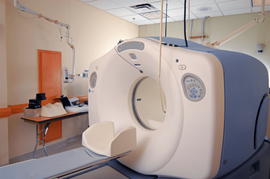
How Does Positron Emission Tomography (PET) Work?
PET imaging technology works using a scanning device (a machine with a large central opening) that detects photons (subatomic particles) emitted from the radiotracer in the organ or tissue being examined.
The radiotracers used in PET scans are created by attaching a radioactive atom to chemical compounds that the target organ or tissue naturally uses in its metabolism.
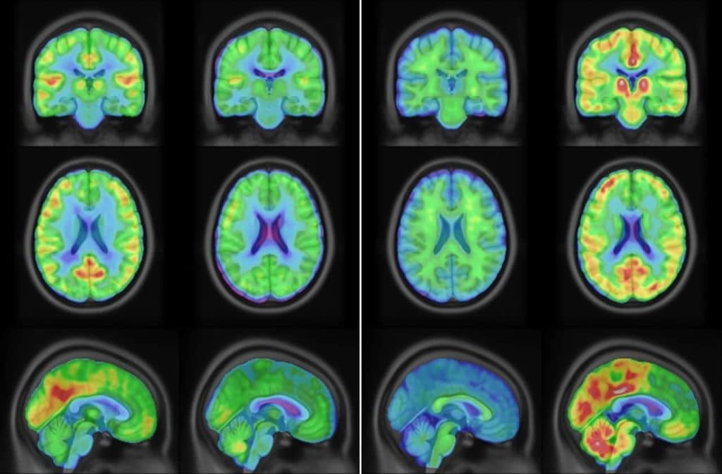
For example, in brain PET scans, a radioactive atom is attached to glucose (blood sugar) to create a radiotracer called fluorodeoxyglucose (FDG). Since the brain relies on glucose for metabolism, FDG is widely used in PET imaging.
Other radiotracers can be used depending on the purpose of the scan. If the focus is on blood flow or tissue perfusion, the radiotracer may be a radioactive form of oxygen, carbon, nitrogen, or gallium.
How is the PET Scans Performed?
- Radiotracer Injection: The radiotracer is injected into a vein through an intravenous (IV) line.
- Scanner Movement: The PET scanner moves slowly over the targeted area of the body.
- Positron Emission: The radiotracer decays, releasing positrons.
- Annihilation Photons Formation: The positrons collide with electrons near the decay site, generating gamma rays known as annihilation photons.
- Photon Detection: The PET scanner detects the annihilation photons, which arrive at detectors simultaneously and 180 degrees apart.
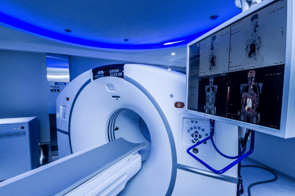
- Computer Analysis: The computer processes the gamma ray data and creates a detailed image map of the organ or tissue.
- Image Interpretation: The brightness of tissues in the image depends on how much radiotracer they absorb, indicating organ or tissue function levels.
How Should a Patient Prepare for a PET Scan?
PET scans are typically performed on an outpatient basis. However, some hospitalized patients may undergo PET imaging for specific treatments. While medical facilities may have their own specific protocols, the general PET scans procedure follows these steps:
- The patient will be asked to remove clothing and wear a hospital gown.
- All jewelry and metallic objects that might interfere with the scan must be removed.
- The patient will be asked to empty their bladder before the procedure.
- Some abdominal or pelvic scans may require a urinary catheter to drain the bladder during the scan.
- An IV line (or two, depending on the study) will be inserted into the hand or arm to administer the radiotracer.
- In some cases, an initial scan may be performed before injecting the radiotracer, depending on the type of study.
- The radiotracer is injected into the IV, and the patient will wait 30 to 60 minutes for it to concentrate in the targeted organ or tissue.
- Once the radiotracer has been absorbed, the scan begins. The PET scanner moves slowly over the area of interest.
- After the scan is complete, the IV line is removed. If a urinary catheter was used, it will also be removed.
How Long Do the PET Scans Take?
The entire PET scans procedure takes approximately two hours. The radiotracer absorption in the body takes about 60 minutes, during which the patient must sit quietly and limit movement. The actual imaging process lasts around 30 minutes. After the scan, the patient will wait while the radiology technician reviews the images to ensure they are clear.
Potential Side Effects of a PET Scans
Generally, a PET scans is considered safe and rarely causes complications. The amount of radiation in the radiotracer is very low and does not stay in the body for long. Patients are advised to drink plenty of water after the scan to help flush out the radioactive material from their system.
However, there are certain risks in specific cases:
- Pregnancy and Breastfeeding: Radiation may be harmful to the fetus or could pass to an infant through breast milk.
- Allergic Reactions: In very rare cases, some individuals may have an allergic reaction to the radiotracer. These reactions are usually mild and can be quickly managed with medication.
- Diabetes: Individuals with diabetes may have difficulty absorbing glucose from the radiotracer, which could affect the accuracy of the results. If you have diabetes, your doctor will provide special instructions on adjusting your diet and medication before the scan.
Final Thoughts
Positron Emission Tomography (PET) is one of the most significant advancements in nuclear medicine, allowing doctors to diagnose diseases with high accuracy, improve treatment plans, and determine the most effective therapeutic approaches. By understanding how PET works, we can appreciate its vital role in early disease detection and management.
Additionally, HSI Center is a leader in biomedical engineering and healthcare solutions, striving to enhance the skills and knowledge of professionals in this critical sector through specialized training programs and expert consultations.
Source: Positron Emission Tomography (PET)