In today’s technological era, Magnetic Resonance Imaging (MRI) stands as one of the most groundbreaking innovations in medical imaging. This advanced technique allows doctors to visualize intricate details of internal tissues without requiring any surgical intervention. While MRI might seem complex to many, it is fundamentally based on straightforward scientific principles that have revolutionized modern medicine.
What Is Magnetic Resonance Imaging (MRI)?
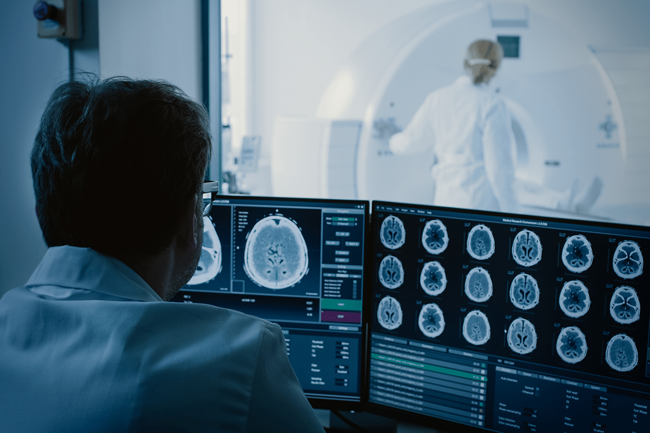
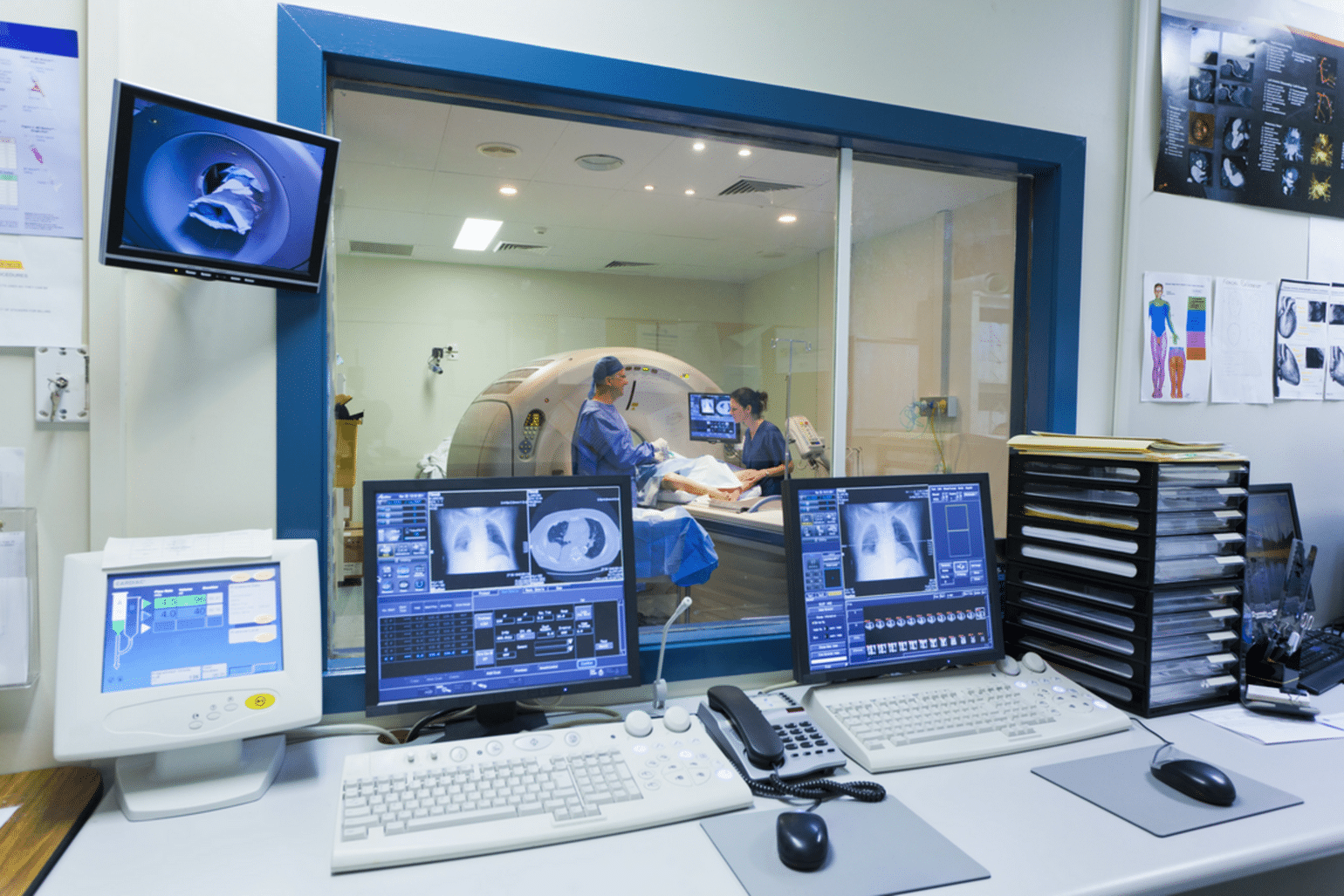
Magnetic Resonance Imaging (MRI) is a non-invasive medical imaging technique that utilizes strong magnetic fields and radio waves to generate highly detailed images of the body’s organs and tissues. Unlike X-rays or CT scans, MRI does not use ionizing radiation, making it a safer option, especially for patients requiring frequent monitoring.
This technique is based on a physical phenomenon known as nuclear magnetic resonance (NMR). It works by stimulating hydrogen atoms in the body using a powerful magnetic field. When exposed to radio waves, these atoms emit signals, which are then captured and transformed into high-resolution, three-dimensional images.
Key Principles of MRI
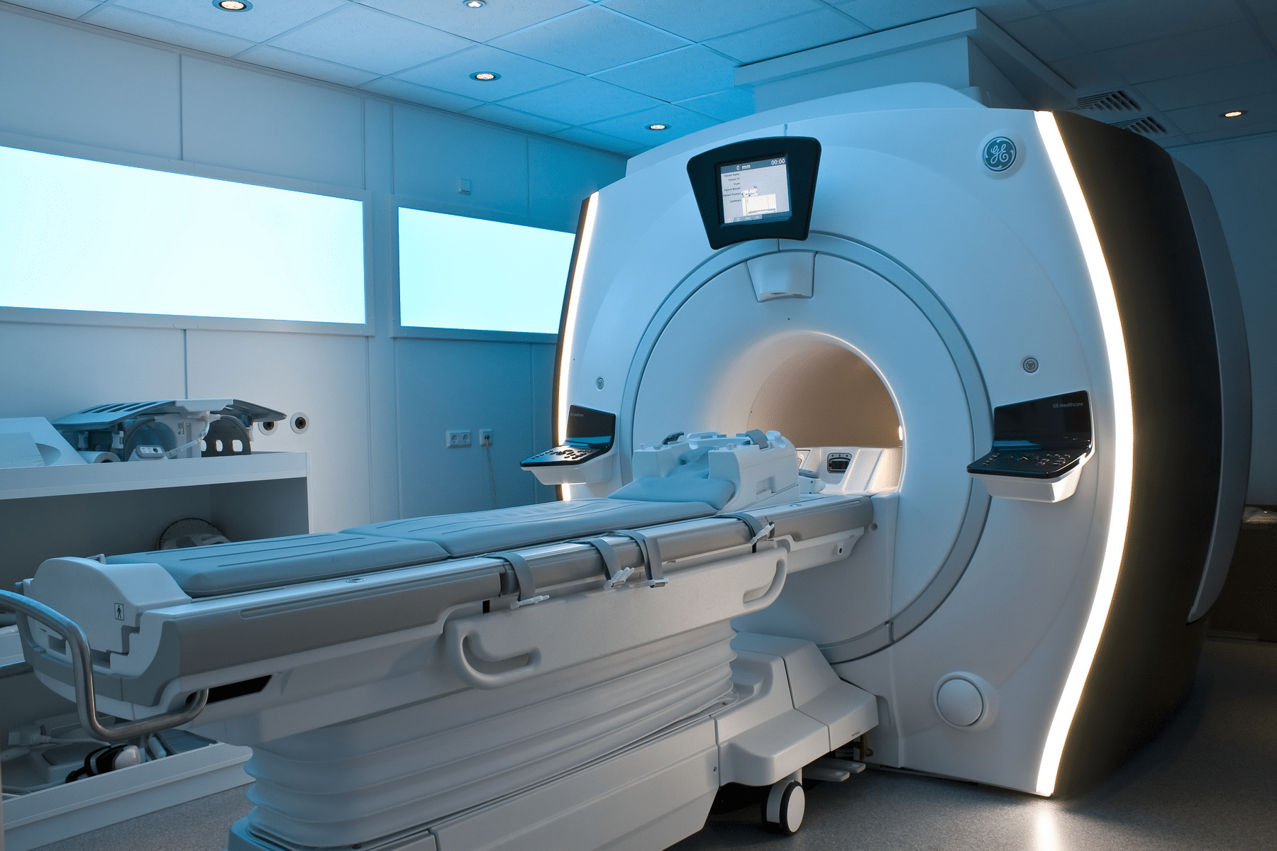
To understand how MRI functions, it is essential to grasp some fundamental principles, Firstly, strong magnetic field, one of the core components of this technology is the use of a powerful magnetic field. When a patient enters the MRI machine, their body is subjected to an extremely strong magnetic force, which alters the orientation of hydrogen atoms in their tissues. The scanner detects signals emitted by these atoms after exposure to the magnetic field.
In addition to radio frequency pulses, after the hydrogen atoms align with the magnetic field, the system of this technology emits radio frequency pulses. These pulses excite the hydrogen atoms, causing them to release energy, which is detected and processed by the scanner. Adding to signal detection and image processing that specialized sensors capture the energy signals emitted by hydrogen atoms. These signals are then processed by a computer to generate highly detailed, multi-dimensional images of the body’s internal structures.
Finally, tissue differentiation that one of the most remarkable features of this technology is its ability to distinguish between different tissue types. The rate at which hydrogen atoms return to their normal state varies depending on the tissue type, enabling the scanner to differentiate between muscles, fat, and organs with remarkable precision.
Key Advantages of MRI in Medical Imaging
Magnetic Resonance Imaging is a powerful and non-invasive medical imaging technique that offers numerous benefits, making it an essential tool for diagnosing various medical conditions. Its unique capabilities help doctors detect and assess health issues that may not be visible through other imaging methods. Below are the main advantages of MRI:
- High-Resolution Imaging: One of the standout benefits of MRI is its ability to produce highly detailed and clear images. This precision allows doctors to examine the body’s internal structures with exceptional accuracy, leading to improved diagnosis and more effective treatment plans.
- A Non-Invasive and Safe Technique: the technology is a completely non-invasive procedure, meaning it does not require surgical intervention. This reduces the risk of infection and pain, making it a safer alternative for patients. Additionally, since MRI does not use ionizing radiation, it is considered safe even for pregnant women in certain cases.
- Suitable for Children and Elderly Patients: Because MRI does not involve radiation exposure or invasive procedures, it is an excellent option for both children and elderly patients. This makes it a preferred imaging technique for diagnosing a wide range of conditions in these vulnerable groups.
- Versatile Medical Applications: MRI is capable of imaging a wide variety of tissues and organs, making it useful across numerous medical specialties, including oncology, cardiology, neurology, and orthopedics.
Applications of MRI in Medicine
Magnetic resonance imaging is widely used in the diagnosis and treatment of various medical conditions. Here are some of its primary applications:
1) Brain and Nervous System Imaging
Magnetic resonance imaging plays a crucial role in diagnosing neurological disorders such as brain tumors, multiple sclerosis, strokes, and traumatic brain injuries. It also helps detect changes in the brain associated with neurodegenerative diseases like Alzheimer’s disease.
2) Spine and Nerve Disorders
Magnetic resonance imaging is commonly used to assess conditions affecting the spine and nervous system, including herniated discs and nerve inflammations. The high-resolution images help doctors evaluate the vertebral discs and spinal cord, facilitating precise treatment planning.
3) Cardiac and Vascular Imaging
Magnetic resonance imaging provides detailed insights into heart health by detecting weakened heart muscles and identifying vascular issues such as aneurysms or blocked arteries. This is particularly valuable for diagnosing cardiovascular diseases without the need for invasive procedures.
4) Internal Organ Examination
Magnetic resonance imaging is a vital tool for examining soft tissues such as the liver and kidneys. It aids in diagnosing conditions like tumors, hepatitis, and chronic kidney diseases with high accuracy.
5) Joint and Soft Tissue Assessment
Magnetic resonance imaging is frequently used in orthopedic and sports medicine to evaluate joint injuries, ligament tears, and tendon damage. It is also effective in diagnosing conditions like arthritis and soft tissue disorders, ensuring optimal treatment strategies.
Why Is MRI Considered a Non-Invasive Medical Imaging Technique?
Magnetic resonance imaging (MRI) is widely recognized as a non-invasive medical imaging technique. But what makes it non-invasive? The primary advantage of MRI lies in its ability to generate highly detailed images of internal organs without the need for surgical procedures. Unlike other imaging techniques such as X-rays or computed tomography (CT) scans, MRI does not rely on harmful radiation. This makes it a safer option, allowing for repeated use without concerns about radiation exposure.
Preparation for MRI Scans
Although MRI is a non-invasive and generally safe procedure, patients need to follow specific preparatory steps to ensure accurate imaging and safety:
- Removing Metal Objects: Due to the strong magnetic field used in MRI, patients must remove all metallic items, including jewelry, watches, and glasses.
- Disclosing Medical History: It is crucial to inform the doctor about any implanted medical devices such as pacemakers or any known allergies to contrast materials that might be used in some scans.
- Fasting Requirements: In certain cases, fasting may be required before the scan, especially when contrast agents are involved.
Limitations and Challenges of MRI
Despite its numerous benefits, Magnetic resonance imaging does come with some limitations and challenges that should be considered:
- Time-Consuming Process: MRI scans generally take longer compared to X-rays or CT scans.
- High Cost: MRI is relatively expensive compared to other diagnostic imaging techniques.
- Suitability Concerns: Individuals with metallic implants, such as pacemakers or artificial joints, may not be suitable candidates for MRI scans due to potential safety risks.
In conclusion, as medical imaging technology continues to evolve, MRI remains a crucial tool in the field. Keeping up with advancements in MRI and biomedical engineering is essential for professionals seeking excellence. HSI provides specialized training programs and innovative consulting solutions to empower healthcare professionals and institutions. Whether you’re looking to enhance your skills in medical device design or healthcare system management, HSI is your ideal partner in achieving success. Join us in shaping a brighter future in the world of biomedical engineering!
Source: National Institute of Biomedical Imaging and Bioengineering at USA