Hybrid Imaging | The Latest Technologies for Disease Detection

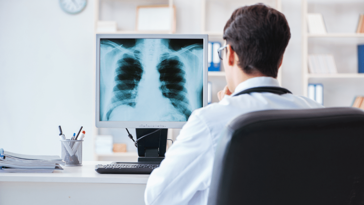
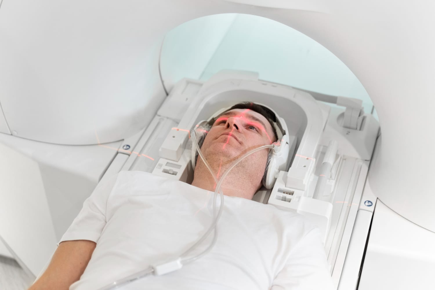
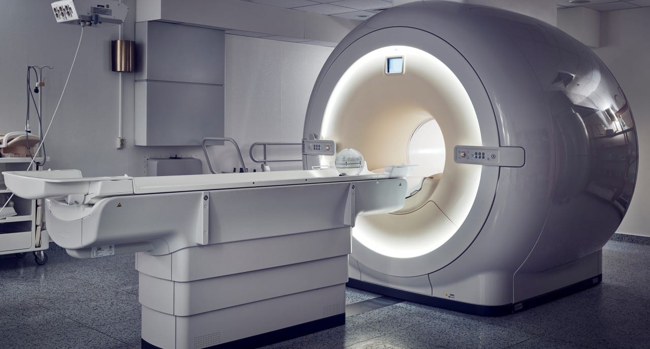
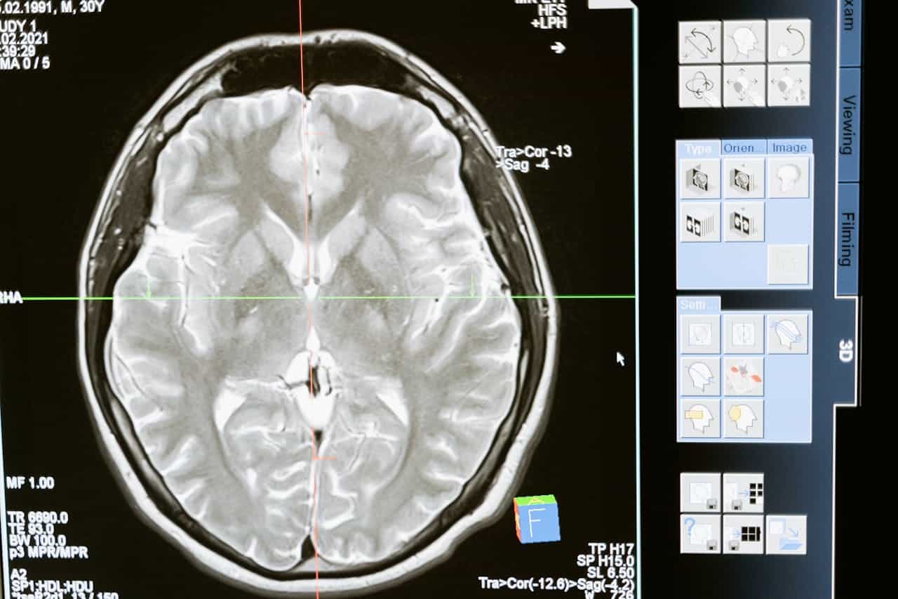
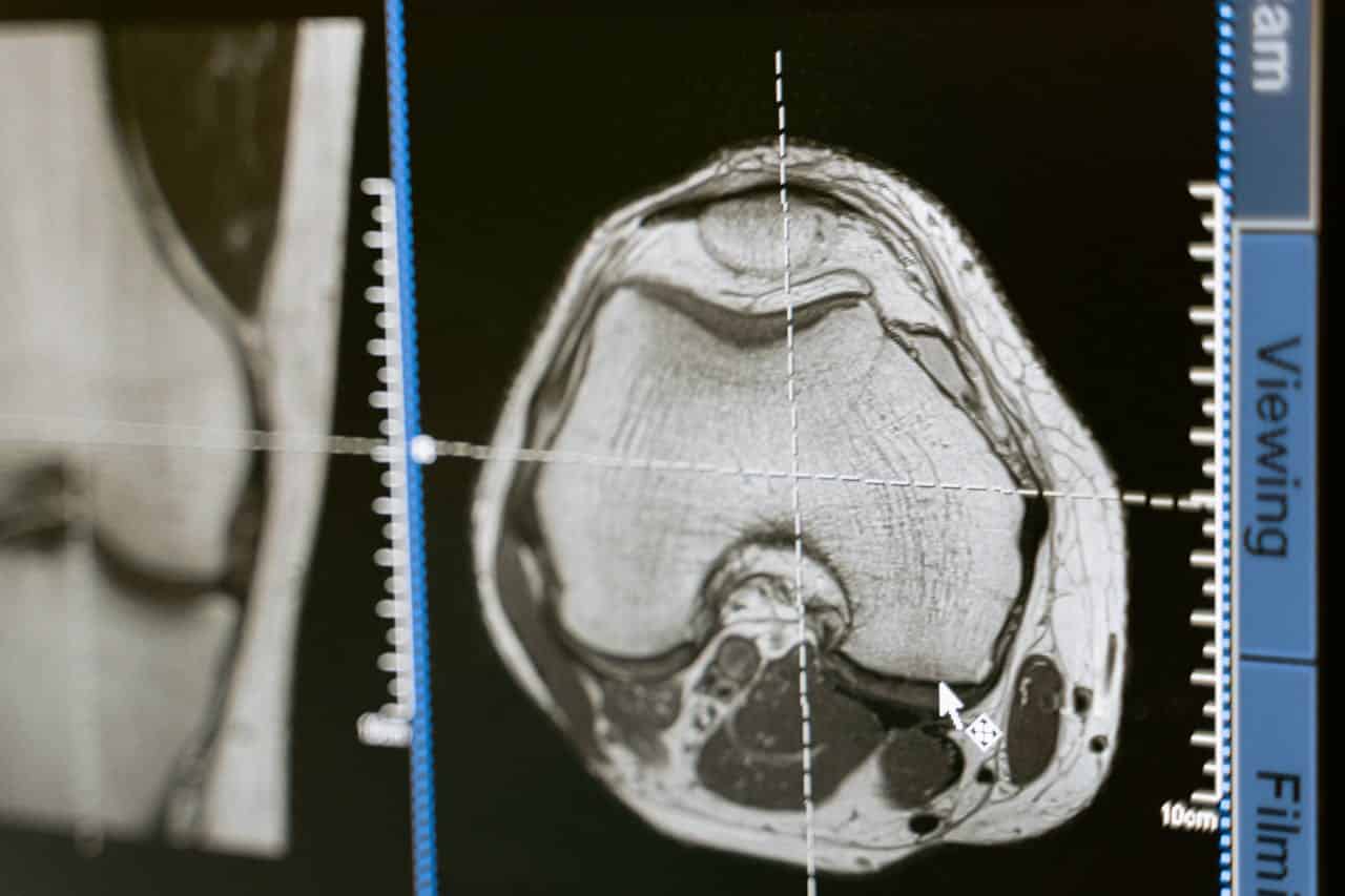
Amid the rapid technological advances in the fields of medicine and biomedical engineering, hybrid imaging has emerged as a revolutionary technique that has transformed the concepts of medical diagnosis like never before. This approach relies on combining multiple imaging modalities such as magnetic resonance imaging (MRI), computed tomography (CT), and nuclear medical imaging, which enables the acquisition of precise and comprehensive images of a pathological condition. Moreover, recent developments in artificial intelligence have contributed to analyzing these images with exceptional precision and speed, making this technology the ideal choice for early diagnosis.

The Remarkable Features of Hybrid Imaging Technology

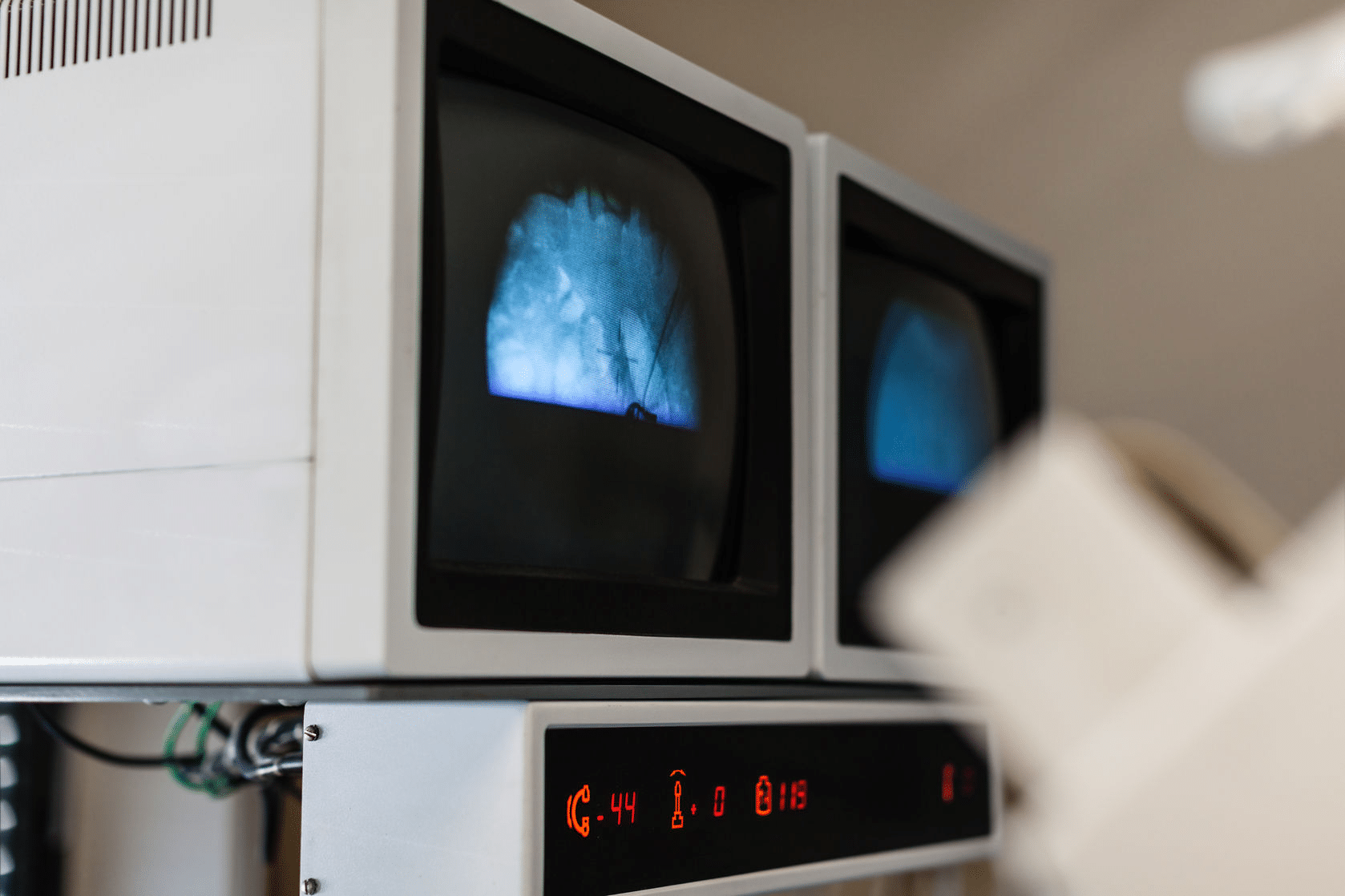
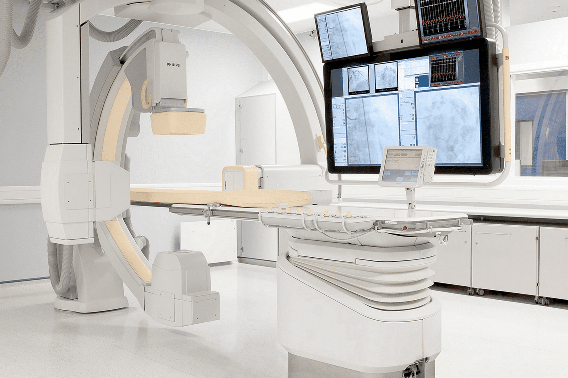
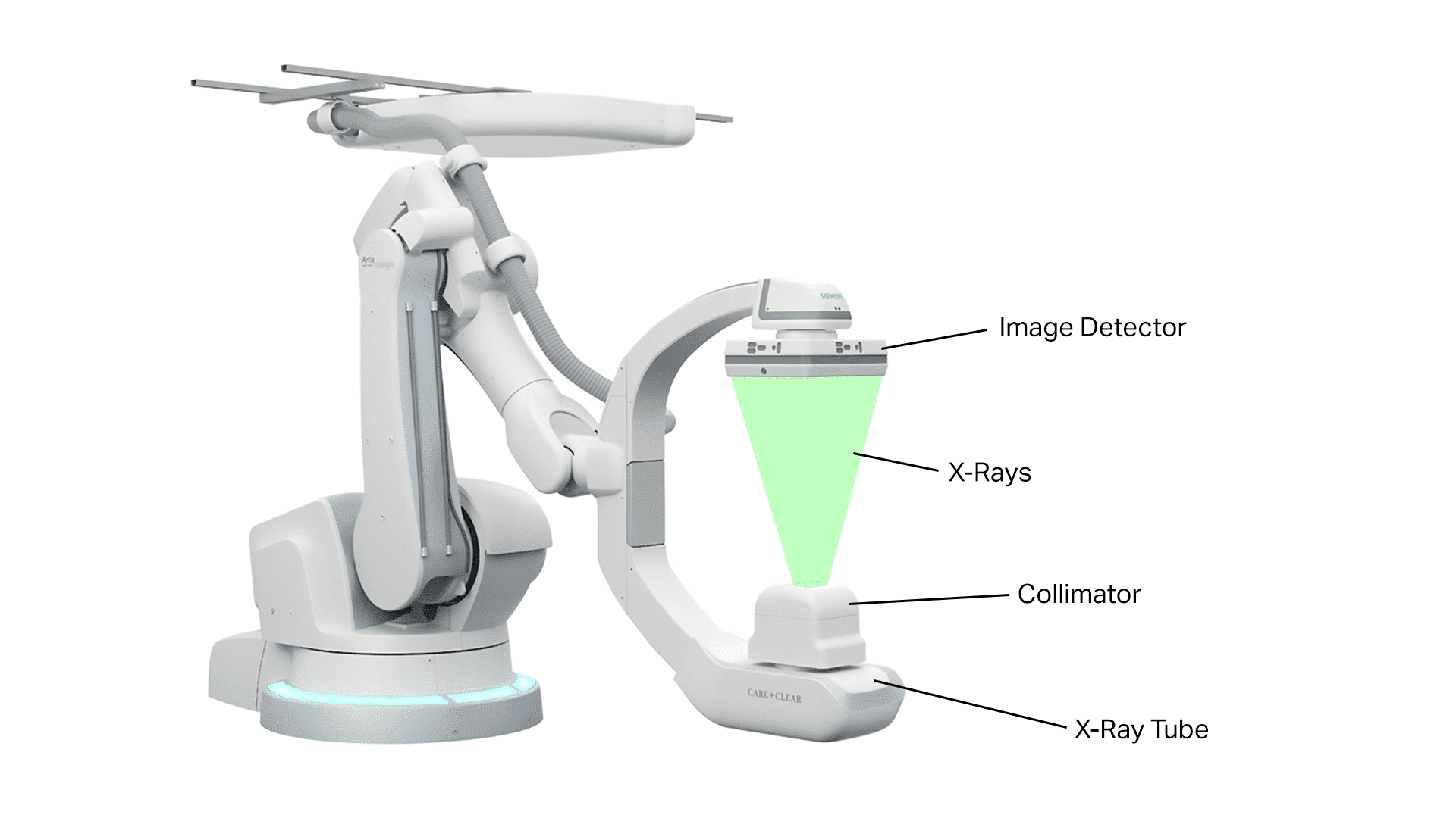
Hybrid imaging is considered an innovative technique that combines the advantages of each imaging modality to provide both quantitative and qualitative information that assists physicians in accurately evaluating a patient’s condition. This technology is used in the detection of tumors, chronic diseases, and functional disorders of vital organs. In addition, the integration of different techniques delivers detailed anatomical and functional data, while artificial intelligence enhances the process of analyzing and interpreting this data in a scientifically reliable manner.

The Importance of Hybrid Imaging in Medical Diagnosis

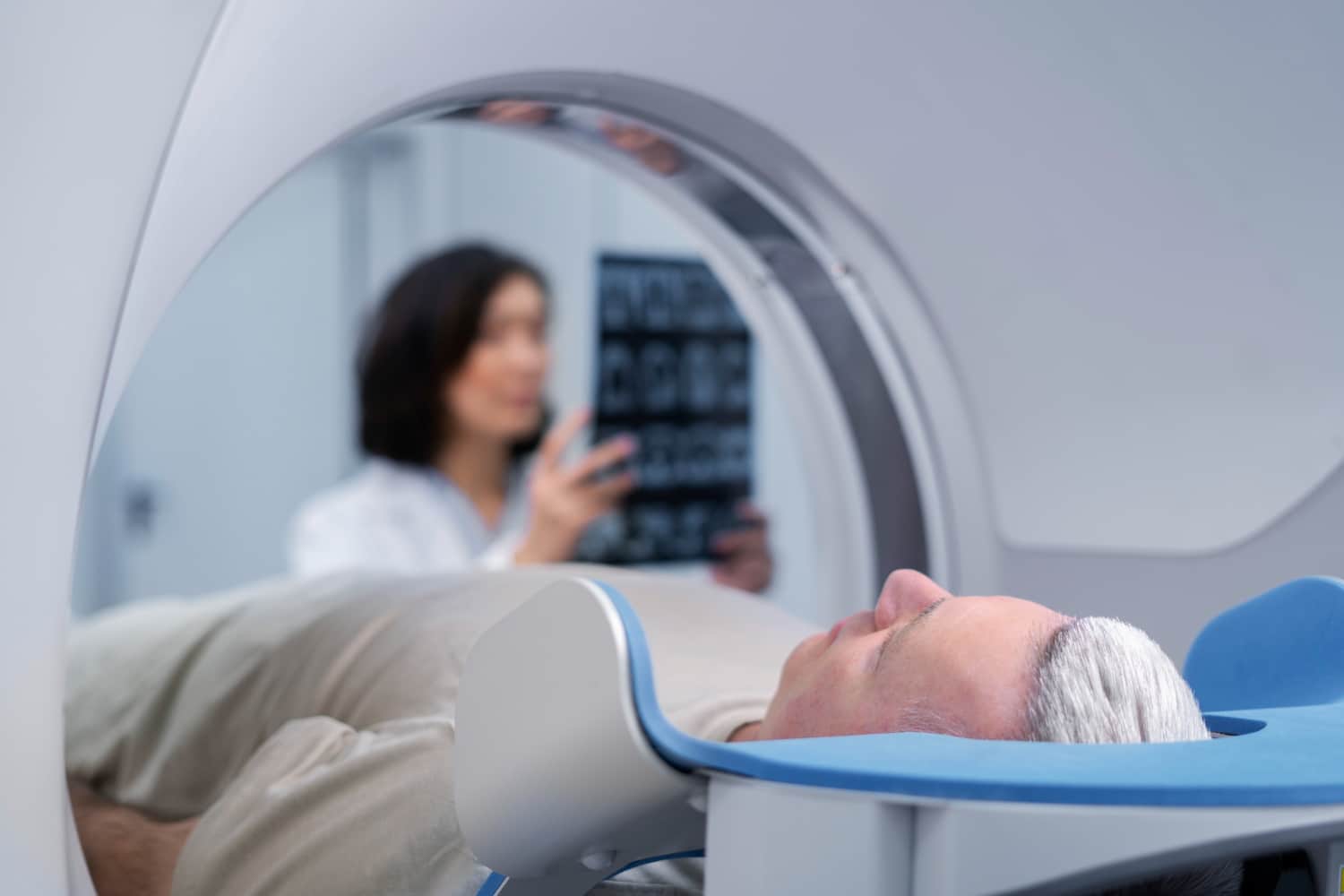
Hybrid imaging plays a vital role in the early and accurate diagnosis of numerous diseases, especially cancerous tumors and cardiovascular and neurological disorders. It provides valuable information that helps doctors in:

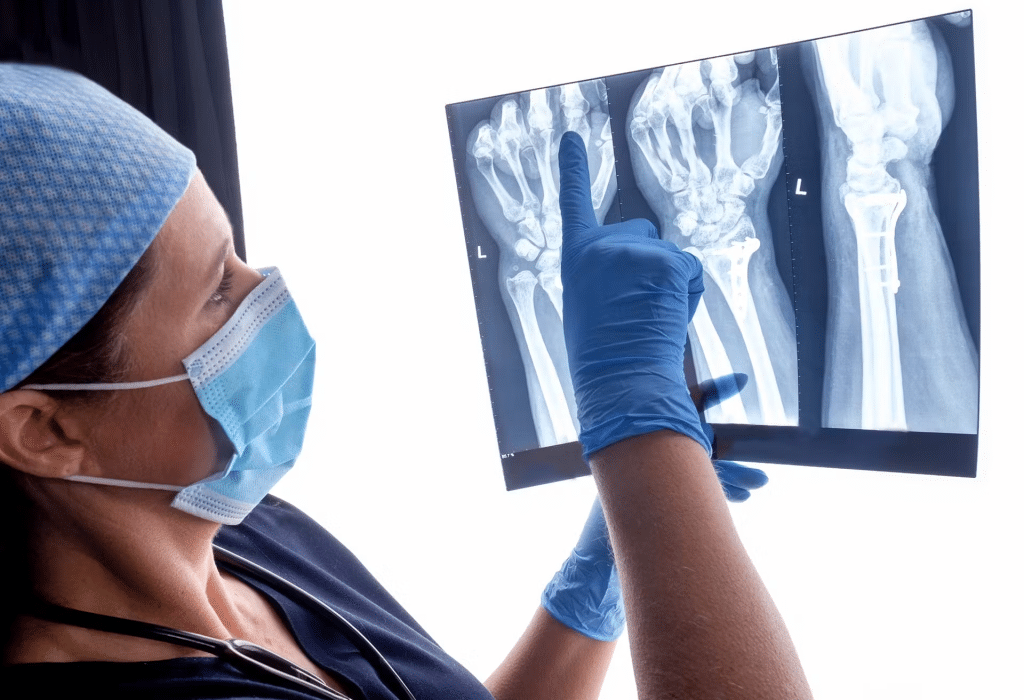
  • Detecting cancerous tumors at early stages: Hybrid imaging can identify small tumors that are difficult to detect using conventional imaging techniques.
  • Determining the extent of cancer spread: It helps in assessing whether the cancer has metastasized to other organs in the body.
  • Evaluating tumor response to treatment: Hybrid imaging can be used to monitor the effectiveness of treatments and determine whether a tumor is responding to therapy.
  • Diagnosing heart diseases: It aids in assessing cardiac functions and identifying areas with insufficient blood supply.
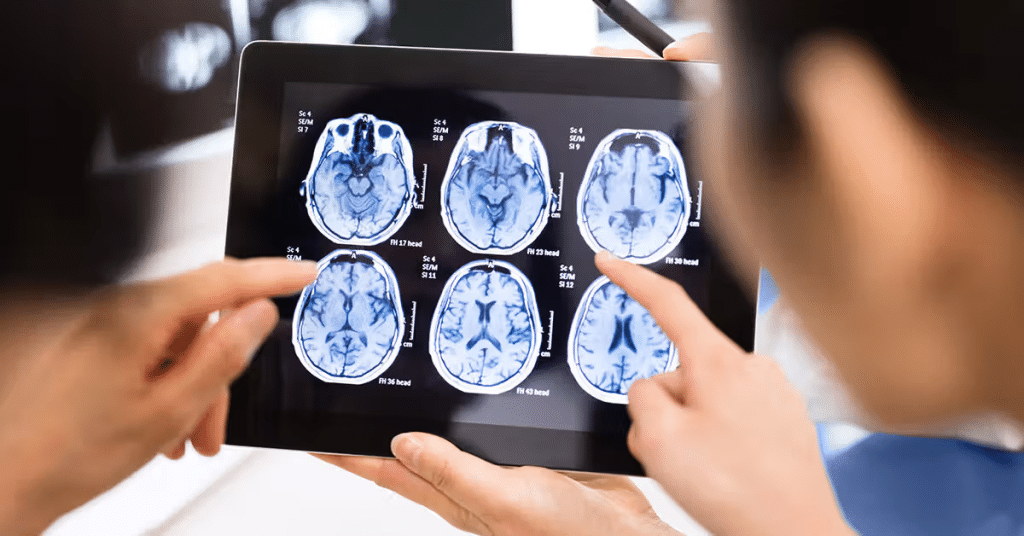
  • Diagnosing neurological diseases: This technology can be used in the diagnosis of conditions
  • such as Alzheimer’s, Parkinson’s, and multiple sclerosis.

Hybrid Imaging

Recent Developments in Imaging Techniques and Artificial Intelligence

With continuous technological progress, hybrid imaging techniques have seen significant advancements in recent years. Modern innovations have enhanced the quality of final images and reduced the time required to obtain results, which has led to an overall improvement in healthcare quality. Artificial intelligence techniques are utilized to process and analyze data in innovative ways, where algorithms are trained on massive datasets of medical images to deliver precise and immediate diagnoses. These applications serve as strong support for traditional techniques and contribute to building an integrated diagnostic system.

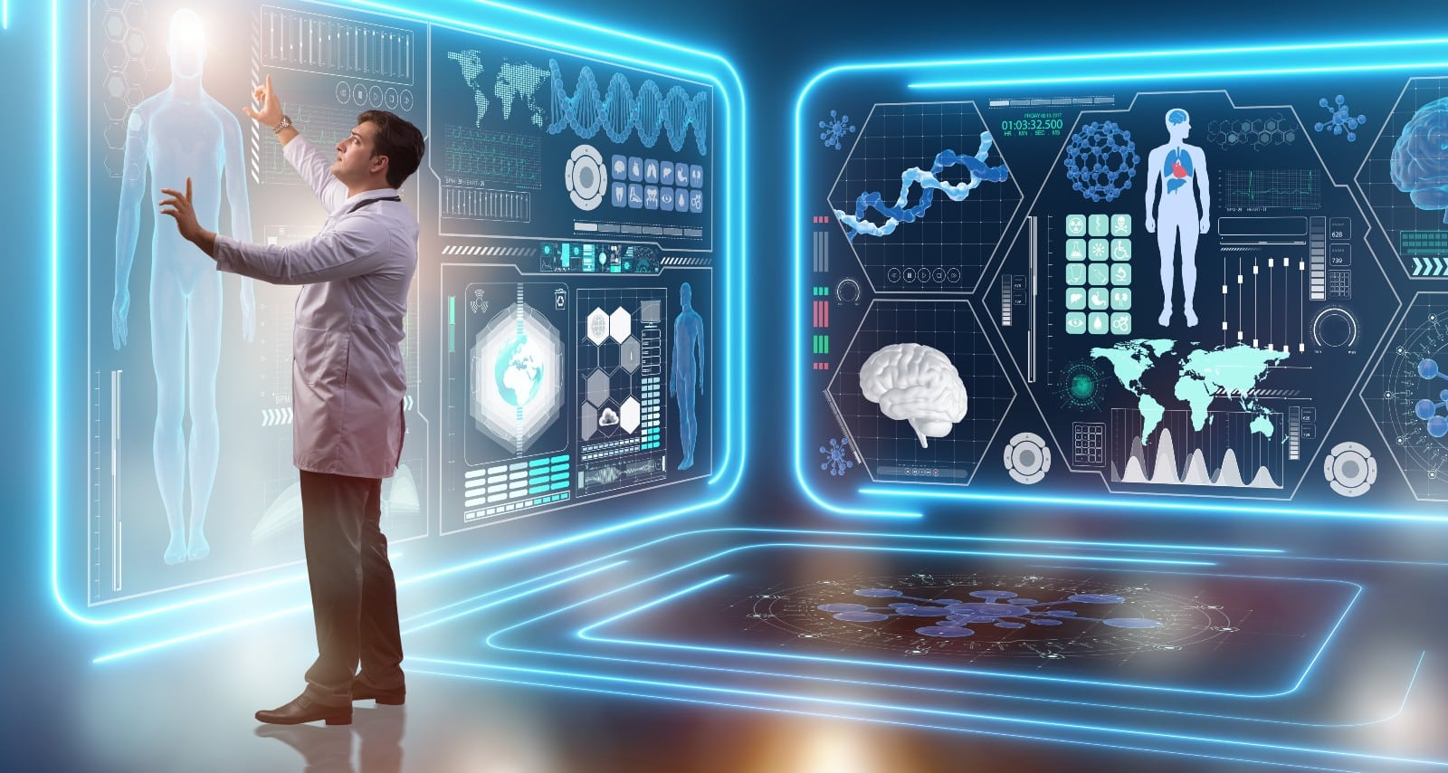
The Role of Artificial Intelligence in Enhancing Medical Diagnosis

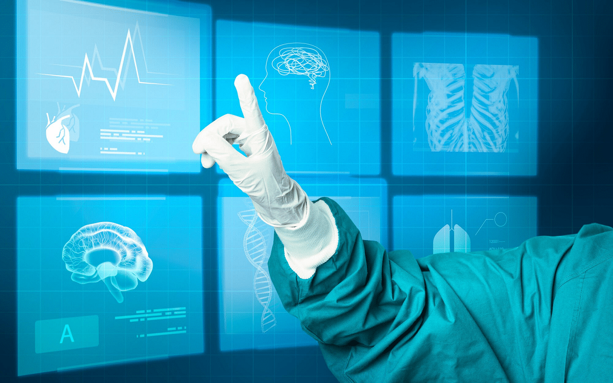
Artificial intelligence is one of the fundamental pillars in the development of hybrid imaging techniques, relying on advanced algorithms to analyze images and detect fine details that are difficult to observe with the naked eye. These algorithms help in precisely determining the characteristics of tumors and tissues and assist in predicting the stages of disease progression more rapidly. By integrating artificial intelligence with hybrid imaging, physicians can achieve early and effective diagnosis, which contributes to improved treatment outcomes and a reduction in potential complications.

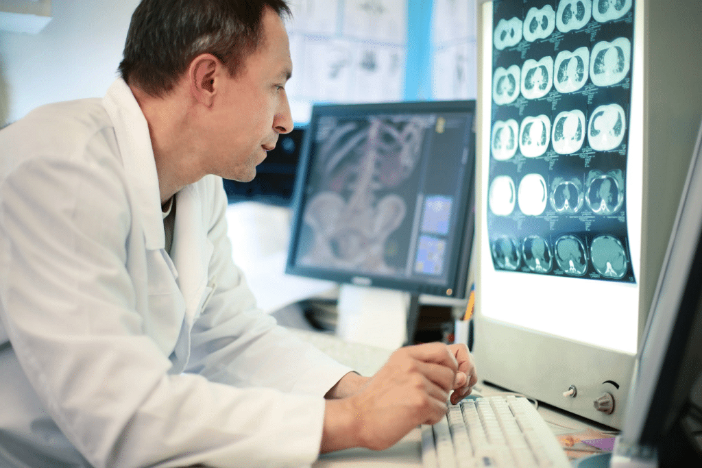
Modern hybrid imaging technologies are vital tools for the early and effective detection of diseases, as they enable doctors to obtain comprehensive data about the patient’s condition. This approach facilitates precise and prompt therapeutic decision-making, thereby enhancing the chances of treatment success. Artificial intelligence plays a pivotal role in analyzing this data and extracting critical biomarkers that may be overlooked by conventional methods, which in turn increases the accuracy and reliability of diagnostic results.

Challenges and Future Development Prospects

Despite the significant achievements of hybrid imaging in the field of medical diagnosis, challenges still remain for researchers and clinicians in the optimal application of this technology. Among these challenges is the need to develop more advanced artificial intelligence algorithms and to provide extensive training datasets to improve diagnostic performance. Moreover, the integration of various imaging techniques represents a complex task that requires high-level coordination between devices and software. Artificial intelligence remains at the forefront of efforts to address these challenges and to achieve promising future developments in medical diagnosis.

A Future Vision for Hybrid Imaging in Medicine

The future of medicine is moving towards an increased reliance on digital technologies and artificial intelligence, opening up new horizons in the field of hybrid imaging. Artificial intelligence is considered a cornerstone for developing diagnostic methods and analyzing data much faster than before, enabling physicians to offer advanced and personalized treatment services to each patient for quicker and better recovery. These technologies contribute to improving survival rates and reducing healthcare costs, while also providing valuable data that supports well-informed therapeutic decisions. Hybrid imaging, assisted by artificial intelligence, shows tremendous potential in enhancing healthcare quality and elevating the standards of medical diagnosis.

Hybrid imaging represents a true revolution in the field of medical diagnosis, thanks to the integration of various imaging techniques and the rapid advancement of artificial intelligence. These technologies provide precise and comprehensive insights that aid in the early detection of diseases and in determining the most appropriate treatment pathways for each case. The synergy between advanced imaging techniques and artificial intelligence holds great promise for improving healthcare quality and achieving outstanding therapeutic outcomes, making this technology one of the most important future tools in medicine. Continuous research and development in artificial intelligence and hybrid imaging remain the key to achieving further breakthroughs and innovations in the world of medical diagnostics.

In conclusion, after exploring hybrid imaging technologies and in light of the rapid advancements in medical imaging and artificial intelligence, if you aspire to become a part of the biomedical engineering field, connect with HSI Center. Through our specialized training and consulting programs, we empower students and professionals to gain both practical and scientific skills, enabling them to keep pace with the latest innovations and actively contribute to improving the quality of healthcare.

Source: Hybrid imaging is the future of molecular imaging – PMC

Fluoroscopy: Real-Time X-ray Imaging in Medicine

Fluoroscopy is one of the most important technologies in modern medical imaging, allowing doctors and specialists to view internal body structures in real-time. This revolutionary technique has transformed diagnostic methods and interventional procedures in medicine, significantly improving diagnostic accuracy and guiding treatments more effectively. In this detailed article, we will explore the working mechanism of this technique, its applications, benefits, associated risks, and the latest advancements in this vital field of medical engineering.

What is Fluoroscopy?

Fluoroscopy is an advanced imaging technique that utilizes X-rays to generate a continuous series of images displayed as a real-time video. This technique is widely used in emergency departments, operating rooms, and diagnostic radiology clinics, enabling surgeons and physicians to monitor the movement of organs and internal structures during procedures or diagnostic examinations. The increasing reliance on fluoroscopy reflects its significance in providing accurate and immediate insights into various medical conditions.

Fluoroscopy

Fundamental Principles of Fluoroscopy

Fluoroscopy is extensively used across multiple medical specialties, enhancing the quality of healthcare. To fully understand this technique, it is essential to delve into the fundamental principles that underpin fluoroscopy, forming its scientific and technical foundation.

X-ray Mechanism

This technique operates on the same principles as conventional X-ray imaging but with the ability to produce sequential images in real time, creating a video-like sequence of still images. The process begins with generating X-rays through a specialized tube, where a high-voltage current is applied. This accelerates electrons toward a metallic target, typically tungsten—a chemical element. Upon collision, X-rays are emitted and directed toward the area of the body being examined.

Image Intensification

The image intensifier in fluoroscopy plays a crucial role in enhancing the clarity of internal details. This device amplifies the signals produced by X-rays passing through the body, resulting in a bright and clear image. Recent advancements have led to the replacement of traditional image intensifiers with flat-panel detectors, which provide high-quality images while minimizing radiation exposure.

Diverse applications of Fluoroscopy

Fluoroscopy has a wide range of medical applications, both in diagnostics and interventional procedures. This system is highly flexible and capable of providing real-time moving images, allowing doctors to monitor physiological changes instantly.

Fluoroscopy

Diagnostic Applications

Fluoroscopy is used in several essential diagnostic procedures, including:

  • Gastrointestinal Studies: Fluoroscopy is employed to assess the digestive tract, including the esophagus, stomach, and intestines. Barium studies, such as barium swallow and barium enema, help evaluate abnormalities or motility disorders.
  • Neuromuscular and Spinal Imaging: This technique is used in diagnostic imaging of the spine, particularly in myelography, which aids in assessing spinal cord conditions.
  • Joint Evaluations: For joint-related issues, fluoroscopy is also used in arthrography to evaluate abnormalities or injuries affecting the joints.

Interventional Applications

This technique is a cornerstone in medical engineering for interventional procedures, providing live imaging during operations, which contributes to:

  • Guiding Surgical Procedures: Fluoroscopy assists in catheter placement during cardiac procedures, such as coronary angiography, ensuring precise positioning and minimizing procedural risks.
  • Interventional Treatments: It is utilized in therapeutic interventions, including stent placement, bone fixation in fracture surgeries, and the insertion of delicate medical devices.
  • Minimally Invasive Procedures: This technology facilitates minimally invasive procedures requiring small or no surgical incisions, reducing patient discomfort and promoting faster recovery.

Benefits of Fluoroscopy in Medical Practice

Fluoroscopy is a pioneering technique that enhances medical care quality for several key reasons:

1) Enhanced and Accurate Diagnosis

This technology provides dynamic imaging that reveals the functional movements of internal organs, such as diaphragm motion, intestinal activity, and blood flow through vessels. This capability helps in early disease detection, as moving images can uncover functional changes that static images might not reveal.

Fluoroscopy

2) High Precision in Interventional Procedures

By offering live imaging during medical procedures, fluoroscopy enables precise manipulation of medical instruments within the body. This reduces the likelihood of surgical complications and improves success rates in procedures like stent placement and fracture fixation.

3) Reduced Surgical Invasiveness

This technique allows for numerous minimally invasive procedures, reducing the need for major surgeries. Minimally invasive techniques are less painful, promote faster recovery, and lower the risk of post-operative infections, making them a significant advancement in modern medicine.

Risks and Safety Considerations in Fluoroscopy

Despite the significant benefits of fluoroscopy, there are certain risks associated with its use that must be carefully considered.

Fluoroscopy

Radiation Exposure

Radiation exposure is one of the primary concerns when using fluoroscopy. Prolonged procedures may increase the amount of radiation a patient is exposed to. The level of exposure depends on the duration of the procedure and the examined area. To mitigate these risks, advanced techniques such as pulsed fluoroscopy and dose-reduction algorithms have been developed to minimize radiation exposure.

Contrast Media Reactions

Contrast media are often used during this technique to enhance the visibility of internal structures. However, some patients may have allergic reactions to these substances, ranging from mild skin rashes to severe anaphylactic responses. To manage these risks, pre-procedure screening and emergency medications are utilized when necessary.

Occupational Radiation Exposure

Medical professionals performing frequent fluoroscopy procedures are at risk of occupational radiation exposure. Strict protective measures, such as wearing personal protective equipment (e.g., lead aprons and thyroid shields), are essential. Additionally, regular monitoring of radiation levels ensures the safety of doctors and healthcare staff, helping to prevent potential long-term risks.

Recent Advances in Fluoroscopy Technology

The field of fluoroscopy has seen significant advancements in recent years, leading to improved image quality and reduced radiation risks. Traditional systems have been replaced with digital fluoroscopy using flat-panel detectors, which enhance image resolution while minimizing radiation exposure. This digital system provides high-definition images and allows for post-processing adjustments to refine diagnostics. Additionally, it enables seamless image sharing among medical teams for quick consultations and decision-making.

Furthermore, the integration of cone-beam computed tomography (CBCT) with fluoroscopy now offers detailed 3D imaging for complex anatomical assessments. This technology is widely used in dentistry, orthopedic surgeries, and interventional procedures requiring precise localization.

Fluoroscopy

Moreover, combining this technique with other imaging modalities such as ultrasound and magnetic resonance imaging (MRI) has led to the development of hybrid imaging systems. These systems provide both anatomical and functional insights, enhancing diagnostic accuracy and guiding interventional procedures more effectively, particularly in oncology and tumor treatments.

The Future of Fluoroscopy

The future of this technique looks promising, with continuous research and technological advancements aimed at improving procedural efficiency and reducing radiation-related risks.

Integration of Artificial Intelligence

Artificial intelligence (AI) is expected to play a pivotal role in fluoroscopy’s evolution. AI algorithms can analyze real-time images, detecting anomalies that may be overlooked by the human eye. Additionally, AI-driven techniques can optimize radiation dose reduction strategies, enhancing patient and healthcare worker safety.

Portable Fluoroscopy Systems

Research and development efforts are currently focused on creating portable fluoroscopy units that can be easily transported to emergency departments or remote areas. These mobile units will enable rapid diagnostic imaging and treatment in locations with limited medical infrastructure, thereby expanding access to essential healthcare services.

Exploring Radiation-Free Alternatives

Scientists are actively exploring non-ionizing imaging alternatives such as optical coherence tomography (OCT) and photoacoustic imaging. While these alternatives are still in experimental stages, they hold great potential for reducing radiation-related risks in this technique, potentially revolutionizing the future of medical imaging.

In conclusion, we live in an era where cutting-edge fluoroscopy technologies are transforming medical diagnostics and interventions. However, behind every advanced imaging system stands a well-trained professional who makes critical decisions. This is where HSI Healthcare Training & Consultancy comes in. Join HSI today—not just to keep up with medical advancements, but to lead them. Because in the world of healthcare, history is not just written by machines but by those who master their language. Are you ready to shape the future of medical imaging?

Source: Real-Time Imaging and Its Impact on Modern Medicine

Radiology 101: The Key Imaging Techniques

Radiology is a field that has revolutionized the way medical conditions are diagnosed and treated. In this comprehensive guide, we will dive into the fundamentals of Radiology 101, explore the various imaging methods, the science behind them, and their crucial role in modern healthcare. Whether you are a medical student, a patient seeking complete knowledge of radiology to feel reassured during imaging procedures, or simply someone interested in medical technology, this article will provide you with a solid foundation in medical imaging techniques.

Overview of Medical Imaging Techniques

Radiology 101 includes a wide range of medical imaging techniques essential for diagnosing and monitoring various medical conditions. Understanding these techniques and their applications can help you appreciate the role of this type of rays in healthcare.

What is Radiology 101?

Radiology is the medical specialty that uses medical imaging techniques to diagnose and treat diseases within the human body. It involves the use of different imaging techniques to visualize the internal structures and functions of organs, tissues, and bones. This field has made tremendous progress over the years, enabling healthcare professionals to make more accurate diagnoses and develop precise treatment plans.

أساسيات الأشعة 101 تقنيات التصوير الطبي

What are the Basic Principles of Radiology?

Radiology relies on the principles of ionizing and non-ionizing radiation, along with advanced computer technology. These principles have led to the development of various imaging techniques, each serving its purpose in diagnosing diseases. In the following sections, we will explore these imaging methods in more detail.

X-rays

X-ray imaging, also known as radiography, is one of the oldest and most widely used techniques in radiology. It involves using X-ray beams to create images of the inside of the body. X-rays are an essential tool for examining the skeleton, detecting fractures, and identifying abnormalities in the chest and abdomen.

How Do X-rays Work?

  • Ionizing radiation is used to create images of bones and other dense structures.
  • They are commonly used to detect fractures, infections, and lung diseases.
  • When X-rays pass through the body, they are absorbed differently by various tissues.
  • Dense tissues, such as bones, absorb more X-rays, which appears white on the X-ray film.
  • On the other hand, soft tissues allow X-rays to pass through, making them appear darker on the film.
  • This contrast helps radiologists determine the condition and make an accurate diagnosis.

X-rays

Computed Tomography (CT)

Computed Tomography (CT), often referred to as CT scanning or axial CT scanning, is a type of medical imaging technique that allows for detailed three-dimensional images of the body, providing valuable insights into both anatomy and pathology.

CT combines X-ray images taken from different angles to produce detailed cross-sectional images. It is particularly useful for diagnosing complex conditions such as head injuries, tumors, and internal bleeding.

How does CT produce a three-dimensional view?

  • CT scanners work by capturing a series of X-ray images from different angles around the body.
  • These images are then processed by a computer to create cross-sectional slices, similar to cutting a loaf of bread.
  • The result is a three-dimensional representation of the scanned area, offering a more comprehensive view of the internal structures.

Computed Tomography (CT)

Magnetic Resonance Imaging (MRI)

Magnetic Resonance Imaging (MRI) is a non-invasive imaging technique that provides detailed images of soft tissues, such as the brain, spinal cord, joints, muscles, and organs. Unlike X-rays and CT, which use ionizing radiation, MRI relies on magnetic fields and radio waves. Since MRI does not use radiation, it is considered a safer option for certain patients, including pregnant women and children.

How does MRI work without radiation?

MRI machines use powerful magnets to align hydrogen nuclei in the body. Then, radio waves are applied, causing these nuclei to emit signals. These signals are captured and converted into detailed images by a computer.

Ultrasound

Ultrasound, commonly associated with pregnancy monitoring, is a versatile imaging technique that uses high-frequency sound waves to create images of different parts of the body. It is particularly useful for early detection of cancer, heart diseases, internal medicine, vascular conditions, and a variety of other medical conditions.

Ultrasound can be used for other purposes and on various body areas, including breast ultrasound, pelvic ultrasound, vascular ultrasound, and musculoskeletal ultrasound. It is effective in detecting masses and types of abnormalities within the body. Ultrasound is an important diagnostic tool, often used complementarily alongside other medical imaging techniques.

How does ultrasound produce images?

  • Ultrasound relies on the principle of sound wave reflection.
  • A transducer sends high-frequency sound waves into the body, which bounce back from different tissues and return as echoes.
  • The time it takes for these echoes to return is processed by a computer to create dynamic images.

Ultrasound

Nuclear Medicine

Nuclear medicine is a unique branch of radiology that focuses on the functional aspects of the body at the cellular level. Unlike other medical imaging techniques, it involves injecting radioactive materials known as radiopharmaceuticals, which are then tracked within the body. Nuclear medicine techniques, such as PET and SPECT scans, provide functional information about organs and tissues, making them powerful tools for diagnosing conditions such as cancer, heart disease, and thyroid disorders.

How do radiopharmaceuticals reveal cellular activity?

  • Radiopharmaceuticals emit gamma rays, which can be detected by special cameras called gamma cameras.
  • By monitoring the distribution of these radioactive substances,
  • nuclear medicine specialists gain insights into cellular functions.

The Role of Radiology in Healthcare

Radiology plays a pivotal role in modern healthcare. It not only helps diagnose diseases but also guides surgical interventions, monitors treatment effectiveness, and assists in surgical procedures.

Radiologists work closely with doctors, surgeons, and other specialists to provide accurate diagnoses and clear treatment plans. Their expertise contributes to improving patient outcomes, making radiology an indispensable component of the medical field.

Applications of Radiology

Radiology is integral to diagnosing a wide range of medical conditions, from acute injuries to chronic diseases. Below are some key applications:

  1. Trauma and Emergency Care: X-rays and CT scans quickly identify fractures, internal injuries, and bleeding, enabling rapid treatment.
  2. Neurology: MRI and CT scans are used to diagnose brain tumors, strokes, and spinal cord abnormalities.
  3. Cardiology: Echocardiograms (ultrasound of the heart) assess heart function and detect heart conditions.
  4. Oncology: CT scans, MRI, and PET scans help detect, monitor, and assess tumor growth, evaluate treatment responses, and guide biopsy procedures.
  5. Obstetrics and Gynecology: Ultrasound monitors fetal development and identifies gynecological conditions like ovarian cysts and fibroids.
  6. Gastroenterology: Ultrasound and CT scans evaluate abdominal pain and detect conditions such as gallstones, liver disease, and pancreatitis.

Radiology 101

Preparation for Radiological Exams

Preparation for radiological exams varies depending on the imaging technique used. For example, you may need to fast before a CT scan or fill your bladder for a pelvic ultrasound. The healthcare team will provide specific instructions to ensure accurate results. During the exam, you will be positioned appropriately, and the technician will operate the imaging equipment. It is essential to stay still to obtain clear images.

Benefits and Risks of Radiology

Radiology offers numerous benefits, including precise, non-invasive diagnostic capabilities that aid in the early detection and treatment of various conditions. However, it is important to be aware of potential risks, such as exposure to ionizing radiation in X-rays and CT scans. To minimize these risks and ensure patient safety, the healthcare team follows strict safety protocols.

HSI offers an opportunity for students interested in learning and studying medical imaging techniques and their applications. Through these training courses, participants gain both practical and theoretical knowledge to contribute to the advancement and development of this leading field.

MRI Safety Essentials

The use of Magnetic Resonance Imaging (MRI) scans has become a fundamental diagnostic tool for detecting back pain, sports injuries, and diseases of the brain and heart. MRI machines must be operated by highly trained technicians who are experts in MRI safety and thoroughly familiar with the manufacturer’s guidelines to assess safety protocols and compatibility for each patient during every scan. This article highlights the key MRI Safety Essentials and precautions necessary to ensure patient safety during scans.

MRI Technology

The MRI machine is an advanced technology that uses magnets and radio waves to create detailed images of the body’s internal structures. This process allows for the production of cross-sectional and 3D images of organs and tissues without the use of ionizing radiation.

MRI scans are often used to diagnose conditions affecting muscles, ligaments, the spine, and the abdomen because they provide more detailed images of soft tissues than X-rays or CT scans. While MRI scans are generally safe, the large magnetic fields generated by this advanced technology necessitate multiple safety precautions before, during, and after each scan.

MRI Safety Essentials

MRI Safety Essentials

Because MRI machines generate extremely powerful magnetic fields, even small metallic objects can become projectiles inside the MRI room. This phenomenon, known as the “missile effect,” can pose a danger to individuals nearby and damage the equipment.

As the MRI magnet is always active, no one is allowed to enter the MRI room without proper clearance and authorization from a certified technologist. Metallic objects, such as nail clippers, small knives, and even pens, can cause severe bodily injury if brought into the magnetic field.

To mitigate risks, MRI safety guidelines established by the American College of Radiology (ACR) are strictly followed, including the implementation of designated safety zones. These safety zones are designed to restrict access to areas near the MRI scanner and ensure that only individuals who have been thoroughly screened for metal are allowed near the magnetic field.

Pre-Scan Safety Procedures

All individuals scheduled for an MRI scan must be thoroughly screened to ensure they are free of any items that could pose a risk to themselves or others before entering the MRI room. It is highly recommended that the personnel responsible for patient preparation utilize the standard MRI screening form during the process. Key pre-scan safety measures include:

  • Changing into designated examination clothing, as regular clothing with metallic parts like buttons or wires may be attracted to the magnet, posing a safety risk to the patient and potentially damaging the equipment.
  • Removing jewelry to prevent interference with medical equipment and to ensure a smooth, safe, and metal-free scanning process.
  • Screening for foreign objects or medical devices that might be present in the patient’s body, such as hearing aids, insulin pumps, pacemakers, or other medical devices.
  • Removing all metallic items before approaching the MRI machine, including removable medical devices, jewelry, hairpins, and clothing with metallic fibers.

MRI- Safety

In-Scan Safety Protocols

During the MRI procedure, several measures are taken to ensure patient safety and comfort. Patients are provided with means to communicate with MRI technicians throughout the procedure, which may last between 15 minutes to an hour, depending on the type of scan required. Patients are encouraged to inform the technicians of any discomfort or concerns they may experience during the scan.

To reduce the noise generated by the MRI machine, special techniques are employed, along with the provision of external aids such as earplugs. Patients are positioned carefully within the machine to ensure safety and optimal image quality. For patients experiencing claustrophobia, appropriate assistance is provided to make them feel at ease.

In some cases, the procedure may require the use of a contrast agent, which is a safe solution injected intravenously to enhance image quality. Although the likelihood of an allergic reaction to the contrast agent used in MRI scans is lower compared to that used in CT scans, rare adverse reactions may occur. Patients are advised to inform the medical staff of any known allergies to ensure necessary precautions are taken.

Post-Scan Safety Procedures

MRI safety protocols extend beyond the completion of the scan, with a focus on ensuring patient well-being. Clear instructions are provided regarding post-scan care measures. These include monitoring the body for any immediate or delayed adverse reactions following the scan and adhering to the care guidelines provided by the physician, such as resuming daily activities or following any specific restrictions.

It is crucial to report any unusual symptoms or unexpected reactions to the treating physician promptly within the hours following the scan to address any emerging concerns effectively.

MRI- Safety

Contraindications for MRI Scans

Several contraindications may prevent a patient from undergoing an MRI scan, as they are a critical part of MRI safety protocols. These contraindications are categorized into absolute and potential contraindications. Therefore, one of the most important safety measures in MRI is thoroughly screening the patient to ensure there are no risks that could endanger them. All necessary precautions are taken to guarantee their safety throughout the procedure.

Absolute Contraindications

  • Pacemakers
  • Implantable cardioverter-defibrillators (ICDs)
  • Internal pacemaker wires
  • Clips, such as those for cerebral aneurysms or carotid and aortic arteries
  • Cochlear implants
  • Any implant containing magnets
  • Catheters
  • Pregnancy
  • Possible pregnancy

Potential Contraindications

  • Open wounds on the body
  • Permanent makeup (e.g., eyeliner or lip tattoos)
  • Dentures (to be removed)
  • Hearing aids (to be removed before the scan)
  • Contact lenses
  • Prosthetic limbs
  • Joint replacements
  • Metal plates inside the body
  • Spinal or ventricular shunts
  • Artificial heart valves
  • Insulin pumps or other medication devices
  • Neurostimulators or bone growth stimulators
  • Vascular stents or filters
  • Electrodes (on the body, head, or brain)
  • Intrauterine devices (IUDs) or diaphragms
  • History of prior surgeries

In summary, operating an MRI machine requires rigorous training to ensure that all MRI safety protocols are followed, enabling new operators to perform scans safely. The HSI Center for Healthcare Training & Consultation offers specialized medical courses providing both practical and theoretical training in this field. Upon meeting all requirements, participants are awarded certification to operate MRI machines, ensuring that healthcare professionals are trained to the highest standards.

All copyrights reserved to © HSI