The Role of Mammography in Breast Cancer Detection

Mammography, also known as a mammogram, plays a pivotal role in the early detection of breast cancer. It can reveal changes in the breast years before they can be felt by the patient or the doctor. Current guidelines from the American College of Radiology (ACR) and the National Comprehensive Cancer Network (NCCN) recommend annual mammograms for women starting at age 40. Research has shown that annual mammograms help with early cancer detection, improve treatment plans, and increase survival rates. In this article, we will explore the importance of mammograms in breast cancer screening and diagnosis.

What is Mammography?

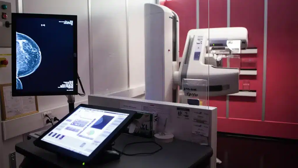
Mammography is a specialized medical imaging technique that uses a low-dose X-ray system to view the internal tissues of the breast. Mammograms assist in the early detection and diagnosis of breast diseases in women.

image shows Mammography in Breast Cancer Detection

Mammography can detect abnormal breast tissue, but it cannot diagnose cancer on its own. It shows irregular areas that might require further testing, such as a breast biopsy, which can confirm whether the tissue is cancerous or benign.

Mammogram developments

Three key advancements in mammography include digital mammography, computer-aided detection (CAD), and breast tomosynthesis.

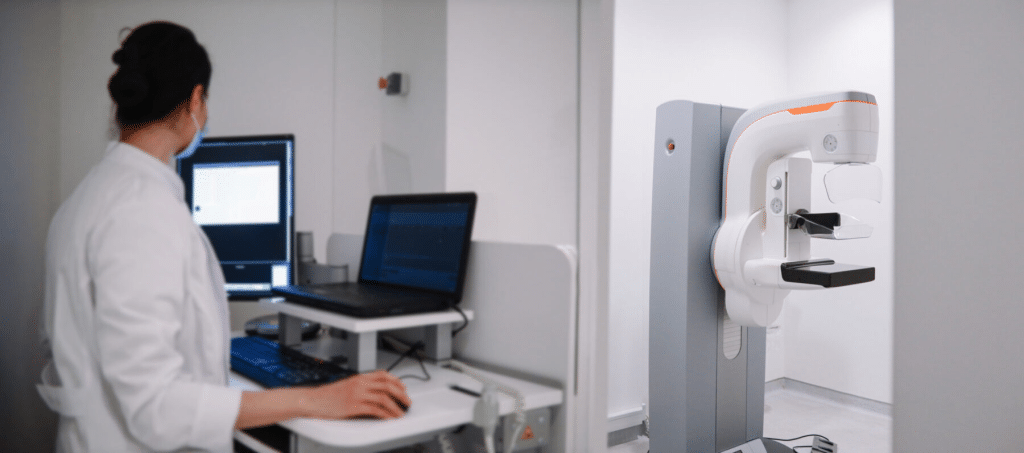
Digital Mammography:

Also known as full-field digital mammography (FFDM), this system replaces traditional film with digital sensors that convert X-rays into breast images. Like digital cameras, it provides clearer images with less radiation exposure. The images can be reviewed by radiologists on computers and stored for long-term access. The patient’s experience during digital mammography is similar to that of conventional mammograms.

Computer-Aided Detection (CAD):

CAD systems analyze mammogram images to identify areas of unusual density, mass, or calcifications that might indicate cancer. These areas are highlighted for the radiologist to review more carefully.

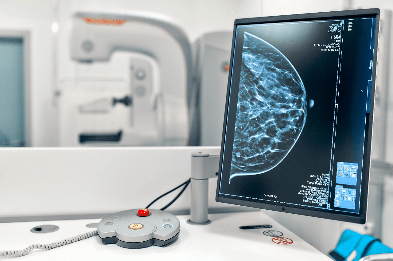
Breast Tomosynthesis:

Also known as 3D mammography or digital breast tomosynthesis (DBT), this technique captures multiple images of the breast from different angles, which are then reconstructed into a 3D image. Similar to a CT scan, the process provides more detailed, layered images.

Mammography in Breast Cancer Detection

Breast tomosynthesis can:

  • Detect small cancers that might be hidden in traditional mammograms
  • Reduce unnecessary biopsies or additional tests
  • Improve the detection of multiple tumors
  • Provide clearer images of dense breast tissue
  • Offer more accurate information about tumor size, shape, and location

Important Note: Mammograms expose patients to a small amount of radiation. However, research indicates that the benefits of early detection far outweigh the risks. The radiation dose is comparable to 18 weeks of natural environmental exposure, and modern mammography machines use minimal radiation to maintain high-quality imaging.

Types of Mammograms

Mammography can be categorized into two types:

Screening Mammogram:

This test is performed on women with no symptoms of breast cancer to detect tumors too small to be felt. It can identify cancers as tiny as a grain of rice. For women over 50, screening mammograms are the most effective method for early breast cancer detection.

Diagnostic Mammogram:

This type is used to investigate symptoms such as lumps, nipple discharge, or abnormalities found in a screening mammogram. Radiologists may take additional images to get a more detailed view of the suspicious area.

an-image-showing-the-results-of-a-mammogram

The Importance of Mammograms in Cancer Detection

Mammograms reduce the risk of death from breast cancer by helping doctors find cancer early, when it is most treatable. They detect various types of breast cancer, including invasive ductal carcinoma and invasive lobular carcinoma. Early detection provides more treatment options and increases the likelihood of successful outcomes. Benefits of Screening Mammograms:

1- Increased Survival Rates:

Studies show that for every 1,000 women screened biennially from age 50 to 74, approximately 8 lives are saved.

2- Improved Early Detection:

Mammograms can find cancers too small to be detected by touch. Detecting cancer early significantly improves the chances of effective treatment.

3- Better Treatment Outcomes:

Cancers detected early are more likely to be smaller and easier to treat, often requiring less aggressive therapies and offering a higher quality of life during and after treatment.

Conclusion:

Mammography plays a crucial role in the early detection of breast cancer by identifying abnormalities before symptoms appear. It remains the most reliable tool for routine breast cancer screening, helping save lives through early detection. If you are interested in learning more about radiology, HSI Center offers specialized training programs in diagnostic imaging, including mammography, led by experienced professionals in the medical engineering field.

Source: The Vital Role of Mammograms in Early Cancer Detection

X-Ray Imaging and Disease Diagnosis

Before the late 19th century, doctors relied on stethoscopes to diagnose lung diseases and physical touch to identify bone fractures. However, the German physicist Wilhelm Röntgen discovered X-rays and their use in imaging. Initially, the use of X-ray imaging was merely a side demonstration, but it quickly spread worldwide and became a fundamental aspect of medical diagnosis.

What is X-ray imaging?

X-ray imaging uses invisible electromagnetic energy beams to produce images of internal tissues, bones, and organs on film or digital media. Standard X-rays are performed for various reasons, including diagnosing tumors, bone injuries, dental examinations, and aiding in detecting a wide range of injuries, disorders, and diseases.

Historical_X-ray

X-Ray Imaging are performed using external radiation to produce images of the body, its organs, and other internal structures for diagnostic purposes. X-rays pass through the body’s structures onto specially treated plates, similar to camera films or digital media. The denser the structure, the whiter it appears on the film.

How do X-rays work?

Most people refer to X-ray imaging as a regular radiographic image (a single film or photo). However, X-rays are also a type of radiation used by imaging devices to create images. X-rays are also used in other types of medical imaging, such as computed tomography (CT) scans, to obtain multiple images that computers interpret to create three-dimensional pictures.

When the body is exposed to X-rays, different parts of the body allow varying amounts of X-rays to pass through. Soft tissues like blood, skin, fat, and muscles allow most X-rays to pass through, appearing dark gray on film or digital media. On the other hand, bones or tumors, which are denser than soft tissues, allow fewer X-rays to pass through and appear white on X-rays. When a bone fracture occurs, the X-ray beam passes through the broken area, which appears as a dark line on the white bone in the X-ray.

X-Ray Imaging with contrast material

Some X-Ray Imaging use contrast material (also called contrast agents or dyes). These make certain structures in the body, such as blood vessels, easier to see.

Contrast material can be in liquid, powder, or tablet form, consumed before the X-ray depending on the type of examination. Patients may receive contrast material via:

  1. Oral ingestion.
  2. Intravenous (IV) injection or spinal fluid injection.
  3. Enema.

X-Ray Imaging

How is an X-ray performed?

X-ray examinations can be performed in outpatient clinics or as part of inpatient care in hospitals. Although each facility may have specific protocols, the general steps for performing an X-ray are as follows:

1. Preparation before imaging

The patient is asked to remove any clothing or jewelry that may interfere with imaging the area being examined. If necessary, the patient is provided with a gown. The use of lotions, creams, or perfumes is avoided, as these substances can create shadows on X-rays, leading to inaccurate results. Patients may also need to refrain from eating or drinking several hours before the procedure.

2. Body positioning

The area being examined is carefully placed between the X-ray machine and a plate containing the X-ray film or a specialized image plate. Depending on the type of examination, the patient may need to lie down, sit, or stand.

3. Radiation protection

The parts of the body not being examined are covered with a lead shield to reduce radiation exposure.

4. Directing the X-ray beam

The beam is directed at the area being examined. The patient must remain still to avoid blurry images. Maintaining stillness during the imaging process ensures high-quality results.

5. Capturing images

The radiology technician stands behind a protective window to capture the image. Multiple images may be taken from different angles depending on the area being examined. For example, when imaging the chest, both front and side views are usually taken.

x-ray imaging

What are the types of X-rays?

Several types of X-rays are used to image different areas of the body. Some of the most common types include:

1- Abdominal X-rays

These X-rays help healthcare providers evaluate parts of the digestive system and diagnose conditions such as kidney or bladder stones.

2- Bone X-rays

Patients may undergo bone X-rays if a doctor suspects fractures, joint dislocations, or arthritis. Bone X-rays can also show signs of bone cancer or infections.

3- Chest X-ray

A doctor may request chest X-rays if a patient experiences symptoms such as chest pain, shortness of breath, or a persistent cough.

4- Dental X-rays

Dentists regularly take X-rays of the mouth to check for issues in the teeth, gums, or jaw.

5- Head X-rays

These images help identify skull fractures from head injuries or conditions affecting how skull bones form, such as craniosynostosis.

6- Spinal X-rays

Spinal X-rays can help diagnose spinal curvatures, herniated discs, or other spinal issues.

Other advanced medical imaging techniques using X-rays interpreted by computers include:

  • Bone density scans (DXA)
  • Computed tomography (CT scans)
  • Fluoroscopy
  • Mammography

x ray imaging

When does a patient need X-rays?

  1. Diagnosing tumors.
  2. Detecting bone injuries.
  3. Imaging arteries to examine blood vessels.
  4. CT scans for three-dimensional imaging.
  5. Fluoroscopy to monitor internal movements.
  6. Identifying the causes of symptoms like pain or swelling.
  7. Detecting signs of lung infections.
  8. Locating foreign objects inside the body.
  9. Identifying structural problems in bones, joints, or soft tissues.
  10. Planning and evaluating treatments.

What can X-Ray Imaging show?

X-rays can reveal issues such as:

  • Arthritis.
  • Bone fractures.
  • Changes or abnormalities in bone structure.
  • Spinal disc herniation.
  • Infections.
  • Kidney stones.
  • Spinal curvatures.
  • Dental cavities.
  • Tumors.

Can X-Ray Imaging show cancerous tumors?

X-Ray Imaging can reveal cancerous tumors, but they are not the primary method for detecting or diagnosing cancer. This is because tumors in certain organs may be small, hidden behind other structures (e.g., ribs in chest X-rays), or blend with normal tissues.

In conclusion

X-ray imaging has revolutionized the diagnosis of medical conditions, enabling healthcare professionals to visualize the internal structures of the human body, which has facilitated the detection of various diseases. For healthcare professionals seeking to deepen their understanding and proficiency in the field of medical imaging, the HSI Center for Health Training and Consulting offers specialized courses and consulting services that keep them up to date with the latest advancements in the medical field, helping them provide the highest level of service and care to patients.

Source: Diagnostic X-Ray Procedures

All copyrights reserved to © HSI