X-Ray Imaging and Disease Diagnosis

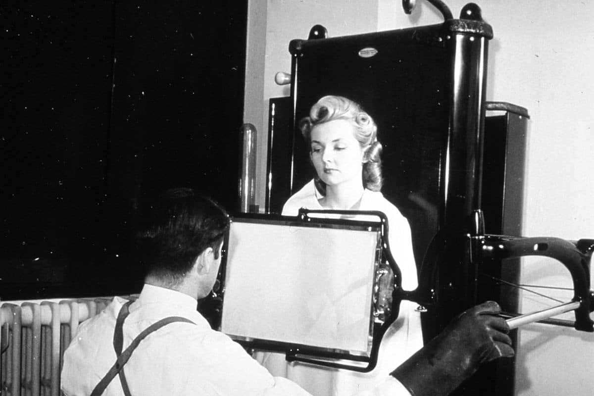
Before the late 19th century, doctors relied on stethoscopes to diagnose lung diseases and physical touch to identify bone fractures. However, the German physicist Wilhelm Röntgen discovered X-rays and their use in imaging. Initially, the use of X-ray imaging was merely a side demonstration, but it quickly spread worldwide and became a fundamental aspect of medical diagnosis.

What is X-ray imaging?

X-ray imaging uses invisible electromagnetic energy beams to produce images of internal tissues, bones, and organs on film or digital media. Standard X-rays are performed for various reasons, including diagnosing tumors, bone injuries, dental examinations, and aiding in detecting a wide range of injuries, disorders, and diseases.

Historical_X-ray

X-Ray Imaging are performed using external radiation to produce images of the body, its organs, and other internal structures for diagnostic purposes. X-rays pass through the body’s structures onto specially treated plates, similar to camera films or digital media. The denser the structure, the whiter it appears on the film.

How do X-rays work?

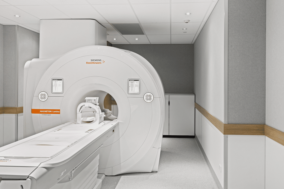
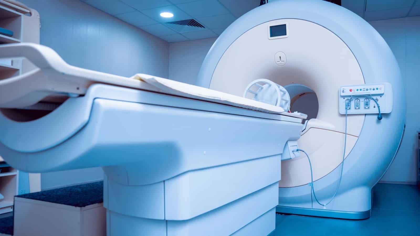
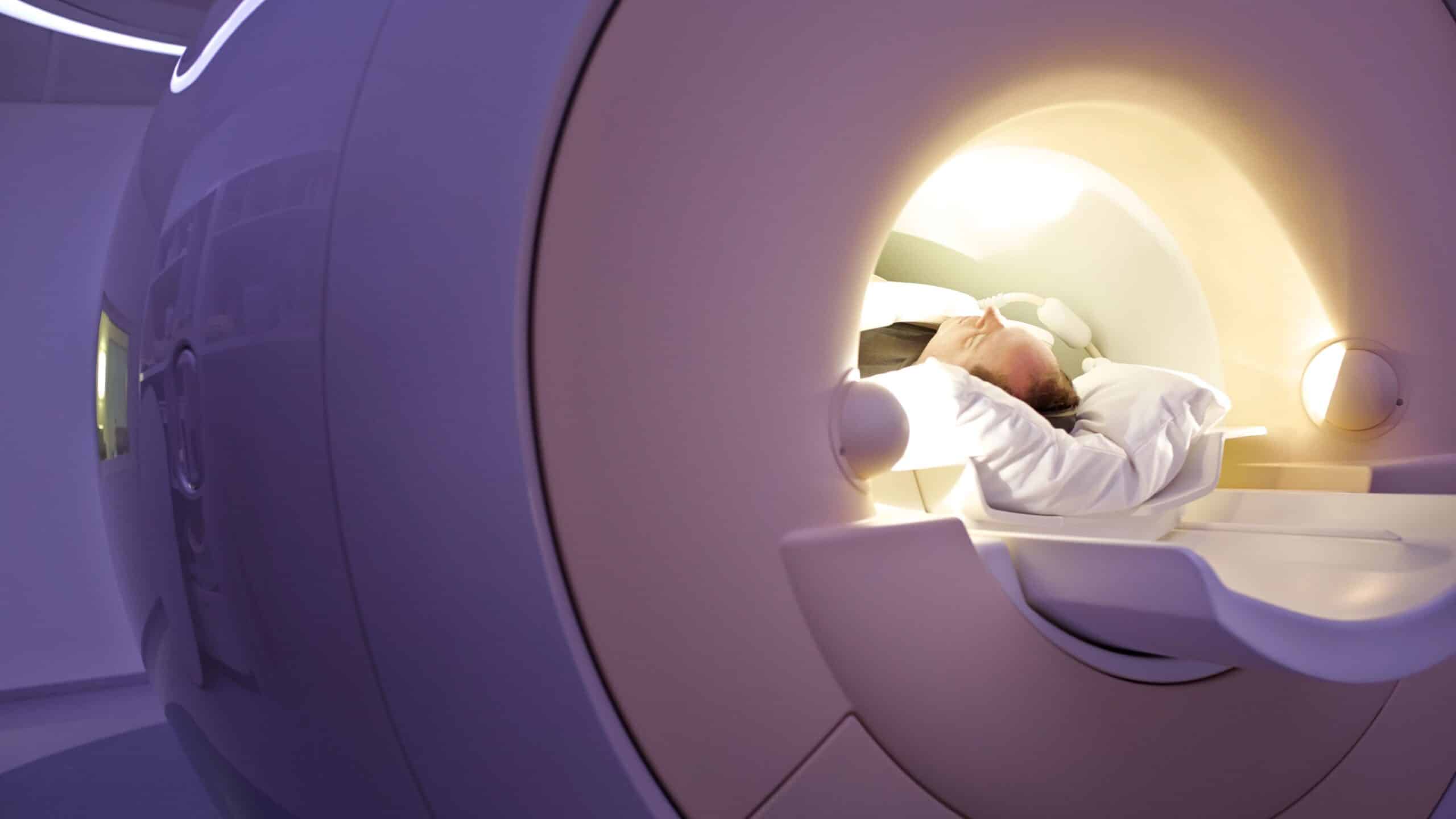
Most people refer to X-ray imaging as a regular radiographic image (a single film or photo). However, X-rays are also a type of radiation used by imaging devices to create images. X-rays are also used in other types of medical imaging, such as computed tomography (CT) scans, to obtain multiple images that computers interpret to create three-dimensional pictures.

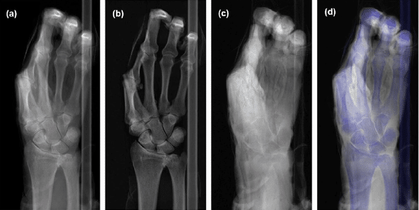
When the body is exposed to X-rays, different parts of the body allow varying amounts of X-rays to pass through. Soft tissues like blood, skin, fat, and muscles allow most X-rays to pass through, appearing dark gray on film or digital media. On the other hand, bones or tumors, which are denser than soft tissues, allow fewer X-rays to pass through and appear white on X-rays. When a bone fracture occurs, the X-ray beam passes through the broken area, which appears as a dark line on the white bone in the X-ray.

X-Ray Imaging with contrast material

Some X-Ray Imaging use contrast material (also called contrast agents or dyes). These make certain structures in the body, such as blood vessels, easier to see.

Contrast material can be in liquid, powder, or tablet form, consumed before the X-ray depending on the type of examination. Patients may receive contrast material via:

  1. Oral ingestion.
  2. Intravenous (IV) injection or spinal fluid injection.
  3. Enema.

X-Ray Imaging

How is an X-ray performed?

X-ray examinations can be performed in outpatient clinics or as part of inpatient care in hospitals. Although each facility may have specific protocols, the general steps for performing an X-ray are as follows:

1. Preparation before imaging

The patient is asked to remove any clothing or jewelry that may interfere with imaging the area being examined. If necessary, the patient is provided with a gown. The use of lotions, creams, or perfumes is avoided, as these substances can create shadows on X-rays, leading to inaccurate results. Patients may also need to refrain from eating or drinking several hours before the procedure.

2. Body positioning

The area being examined is carefully placed between the X-ray machine and a plate containing the X-ray film or a specialized image plate. Depending on the type of examination, the patient may need to lie down, sit, or stand.

3. Radiation protection

The parts of the body not being examined are covered with a lead shield to reduce radiation exposure.

4. Directing the X-ray beam

The beam is directed at the area being examined. The patient must remain still to avoid blurry images. Maintaining stillness during the imaging process ensures high-quality results.
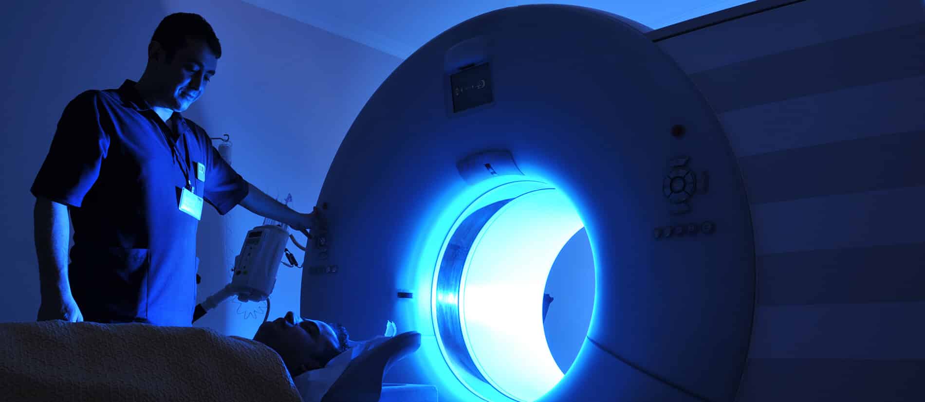
5. Capturing images

The radiology technician stands behind a protective window to capture the image. Multiple images may be taken from different angles depending on the area being examined. For example, when imaging the chest, both front and side views are usually taken.

x-ray imaging

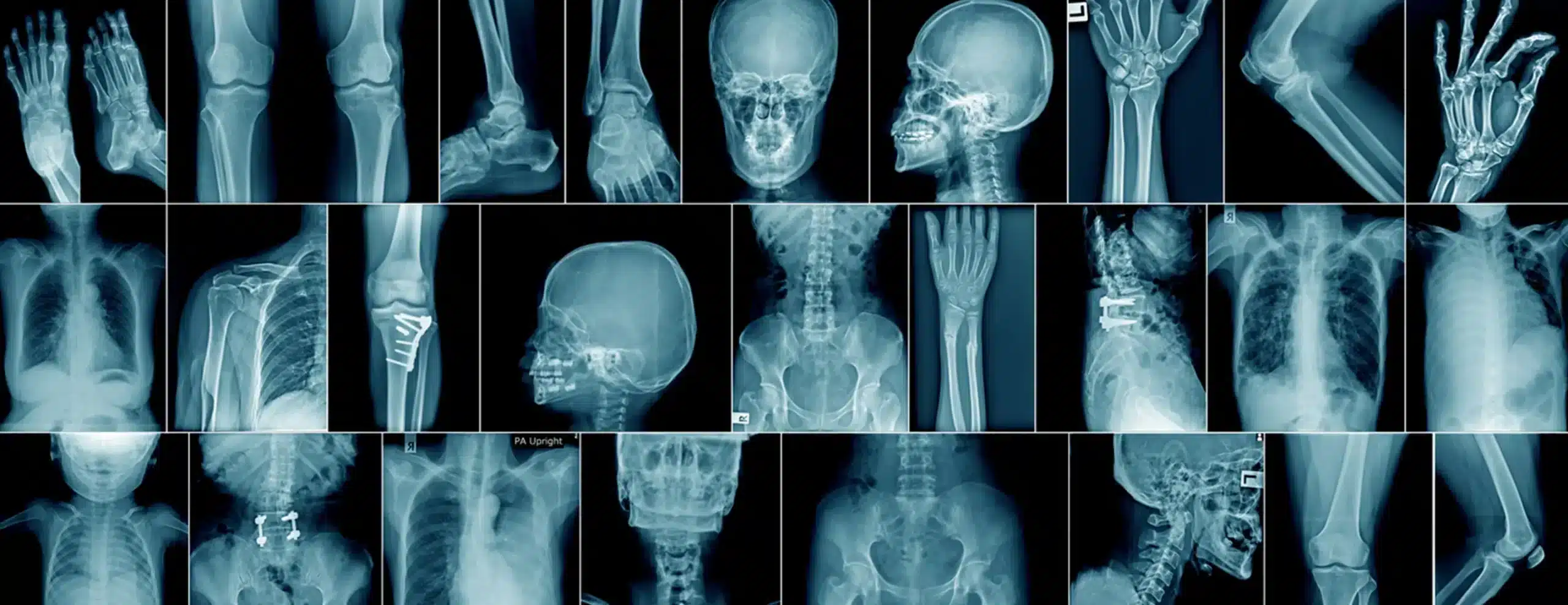
What are the types of X-rays?

Several types of X-rays are used to image different areas of the body. Some of the most common types include:

1- Abdominal X-rays

These X-rays help healthcare providers evaluate parts of the digestive system and diagnose conditions such as kidney or bladder stones.

2- Bone X-rays

Patients may undergo bone X-rays if a doctor suspects fractures, joint dislocations, or arthritis. Bone X-rays can also show signs of bone cancer or infections.
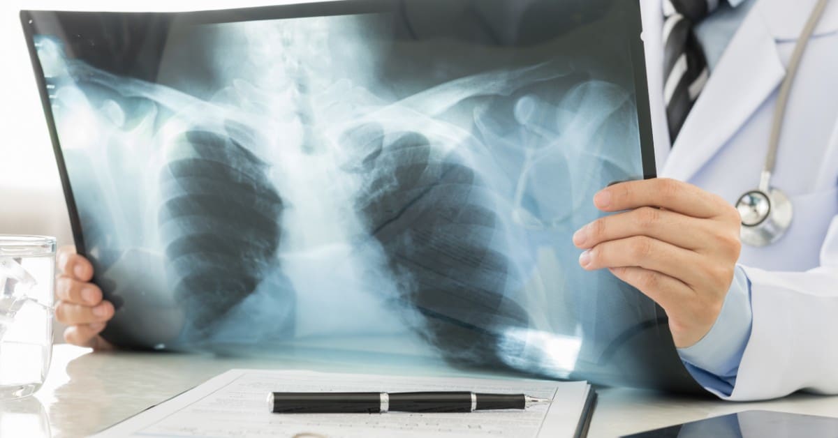
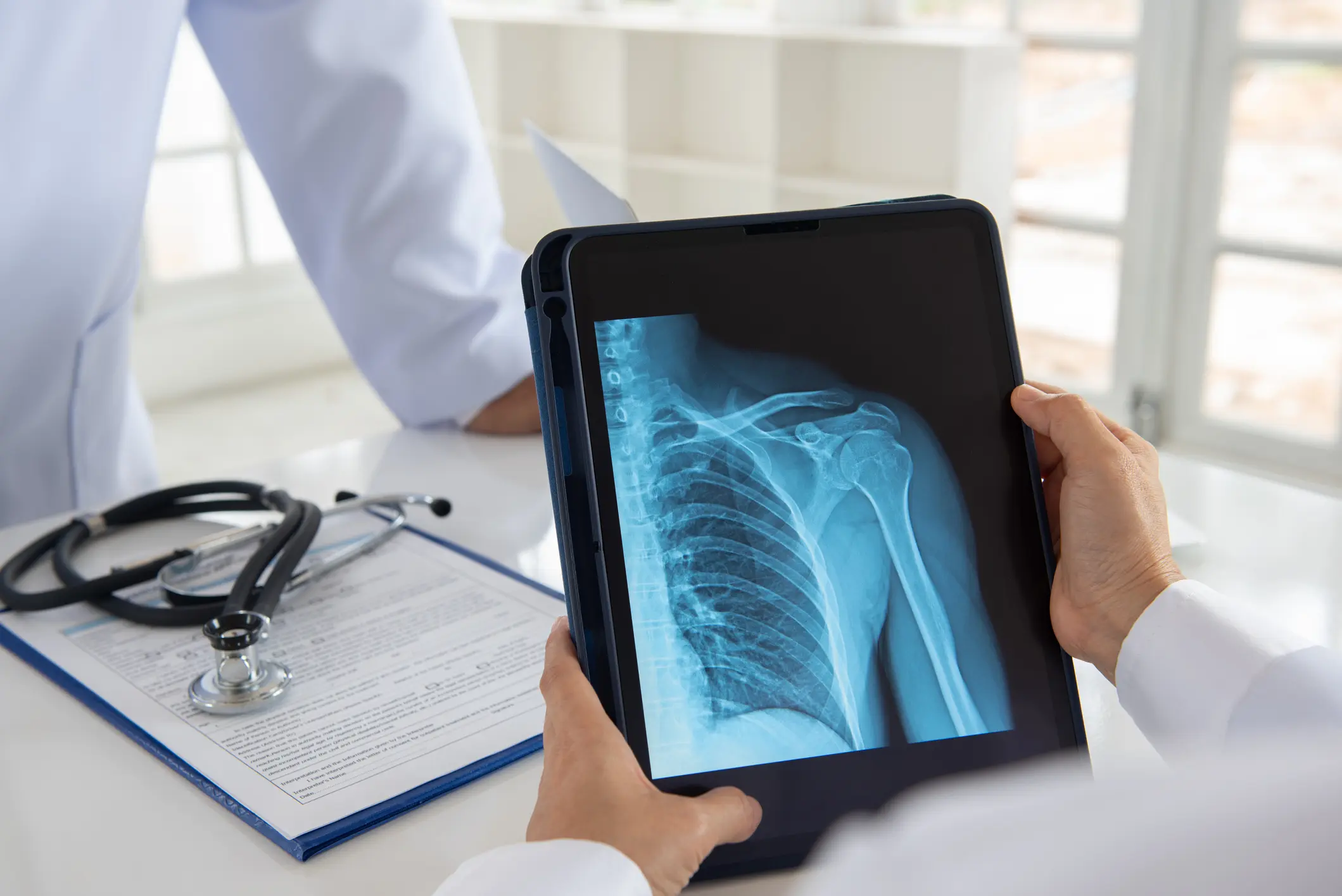
3- Chest X-ray

A doctor may request chest X-rays if a patient experiences symptoms such as chest pain, shortness of breath, or a persistent cough.

4- Dental X-rays

Dentists regularly take X-rays of the mouth to check for issues in the teeth, gums, or jaw.

5- Head X-rays

These images help identify skull fractures from head injuries or conditions affecting how skull bones form, such as craniosynostosis.
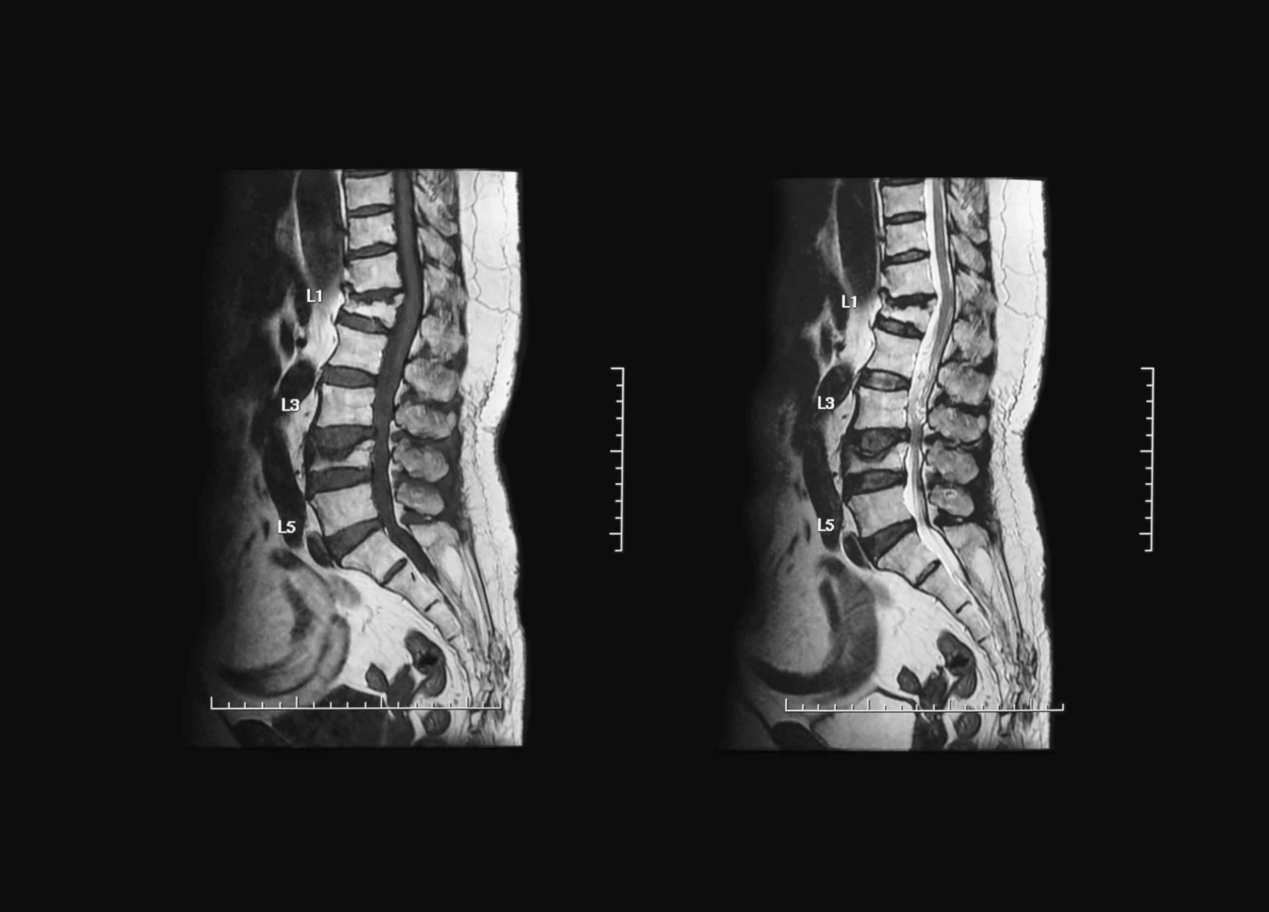
6- Spinal X-rays

Spinal X-rays can help diagnose spinal curvatures, herniated discs, or other spinal issues.

Other advanced medical imaging techniques using X-rays interpreted by computers include:

  • Bone density scans (DXA)
  • Computed tomography (CT scans)
  • Fluoroscopy
  • Mammography

x ray imaging

When does a patient need X-rays?

  1. Diagnosing tumors.
  2. Detecting bone injuries.
  3. Imaging arteries to examine blood vessels.
  4. CT scans for three-dimensional imaging.
  5. Fluoroscopy to monitor internal movements.
  6. Identifying the causes of symptoms like pain or swelling.
  7. Detecting signs of lung infections.
  8. Locating foreign objects inside the body.
  9. Identifying structural problems in bones, joints, or soft tissues.
  10. Planning and evaluating treatments.

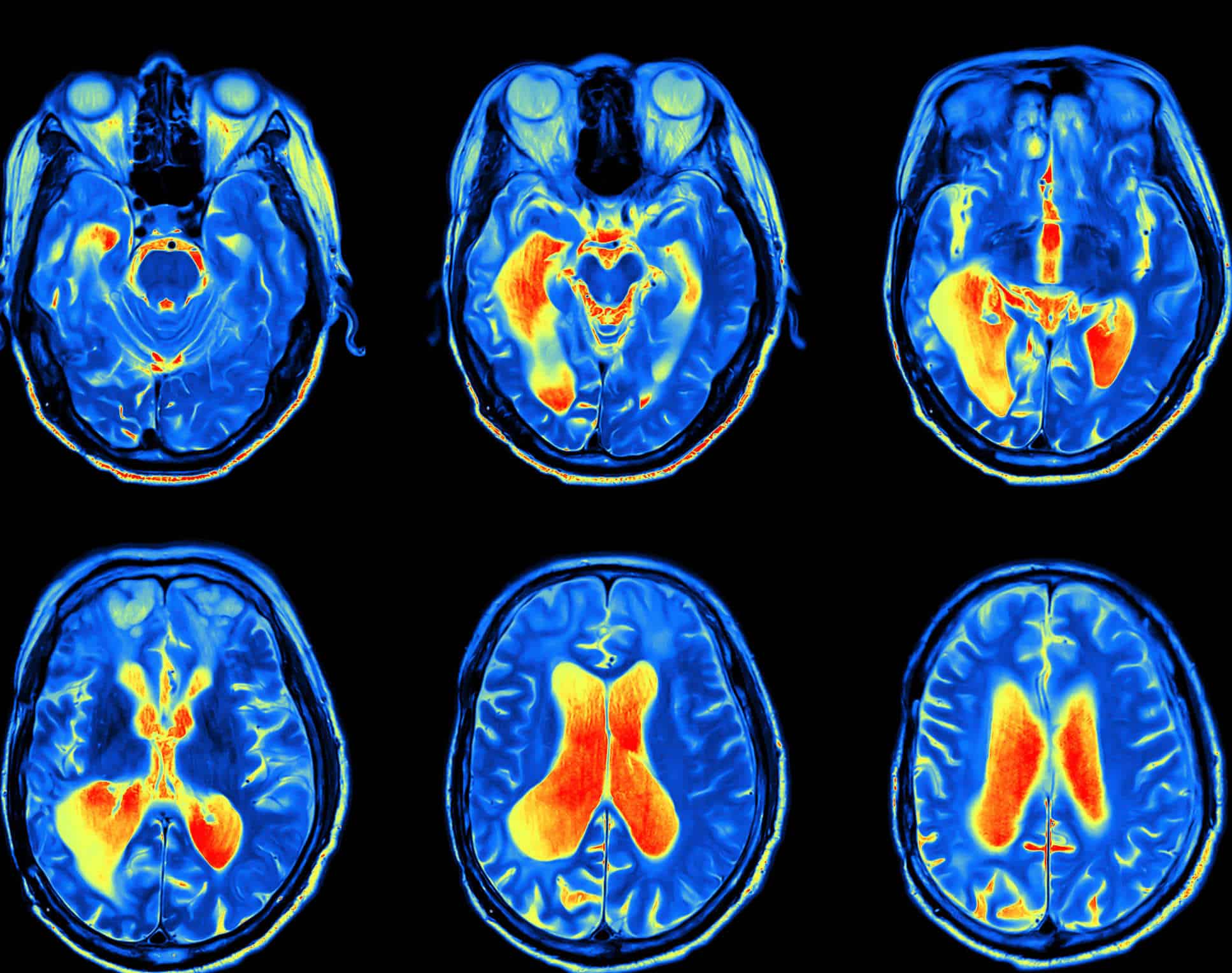
What can X-Ray Imaging show?

X-rays can reveal issues such as:

  • Arthritis.
  • Bone fractures.
  • Changes or abnormalities in bone structure.
  • Spinal disc herniation.
  • Infections.
  • Kidney stones.
  • Spinal curvatures.
  • Dental cavities.
  • Tumors.

Can X-Ray Imaging show cancerous tumors?

X-Ray Imaging can reveal cancerous tumors, but they are not the primary method for detecting or diagnosing cancer. This is because tumors in certain organs may be small, hidden behind other structures (e.g., ribs in chest X-rays), or blend with normal tissues.

In conclusion

X-ray imaging has revolutionized the diagnosis of medical conditions, enabling healthcare professionals to visualize the internal structures of the human body, which has facilitated the detection of various diseases. For healthcare professionals seeking to deepen their understanding and proficiency in the field of medical imaging, the HSI Center for Health Training and Consulting offers specialized courses and consulting services that keep them up to date with the latest advancements in the medical field, helping them provide the highest level of service and care to patients.

Source: Diagnostic X-Ray Procedures

Revolutionizing Early Disease Detection: The Role of MRI in Medical Diagnostics

Magnetic Resonance Imaging (MRI) is a non-invasive diagnostic tool that uses a magnetic field and radio waves to produce detailed images of the inside of the human body. It plays a crucial role in the medical field and is considered a revolution in medical diagnostics. The future of MRI technology will see significant advancements in the clarity of the images it produces, and it is expected to develop further alongside the rise of artificial intelligence. In the following sections, we will explore this technology, its role in early disease detection, and how it will continue to evolve in the future.

الرنين المغناطيسي

MRI Technology
MRI works by exposing the body to a specific magnetic field. This magnetic field exploits the physics of protons, which are abundant in the human body due to the high water content in tissues. When the body is subjected to a strong magnetic field, the hydrogen protons in the tissues align with the field. Then, radio pulses are sent through the body, temporarily altering the orientation of these protons.

After a brief period, the hydrogen protons return to their original state and release signals that are captured by the MRI machine. These signals are used to create detailed images of the internal body structures, clearly distinguishing different tissues and organs and enabling precise identification of diseases.

How MRI Can Be Used for Early Disease Detection

MRI is an effective tool for early disease detection due to the high accuracy of its images of body tissues and organs. It can be used in the early stages of tumor development, increasing the chances of successful treatment. Additionally, MRI is valuable for diagnosing various neurological conditions such as strokes and multiple sclerosis. One of its key roles in neurology is identifying changes in brain tissue even before clinical symptoms appear, significantly aiding early detection and effective treatment.

الرنين المغناطيسي

One of the important uses of MRI is in the early detection of diabetic retinopathy, a complication of diabetes. This is done by creating a digital image of the retina and examining it for early signs of the condition.

MRI is also used for early detection of breast cancer, as traditional imaging methods may not detect the disease in its early stages. Furthermore, MRI can help detect possible artery blockages or narrowing that could lead to heart muscle problems. When it comes to bones, MRI can detect early signs of arthritis and changes in surrounding bone tissue.

Innovative Solutions in MRI Technology

While MRI itself is a revolutionary tool, it is continuously evolving to offer innovative solutions for disease detection and treatment. Some of the key innovations include:

Improved Image Accuracy

MRI imaging has been enhanced using mathematical equations, particularly by applying statistical science to improve the visibility of tumors. One of the methods used is calculating the probability mass function for each image in a given time sequence. This process involves analyzing the distribution of different values within the image, helping to identify changes in tissues and internal organs.

Another technique called Kullback-Leibler divergence is used to determine the best images for detecting tumors. This mathematical measure compares two different values, helping to identify the contrast between healthy tissue and a tumor, making the tumor more visible.

Advanced Image Processing Techniques

To improve image processing, advanced algorithms like GLCM (Gray Level Co-occurrence Matrix) and PNN (Probabilistic Neural Networks) are used to detect different brain tumors. These technologies have greatly advanced medical image processing, making it easier and more accurate for doctors to identify various tumor patterns for accurate diagnosis.

Use of Artificial Intelligence

The rapid growth of artificial intelligence tools in recent years will significantly enhance MRI technology. AI assists in accurately analyzing images and handling large data sets (Big Data) to recognize complex patterns, thereby improving doctors’ ability to diagnose diseases more accurately and quickly.

Challenges Facing MRI Technology

Despite the tremendous advancements in MRI technology and its applications, there are some challenges:

  • High Cost: MRI machines are expensive to purchase and maintain, and operational costs increase when advanced techniques are used.
  • Complexity: Some modern MRI techniques can be complex, requiring high skill levels and a deep understanding of how to operate the equipment.
  • Special Needs Patients: It can be difficult for patients with special needs to undergo MRI scans, as they may struggle to stay still during the examination.
  • Patient Anxiety: Some patients may be fearful of being confined in small spaces for extended periods, requiring doctors to take extra time to reassure them.
  • Long Exam Duration: Some MRI scans take 30 to 90 minutes, which can be uncomfortable, especially for elderly patients.

The Role of Contrast Agents in MRI

Contrast agents are chemicals injected into the body before an MRI scan to enhance image clarity and improve the accuracy of details around tissues and internal organs. Contrast agents play a crucial role in MRI:

  • Increasing contrast to highlight differences between tissues, helping to distinguish between healthy and diseased tissue.
  • Certain liquid agents, such as gadolinium, are used to improve visibility of tissues.
  • Gaseous contrast agents are used in specialized cases, such as lung imaging, to interact with oxygen or carbon dioxide for clearer images.
  • They also clarify blood vessels and examine blood flow for conditions like aneurysms.
  • They assist in detecting tumors.
  • They help examine internal organs like the liver and kidneys for any pathological changes.

Innovative Contrast Agents in MRI

Over time, the contrast agents used in MRI have been developed to provide the best possible image quality and diagnostic accuracy. Some of the innovative contrast agents include:

Gadolinium-based agents, which are the most commonly used in central nervous system imaging.
Iron-based agents, typically used for liver and vascular imaging.
Manganese-based agents, which are highly effective at targeting specific organs for precise imaging.
These agents are essential components of MRI techniques, and specialists choose the appropriate agent for each specific case.

With the tremendous advancements in the medical field over recent years, it is crucial for biomedical engineers to continuously enhance their skills and expertise. This is where platforms like HSI come into play, offering courses and consultations in the medical field to help you advance in your academic journey.

on our HSI website, we offer specialized training courses and consultations in the field of biomedical engineering, led by a team of certified experts and specialists to ensure you receive the best scientific content that combines theory and practical application. For inquiries, please get in touch with us, and we will respond with all the information you need to deepen your knowledge in the field of biomedical engineering.

Source: How Diagnostic Imaging is Revolutionizing Early Disease Detection

All copyrights reserved to © HSI