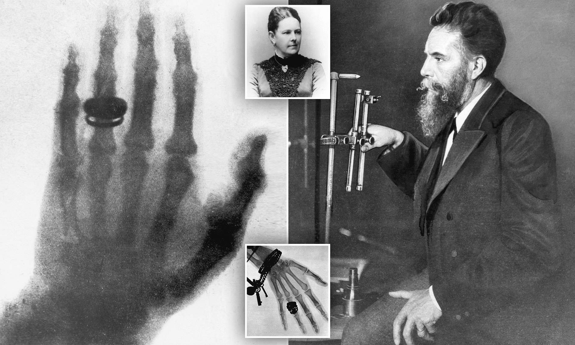
The field of radiology has undergone remarkable transformations since Wilhelm Roentgen’s groundbreaking discovery of X-rays in 1895. Among the many advancements, the development and application of contrast agents in medical imaging stand out as pivotal contributions that have significantly enhanced the diagnostic capabilities of various imaging modalities.
The use of contrast agents in medical imaging has revolutionized diagnostics, greatly improved the visualization of internal structures and enhanced diagnostic accuracy across X-ray imaging, computed tomography (CT), magnetic resonance imaging (MRI), and ultrasound. In this article, we will explore in detail the types of contrast agents, their benefits in medical imaging, and their potential risks.
Historical Development of Contrast Agents
The historical development of contrast agents has been marked by significant milestones. Early contrast materials, such as simple salts and barium sulfate, provided initial glimpses into internal anatomical structures but were limited due to their high toxicity and poor image quality. The development of iodine-based agents in the 20th century revolutionized X-ray imaging by offering lower toxicity and improved clarity. Similarly, the introduction of gadolinium-based agents in the 1980s transformed MRI imaging, providing more precise details. Modern innovations, including nanoparticle-based imaging systems, continue to enhance specificity and safety in diagnostic imaging. Now, let’s explore what contrast agents are and their applications in medical imaging.

What Are Contrast Agents and How Are They Used?
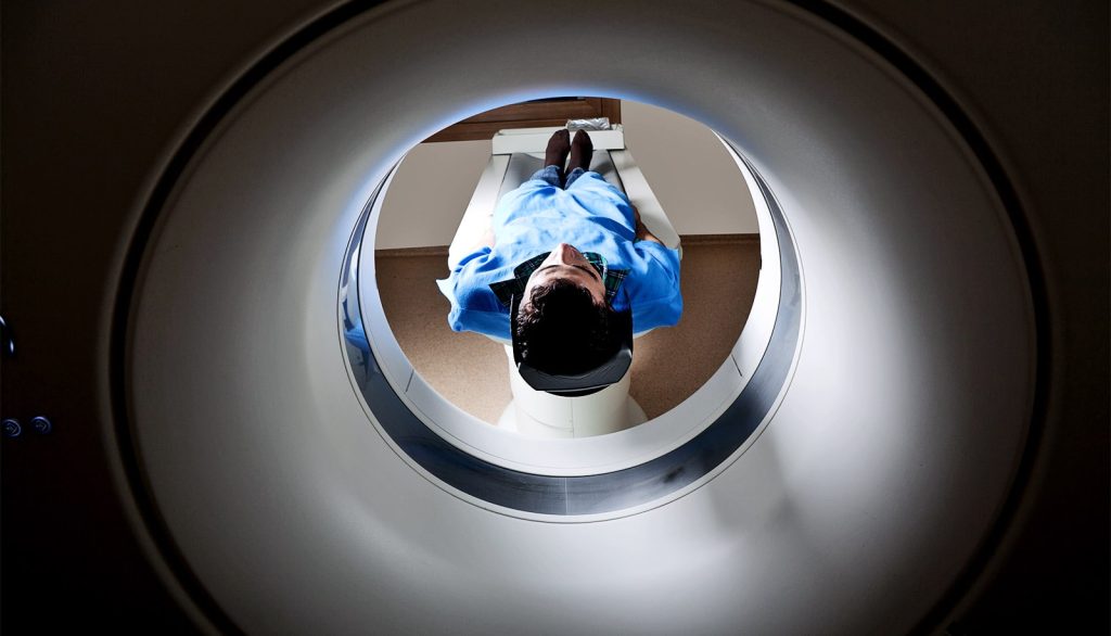
Contrast media (MDC), or contrast agents, are substances used in diagnostic imaging to improve the clarity of high-resolution scans, such as CT and MRI. These agents enhance image quality by highlighting tissue details and potential abnormalities that would otherwise be difficult to detect. Contrast agents improve the visualization of internal structures within the body, facilitating early and accurate diagnosis of a wide range of medical conditions, including vascular diseases, tumors, and neurological disorders. They are also known as contrast media or contrast dyes, but the term “contrast agent” is the most commonly used.
As mentioned, contrast agents are an integral part of various imaging techniques, including X-rays, CT scans, MRI scans, and ultrasound. Each modality utilizes specific types of contrast media designed to improve image quality and diagnostic accuracy. For example, iodine-based contrast agents are primarily used in CT scans and angiographic studies, while gadolinium-based agents are essential for enhancing MRI scans. The introduction of microbubble contrast agents has expanded ultrasound imaging capabilities, enabling real-time, detailed assessments of blood flow and tissue perfusion.
Effects on X-ray Images: Positive, Negative, and Neutral Contrast
Contrast agents in medical imaging enhance the visibility of specific tissues or blood vessels during imaging procedures. They help improve image quality and clarity, allowing for more precise detection and diagnosis of various medical conditions. Contrast agents are categorized into three groups based on their effects on the final images:
- Positive Contrast Agents
- Negative Contrast Agents
- Neutral Contrast Agents
What Are Positive Contrast Agents?
Positive contrast agents increase X-ray attenuation within the body, making tissues or structures containing these agents appear whiter or brighter on X-ray images. These agents are generally radiopaque, meaning they absorb X-rays more readily than surrounding tissues. Increased X-ray absorption leads to greater contrast between the contrast agent and surrounding tissues, making it easier to visualize specific structures or abnormalities.
Examples of Positive Contrast Agents
Common examples of positive contrast agents include iodine-based contrast media and barium sulfate. These agents are widely used in various diagnostic imaging procedures, such as angiography, CT scans, and gastrointestinal tract examinations.
What Are Negative Contrast Agents?
In contrast to positive contrast agents, negative contrast agents decrease X-ray attenuation within the body, making tissues or structures containing these agents appear darker on X-ray images. These agents are radiolucent, meaning they allow X-rays to pass through more easily than surrounding tissues. Reduced X-ray absorption, caused by contrast agents like air or carbon dioxide, creates a distinct contrast between the agent and surrounding tissues, helping highlight specific structures or abnormalities.
Examples of Negative Contrast Agents
Air and carbon dioxide are common examples of negative contrast agents used in imaging. These agents are often utilized for imaging specific areas, such as the gastrointestinal tract, where the presence of gas can provide valuable diagnostic information.
What Are Neutral Contrast Agents?
Water is considered a neutral contrast agent because it is readily available, inexpensive, and harmless to the human body. When used in abdominal imaging, water can be ingested or administered through an enema to aid in visualizing the digestive tract and other structures in the abdomen.
One of the primary benefits of using water as a contrast agent is its natural properties. Water has a density similar to many tissues and fluids in the body, meaning it does not significantly alter the overall appearance of organs and structures being imaged. This natural compatibility helps provide a clear and accurate representation of the area being examined.
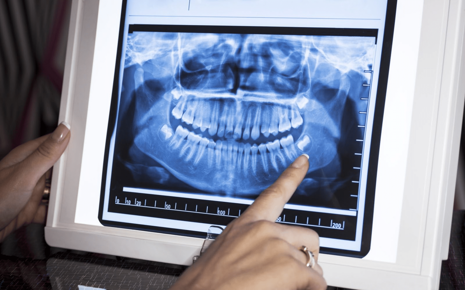
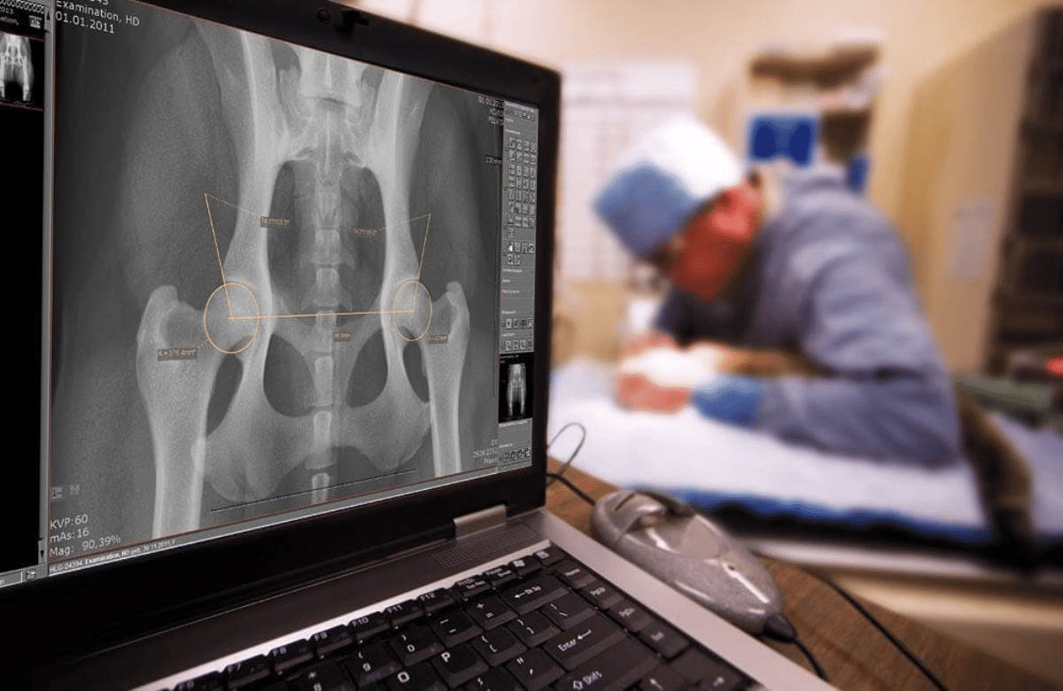
Contrast Agents in X-ray Imaging
All radiographic contrast agents used in X-ray imaging can be classified based on their effects on the resulting images and the type of physical media they utilize. Some also refer to these agents as contrast dyes. In radiology, understanding these classifications is crucial in determining the appropriate iodinated contrast agent for a specific imaging procedure.

In X-ray imaging, contrast plays a critical role in distinguishing between different tissues and structures within the body. The inherent contrast in these images results from the differential attenuation of X-rays and the density variations in anatomical structures. As X-rays pass through the body, they are absorbed or scattered depending on the density of the tissues they encounter. Dense tissues with a high atomic number, such as bones, absorb more X-rays and appear white on the image, whereas less dense tissues, such as muscles or fat, allow more X-rays to pass through and appear darker.
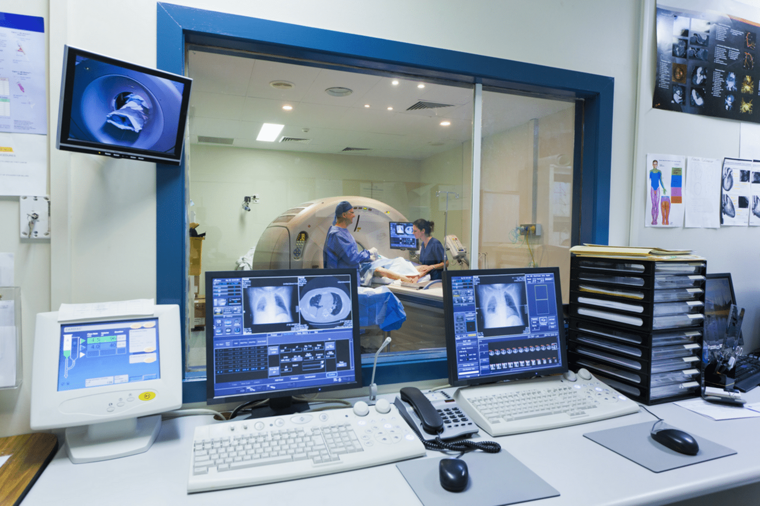
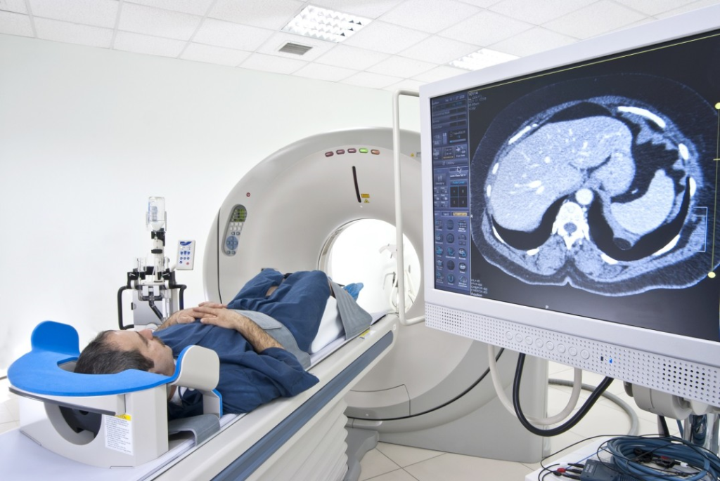
Contrast Agents in Computed Tomography (CT)
One of the imaging modalities that use radiographic contrast fluids is computed tomography (CT). In this case, organic iodine contrast agents are used. Today, advancements in contrast agents have led to the development of substances that are better tolerated by the body and are generally well-accepted, even by patients with severe allergies.
It is important to note that contrast agents are large molecules and are not routinely used medications. They are known for their potential intolerance and may cause unpredictable adverse reactions upon administration.
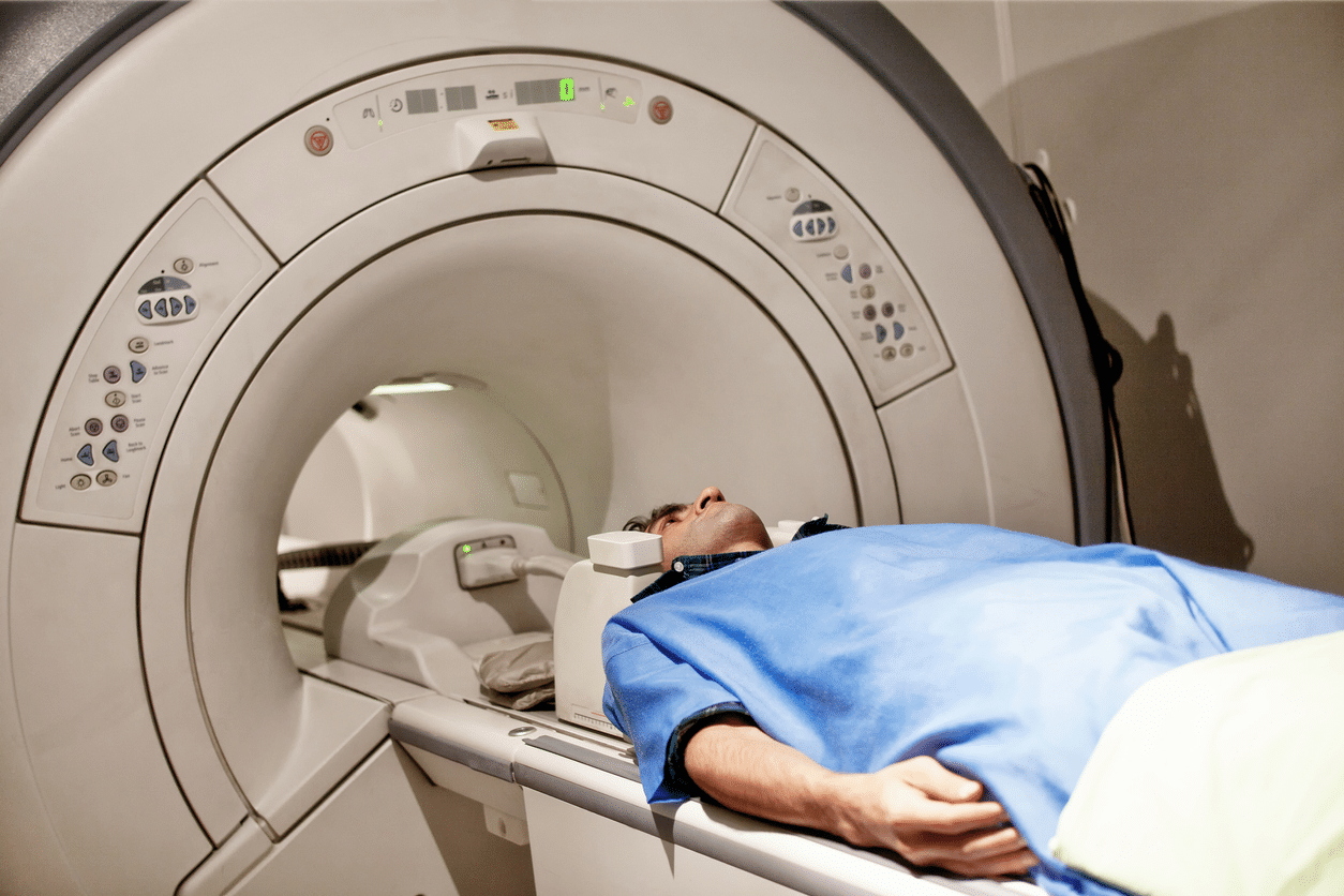
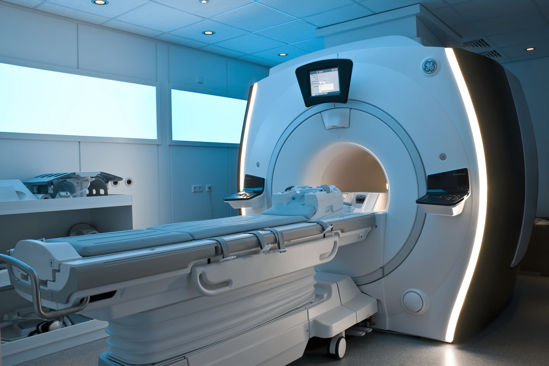
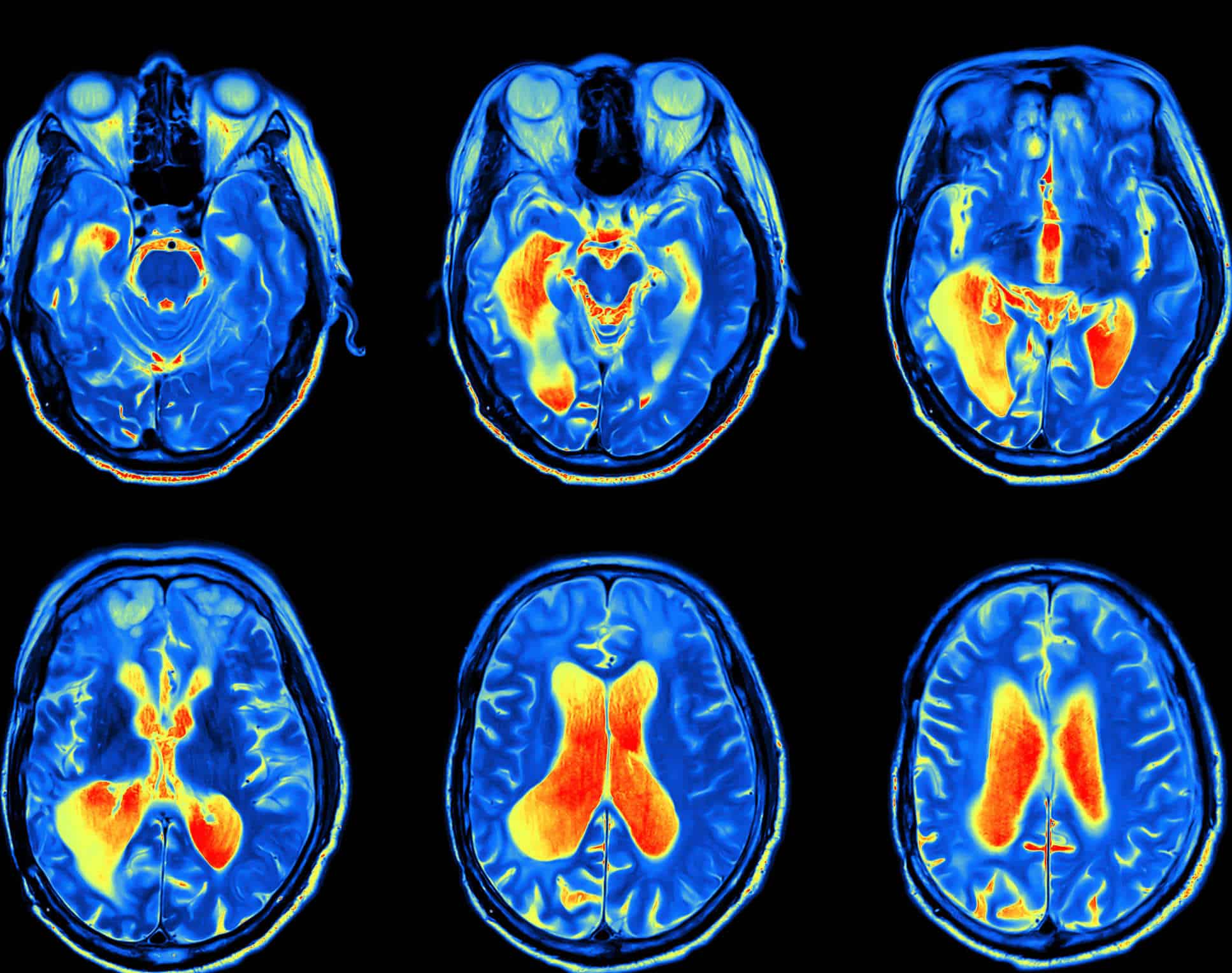
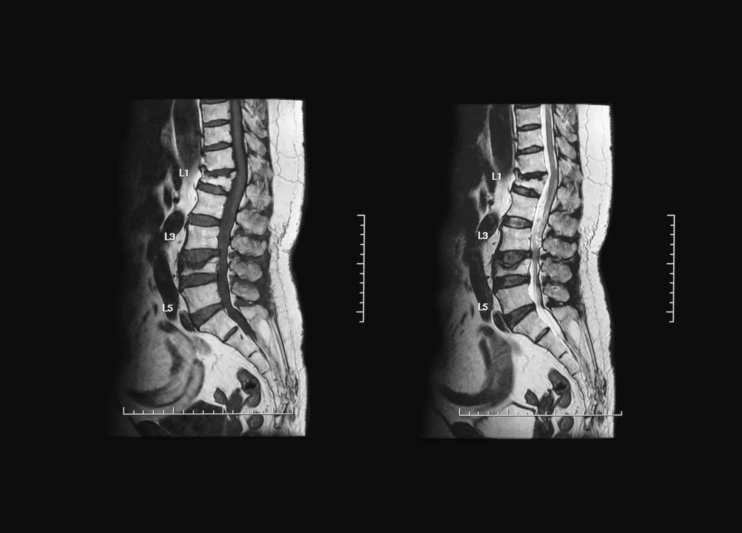
Contrast Agents in Magnetic Resonance Imaging (MRI)
In magnetic resonance imaging (MRI), contrast agents are used, but iodinated organic substances (as in CT scans) are not utilized. Instead, MRI contrast agents are primarily based on gadolinium, an element in the periodic table that belongs to the rare earth metals. Gadolinium is well tolerated by the body and has minimal side effects.
The contrast medium used in MRI is excreted through the kidneys, and thus, the same precautions applied to iodinated contrast agents are considered, including assessing kidney function via plasma creatinine levels before the examination.
Uses of Gadolinium Contrast Agents
Gadolinium-based contrast agents are used for various purposes, including:
- Evaluating suspected abdominal masses to determine whether they are malignant tumors (sarcomas) or benign lipomas.
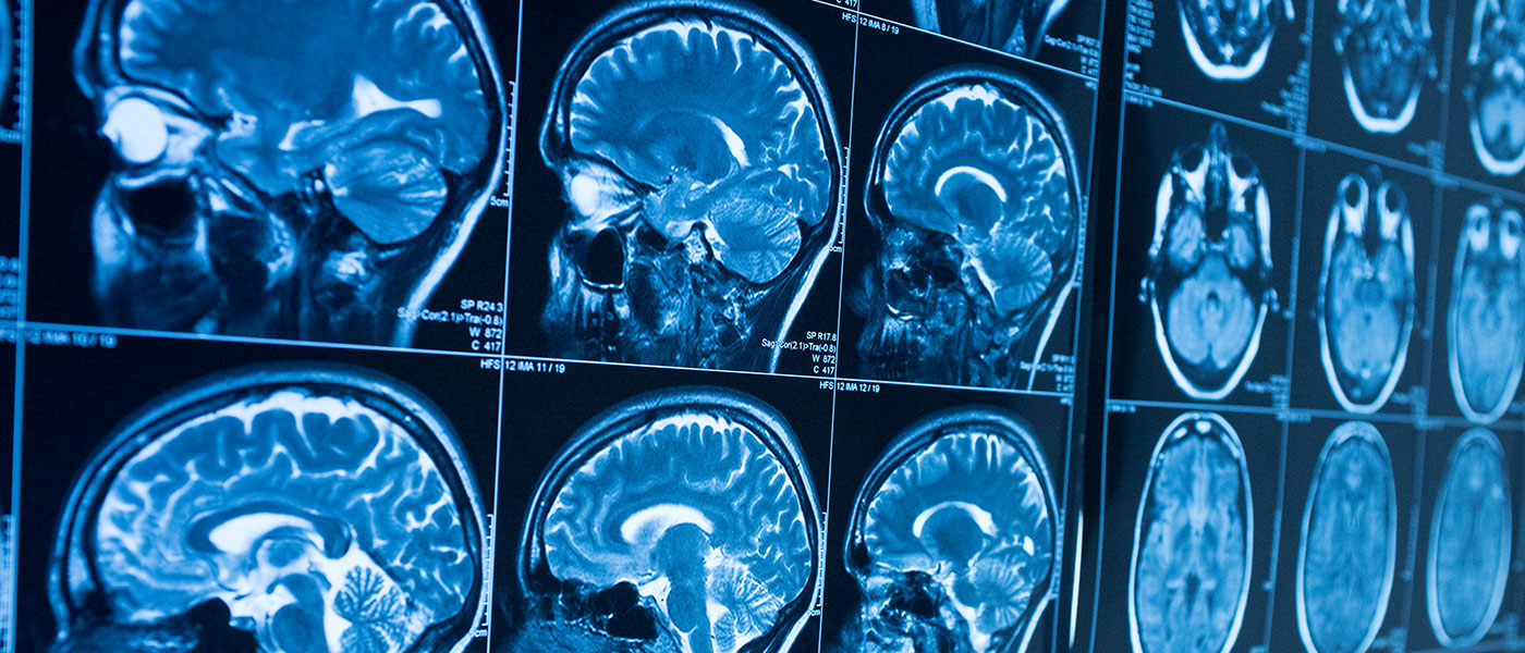
- Studying the brain and central nervous system, including degenerative diseases such as multiple sclerosis.
- Performing MR angiography (angio-MRI) to analyze arterial and venous vascular structures, where contrast agents are injected to enhance visualization.
Nanoparticle Contrast Agents
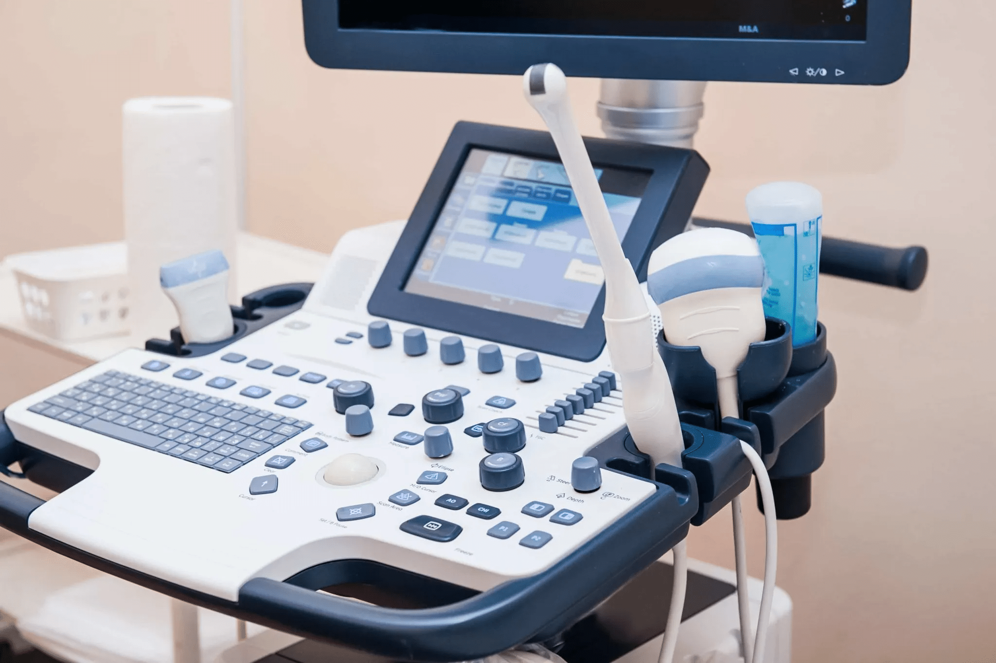
Nanoparticle contrast agents have the potential to revolutionize medical imaging, offering a powerful tool for disease visualization and diagnosis. These unique agents consist of tiny particles ranging from 1 to 100 nanometers, allowing them to interact with specific tissues and organs in the body, enhancing contrast in imaging techniques such as MRI, CT, and ultrasound.
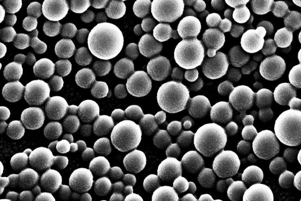
One of the key advantages of nanoparticle contrast agents is their ability to target specific cells or tissues, significantly improving imaging accuracy. This targeted approach not only enhances the visualization of diseased tissues but also reduces the required contrast agent dosage, minimizing potential side effects for patients.
Additionally, the small size of nanoparticles allows them to penetrate biological barriers and accumulate in specific areas, making them ideal for detecting and monitoring diseases such as cancer, cardiovascular diseases, and neurological disorders.
Elimination of Contrast Fluids
With normal kidney function, both gadolinium-based and iodinated organic contrast agents are typically eliminated within a few hours to a maximum of one day.
For contrast agents used in liver imaging, elimination occurs via the liver and bile, followed by excretion through the digestive system.
Side Effects, Considerations, and Safety
Despite their undeniable benefits, contrast agents are not without risks. Adverse reactions to contrast media can range from mild hypersensitivity reactions to severe kidney toxicity, posing significant challenges for physicians.
Understanding the safety profiles of different contrast agents and implementing proper management strategies is essential to minimizing these risks. Therefore, staying updated with the latest literature on contrast protocols is crucial to ensuring optimal patient outcomes.
Some contrast media have specific contraindications, such as:
- Barium contrast: contraindicated in cases of colonic perforation.
- Iodine contrast: contraindicated in severe kidney disease.
- Contrast-enhanced studies are widely used in both X-ray and CT imaging, making them an essential component of modern diagnostic radiology.
Source: Contrast Agents (Radiographic Contrast Agents and Iodinated Contrast Media)