The Role of AI in the Early Detection of Diseases through Medical Imaging

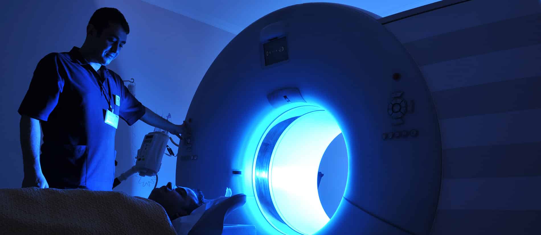
AI’s Role in Early Disease Detection via Medical Imaging
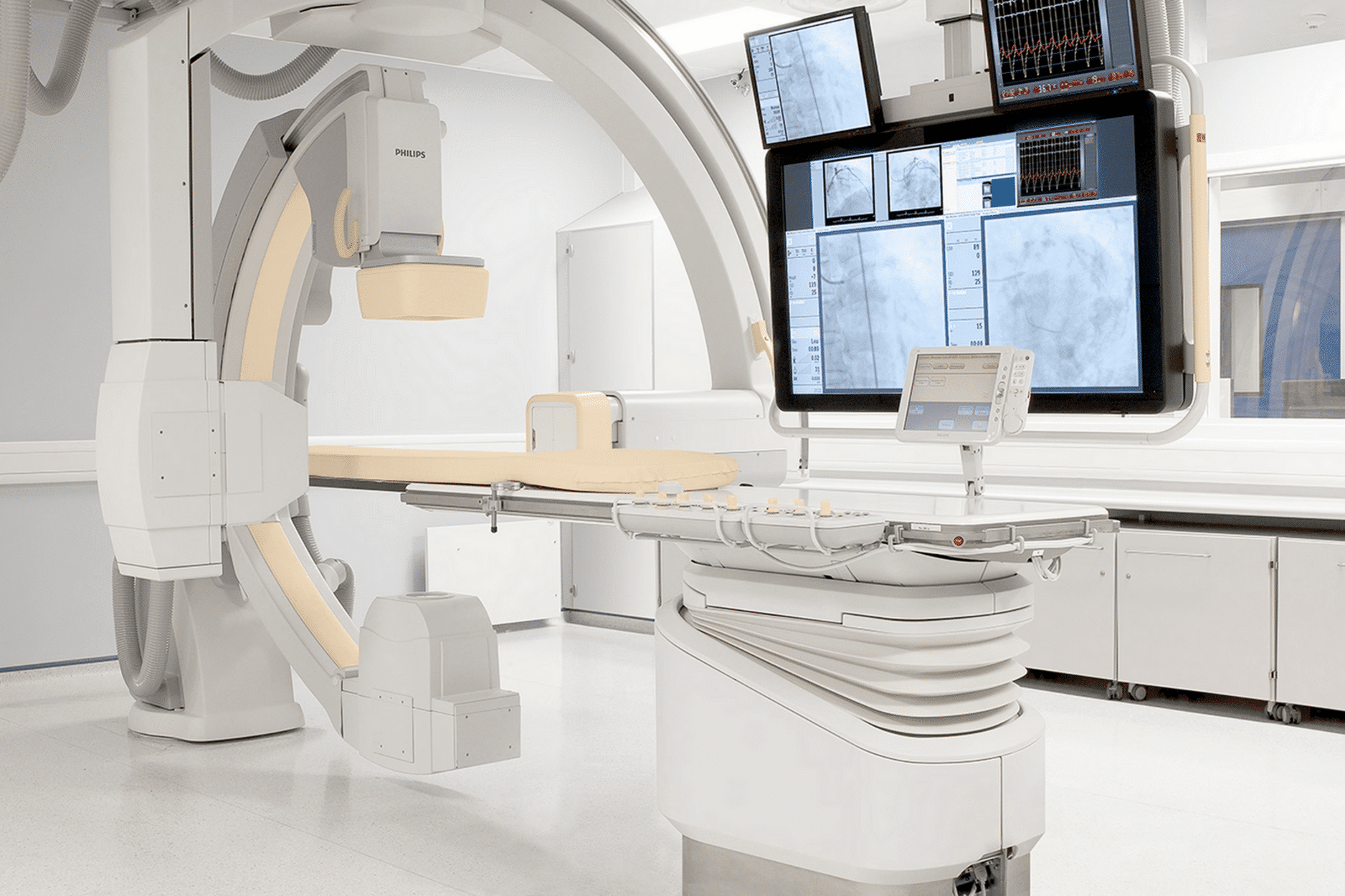
The use of AI in medical imaging represents a major breakthrough in integrating technology into healthcare. This integration is not just a technological advancement, but a qualitative leap in enhancing diagnostic and treatment plans. AI is leveraged in the early detection of diseases through advanced imaging techniques. In this article, we will discuss this role and how it contributes to improving healthcare.

The Importance of AI in Early Disease Detection

We are not exaggerating when we talk about the importance of AI in healthcare and medical imaging. It goes beyond merely improving image quality and processing speed. AI capabilities in medical imaging have evolved from basic diagnostics to aiding in early disease detection. It’s no longer just about faster and clearer images, but also about saving lives through early intervention and providing personalized treatment options.

Early Detection of Diseases

These advancements have had a significant impact on healthcare, especially in fields like oncology and neurology, where timely and accurate diagnosis is critical. More specifically, these innovations have significantly increased the number of patients undergoing screenings, enhancing not only patient outcomes and safety but also productivity and cost-effectiveness.

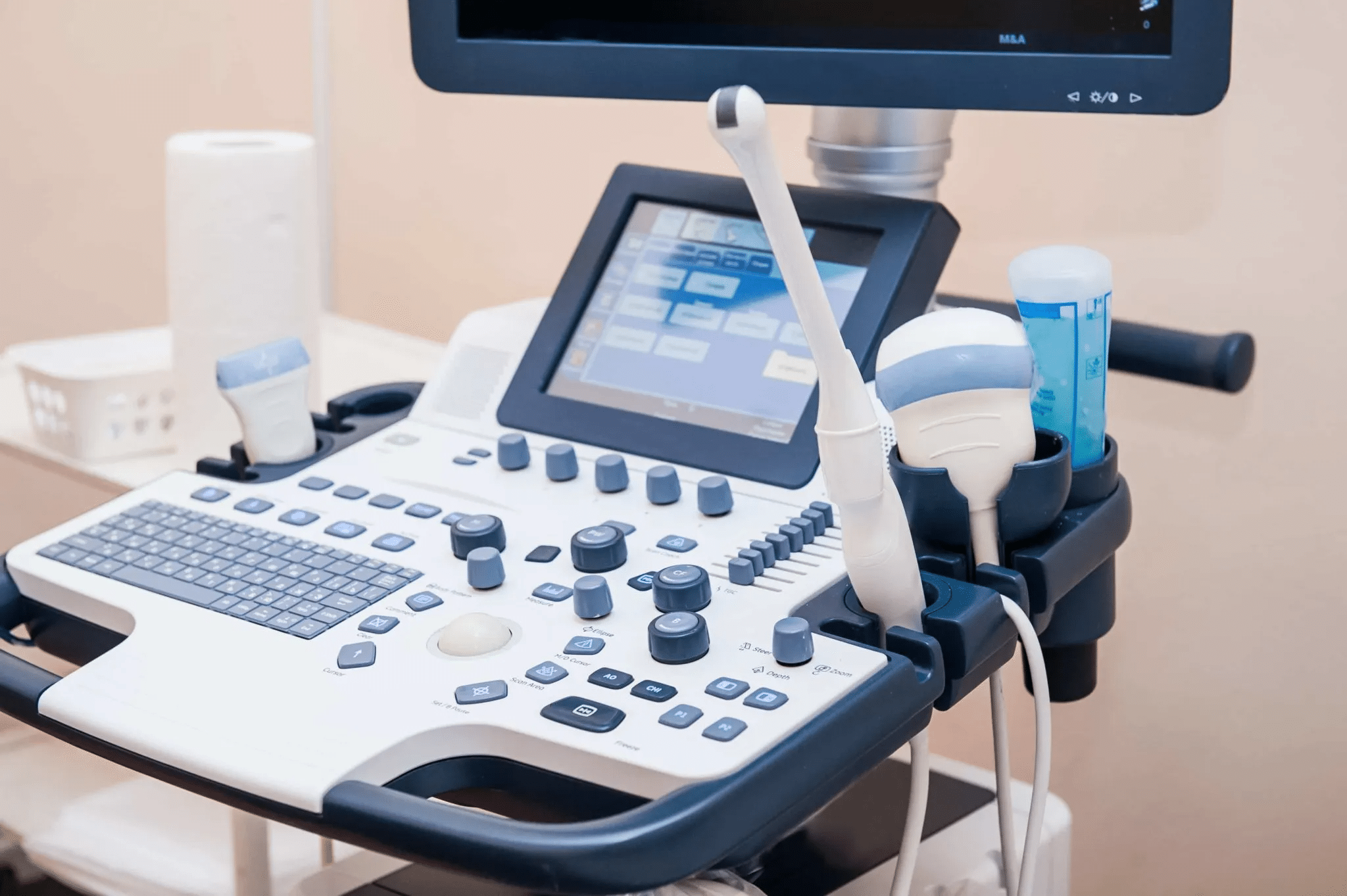
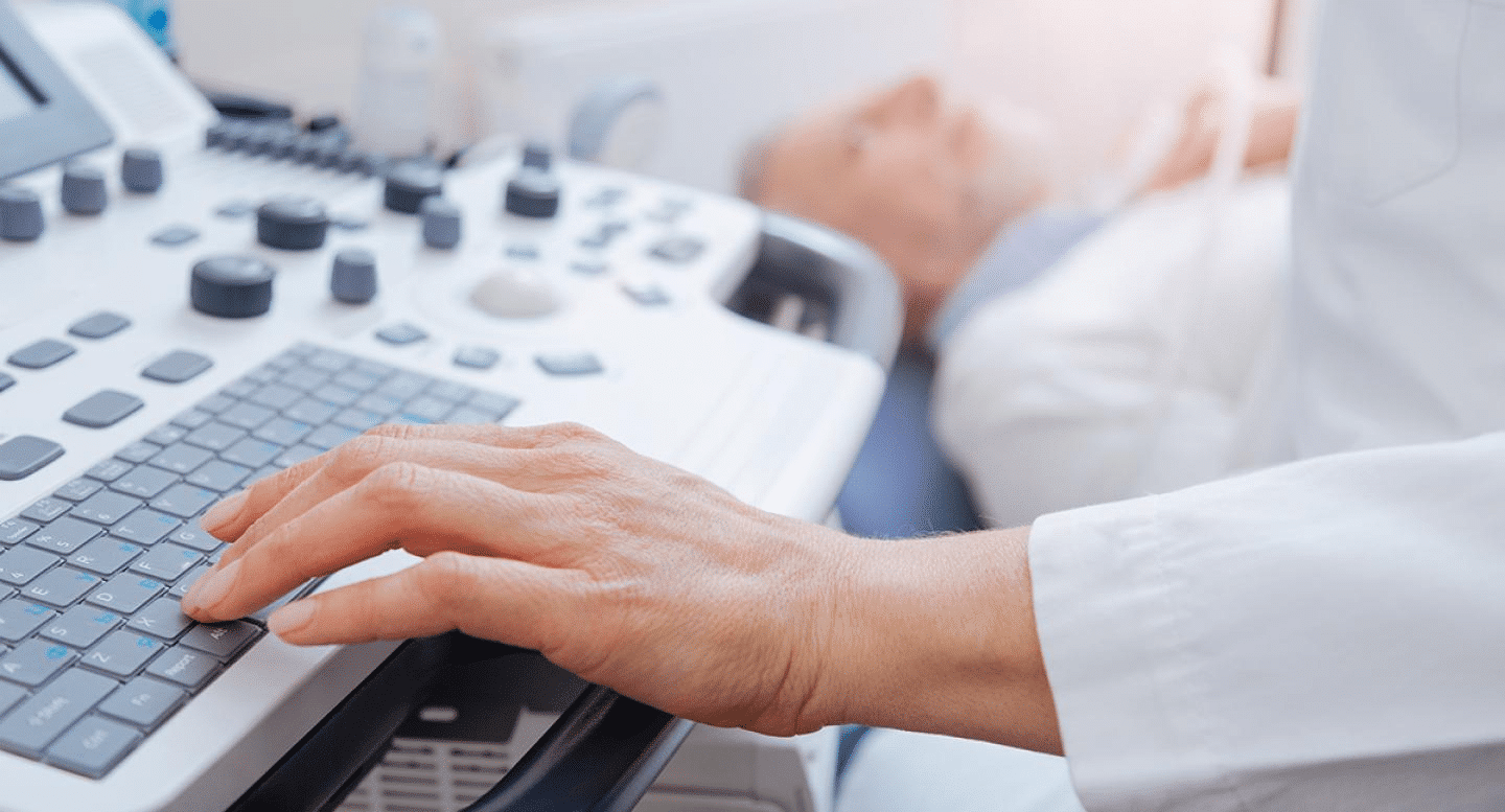
AI Capabilities in Medical Imaging

AI technologies have a multifaceted impact on medical imaging. They improve diagnostic accuracy, reduce human error, and enable early disease detection, ultimately raising the standard of patient care. AI can be utilized in various ways to enhance medical imaging.

The full potential of AI can be realized through the effective use of machine learning and deep learning algorithms, which are now playing a pivotal role in advancing imaging technologies.

AI Improvements in Medical Imaging

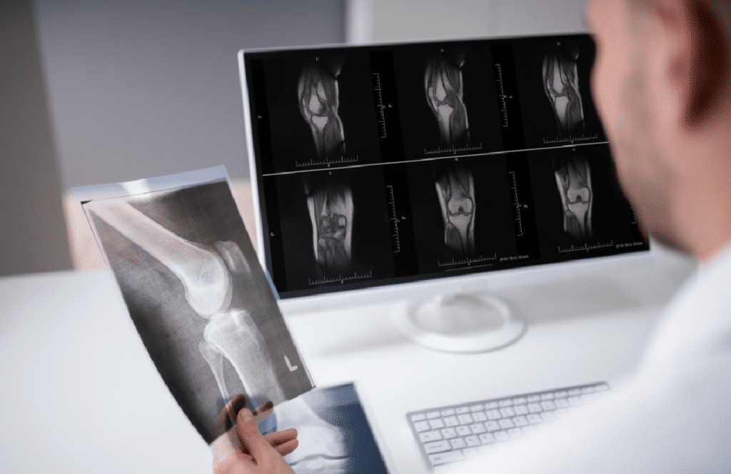
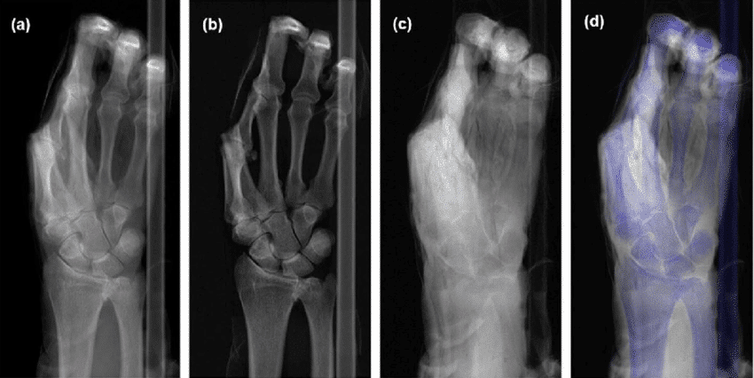
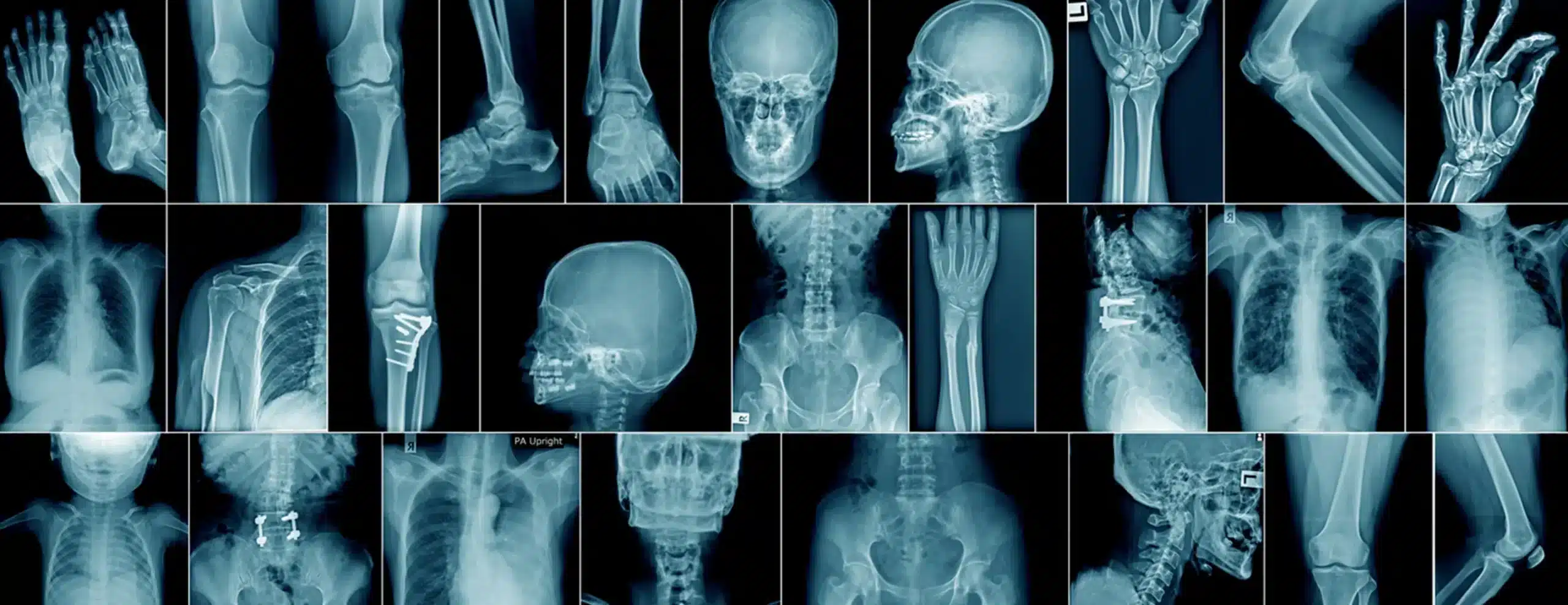
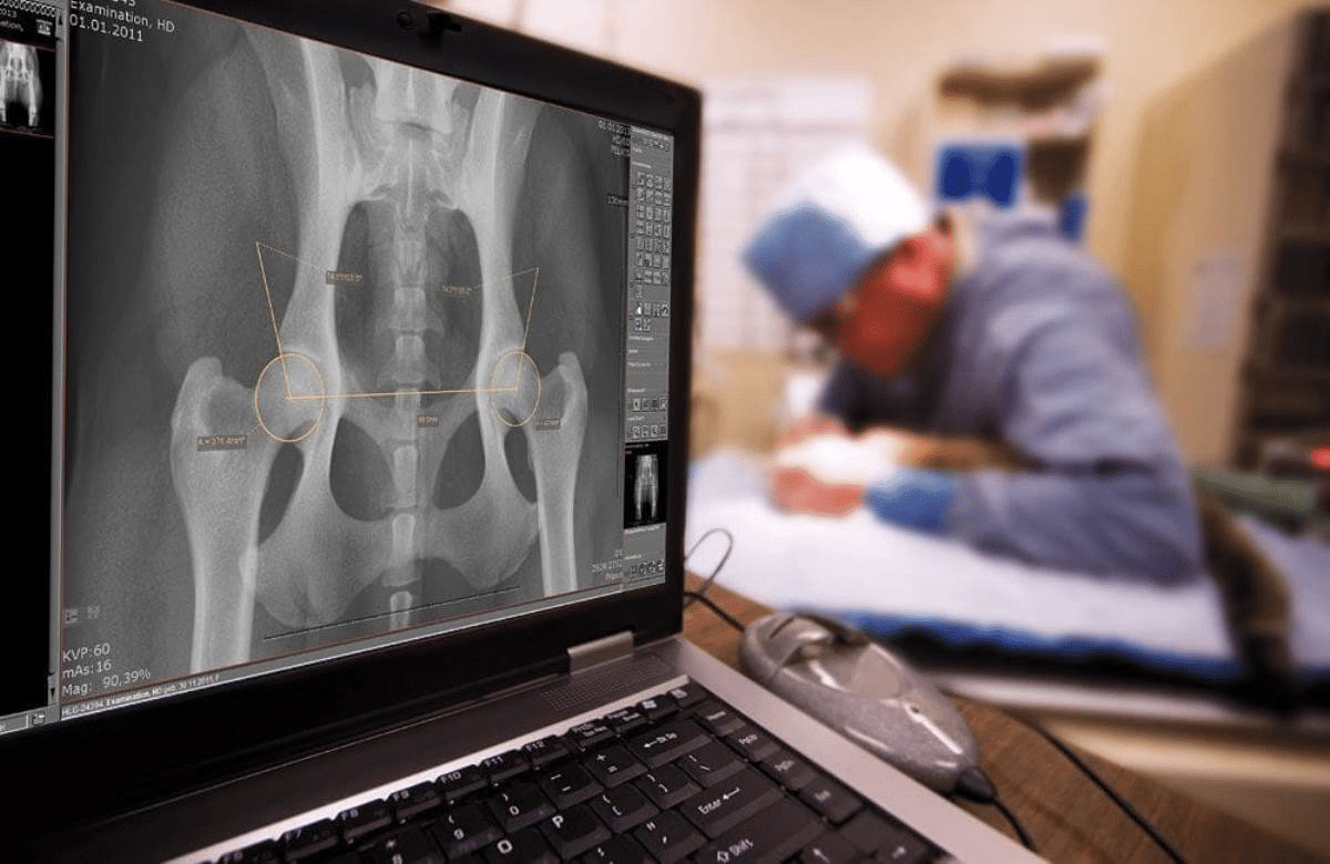
  1. One of the most notable advancements is image quality enhancement. AI algorithms can analyze massive volumes of imaging data, learning to detect subtle patterns and anomalies that may be invisible to the human eye. The increased clarity and resolution of these images have paved the way for more accurate diagnoses, such as the ability to detect fractures.
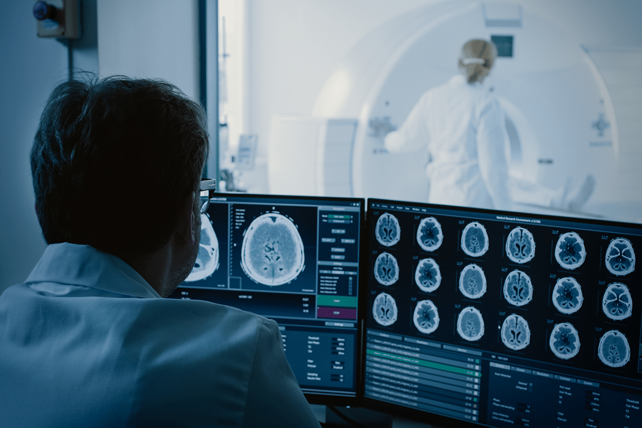
  2. Image processing speed is another area where AI makes a significant difference in early disease detection. In the past, analyzing medical images was time-consuming, which affected the timing of diagnoses and treatment.
  3. Thanks to its ability to quickly process and analyze large datasets, AI significantly reduces the time needed to analyze medical images. As a result, AI systems can now interpret and generate complex images much faster than traditional methods, speeding up the diagnostic process.
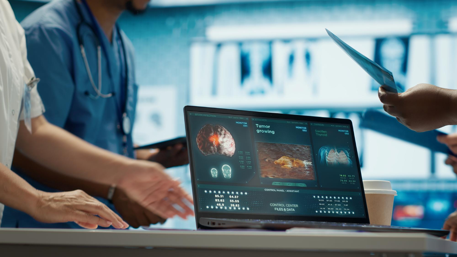
  4. By integrating deep learning algorithms, AI can identify patterns associated with specific diseases, making it a powerful tool for early detection and diagnosis. Through advanced image analysis, AI can detect abnormalities such as tumors in their early stages, greatly enhancing the patient experience.
  5. AI also plays a major role in personalized medicine and preventive healthcare. By analyzing medical imaging data alongside a patient’s medical history, AI can provide personalized diagnostic insights, paving the way for more tailored treatment plans.

Key Applications of AI in Medical Imaging

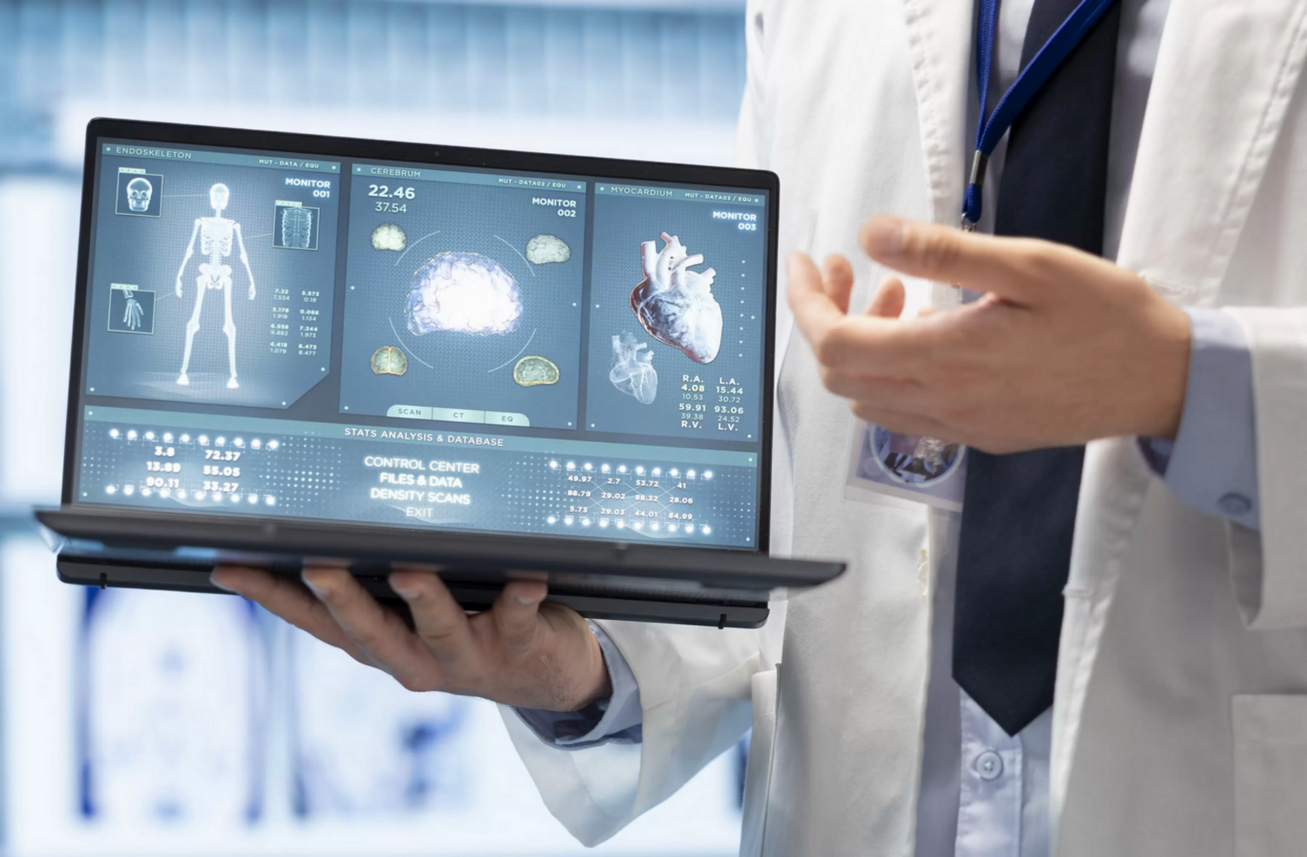
Current AI applications in medical imaging are significantly impacting fields such as radiology, oncology, and neurology. By harnessing AI, medical professionals in these areas can improve the accuracy and efficiency of numerous diagnoses and treat medical conditions more successfully.

These three fields demonstrate how AI applications in medical imaging are not only enhancing current diagnostic and treatment methods but also opening new horizons in personalized medicine. From improving diagnostic accuracy in radiology to aiding in the early detection of cancerous cells and neurological diseases, AI is proving its value in the healthcare sector.

Below are the key fields that benefit from AI in the early detection of diseases.
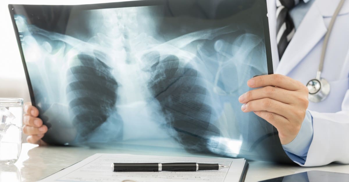
1. Radiology

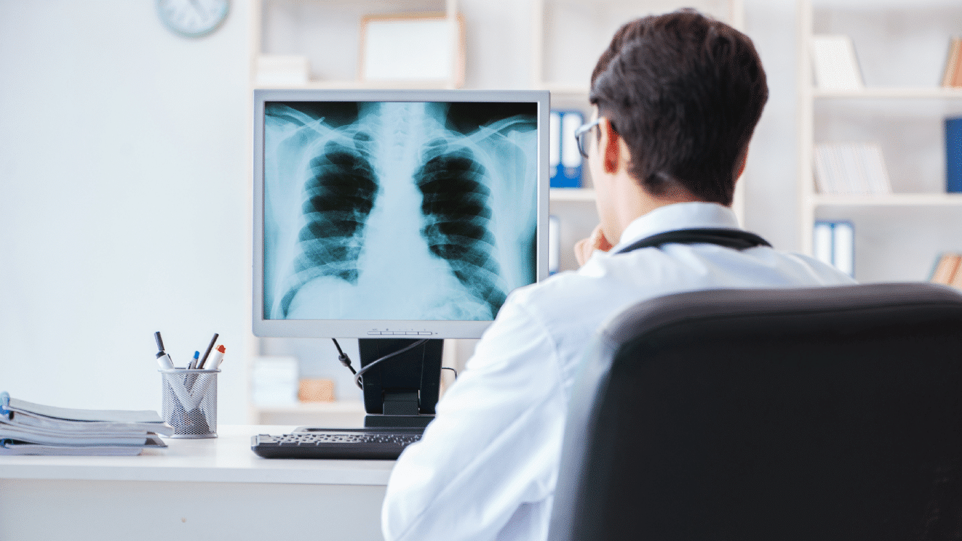
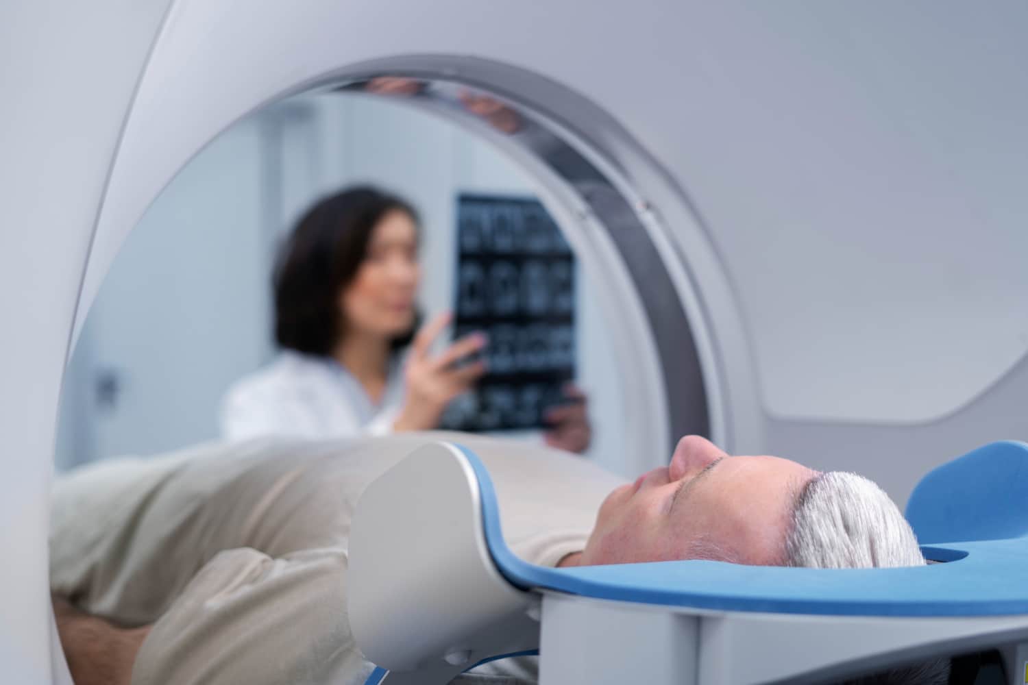
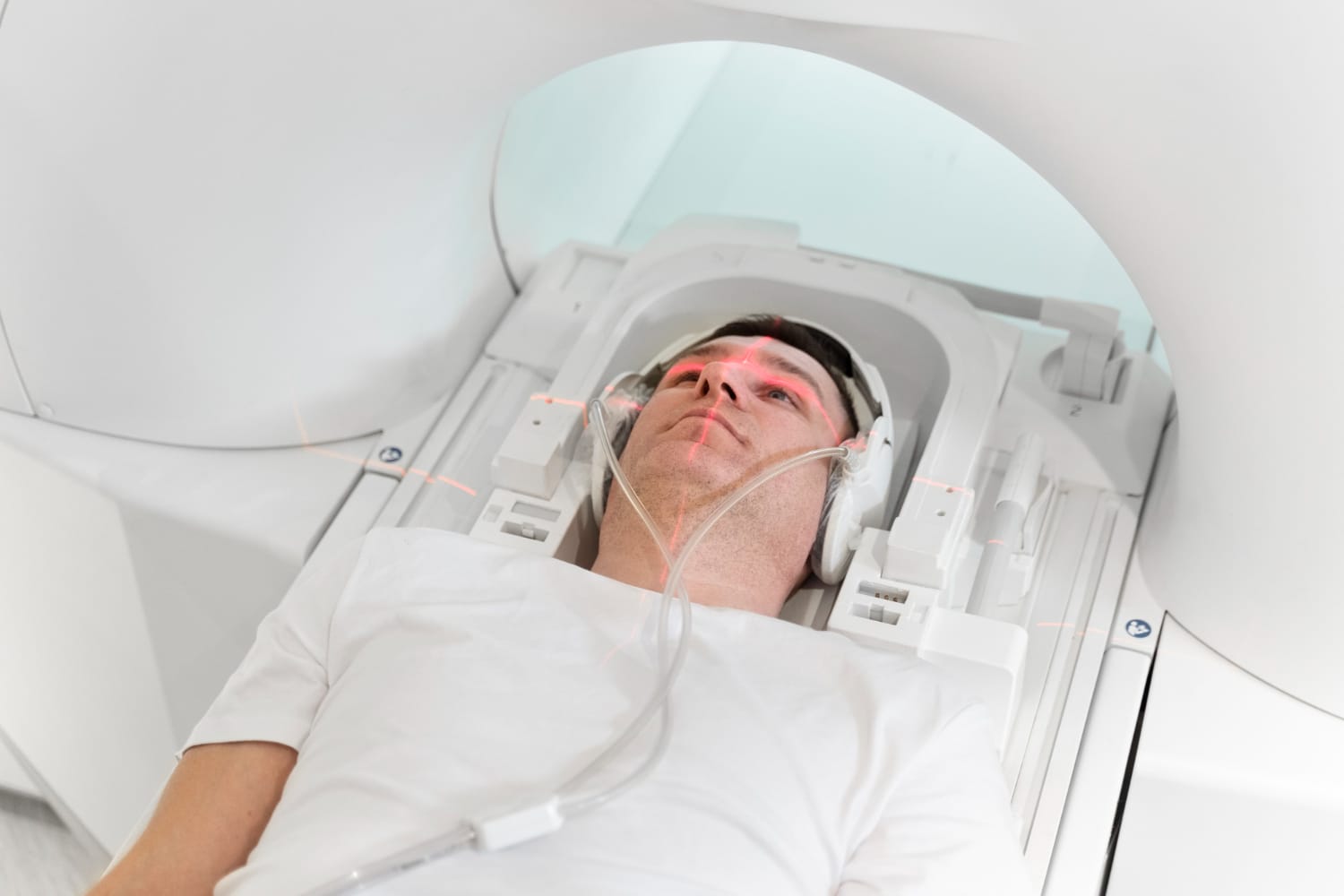
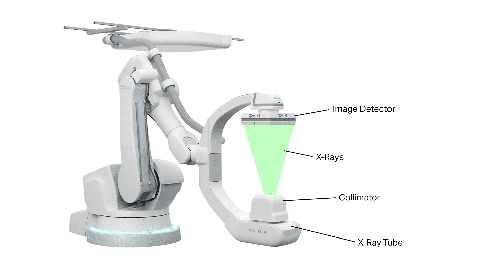
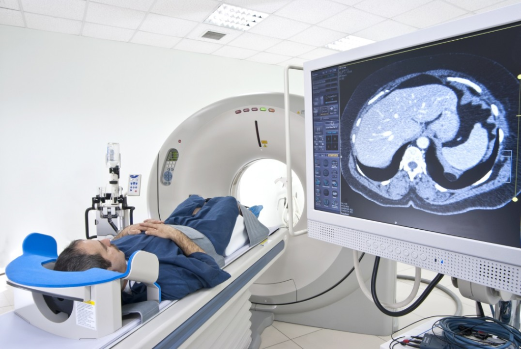
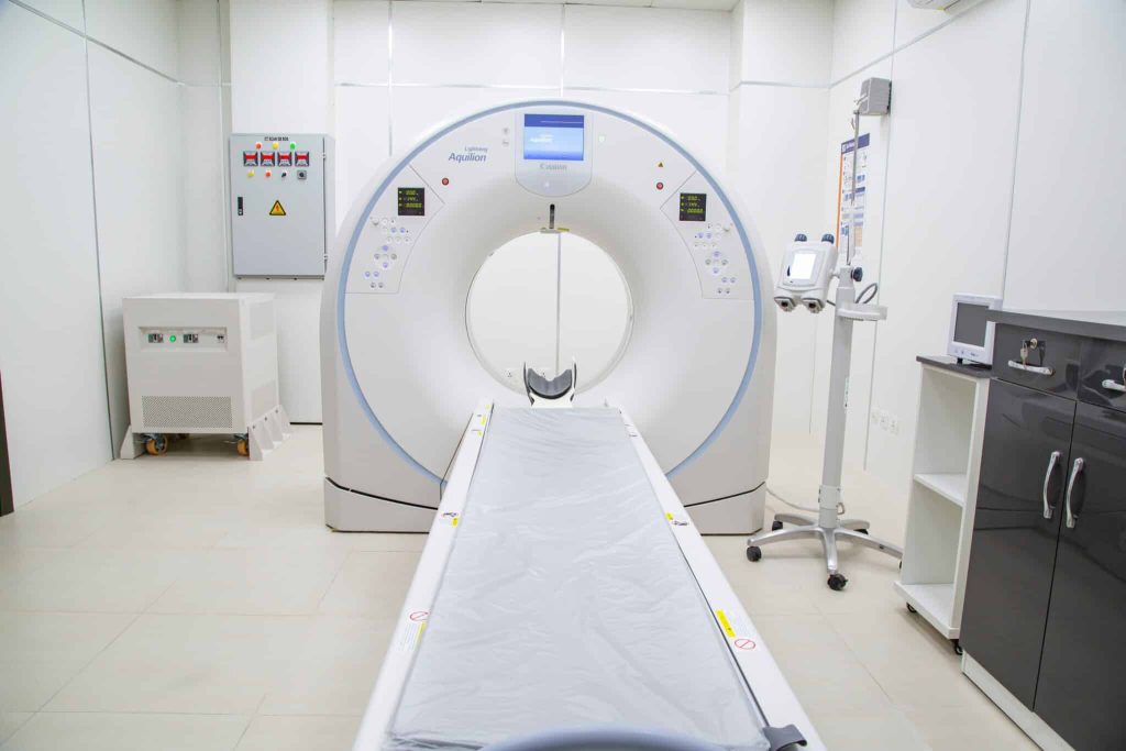
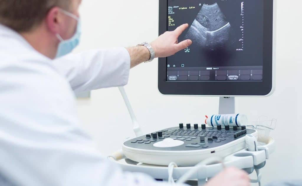
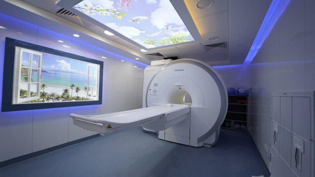
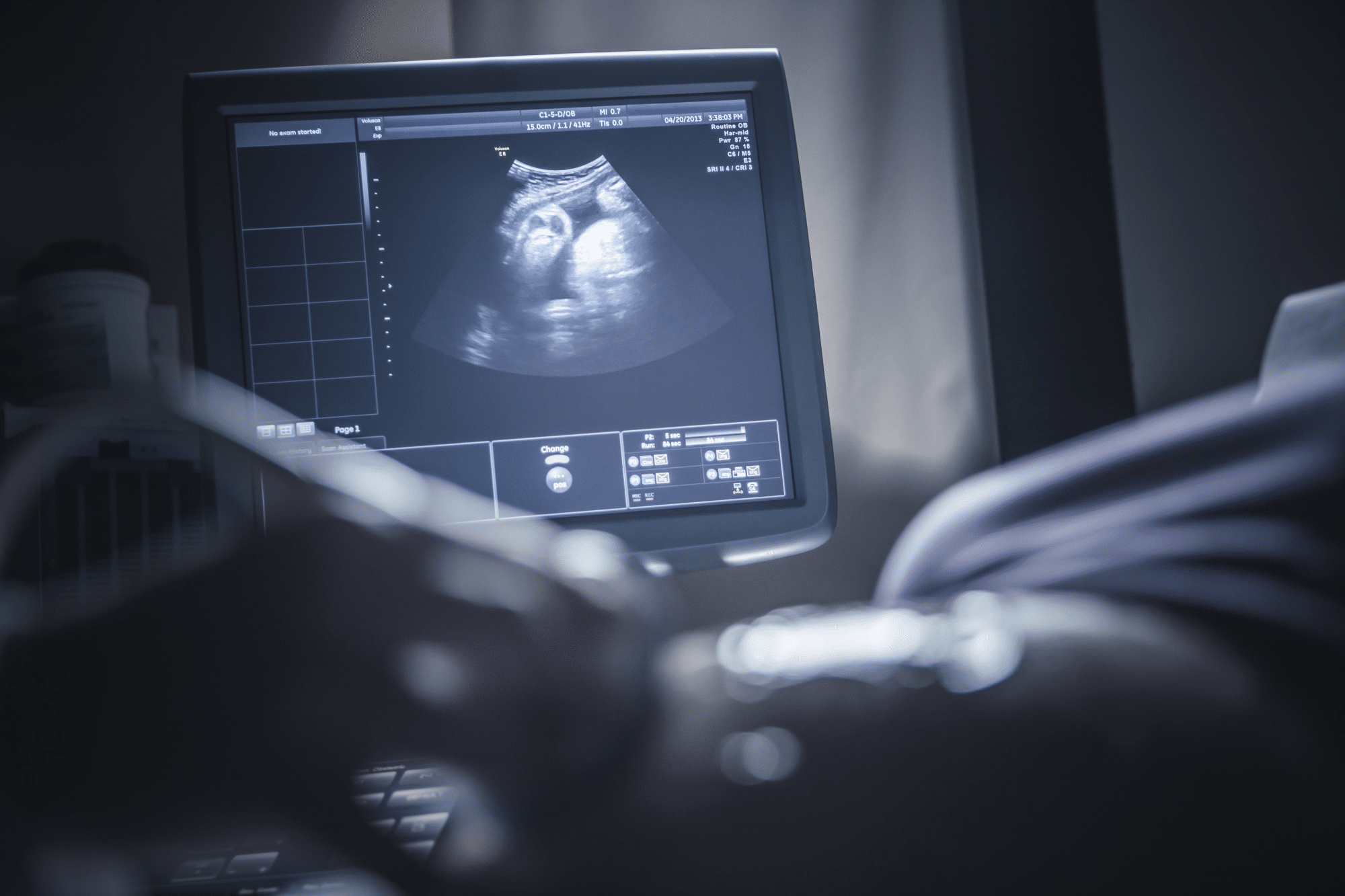
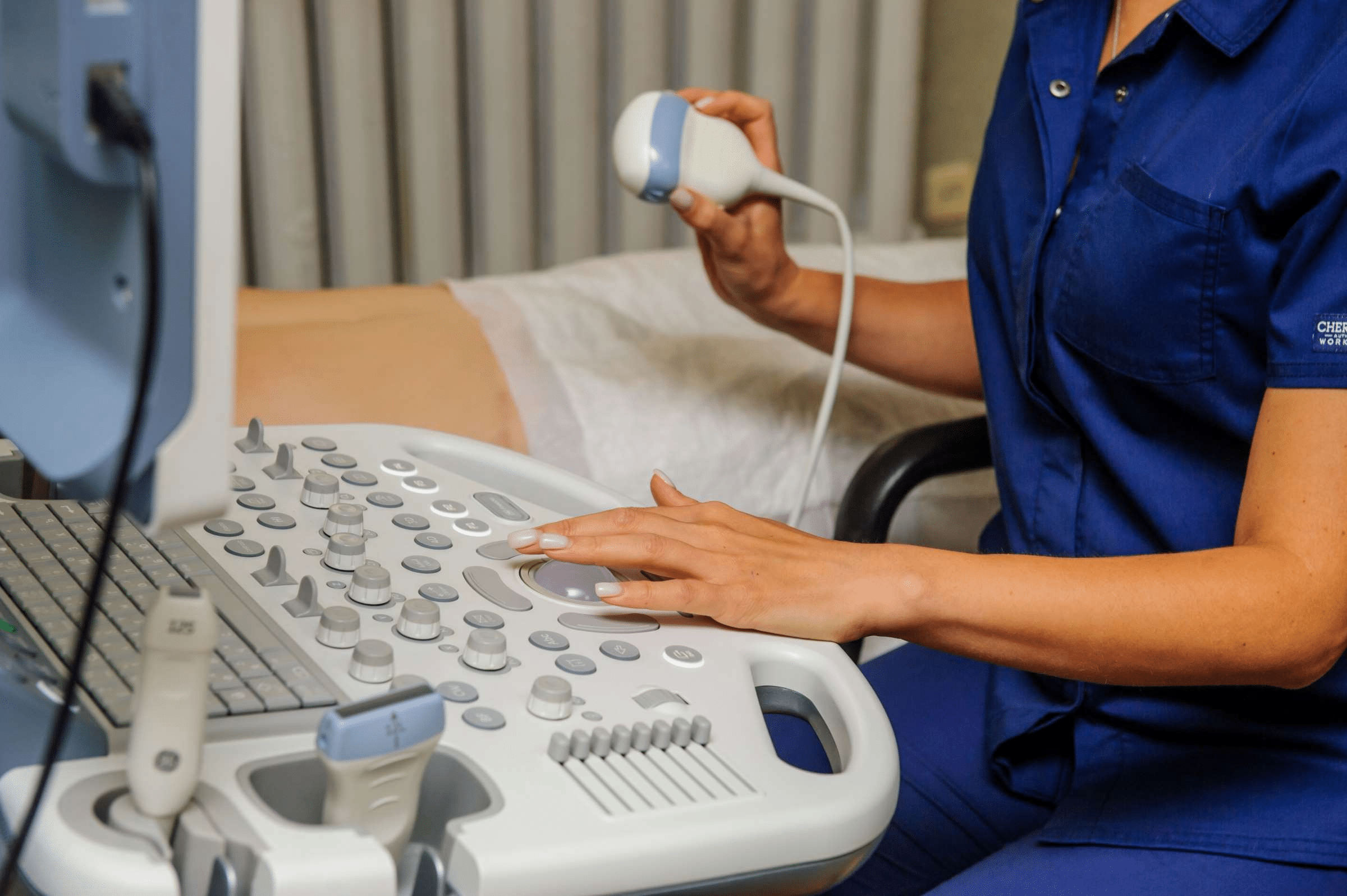
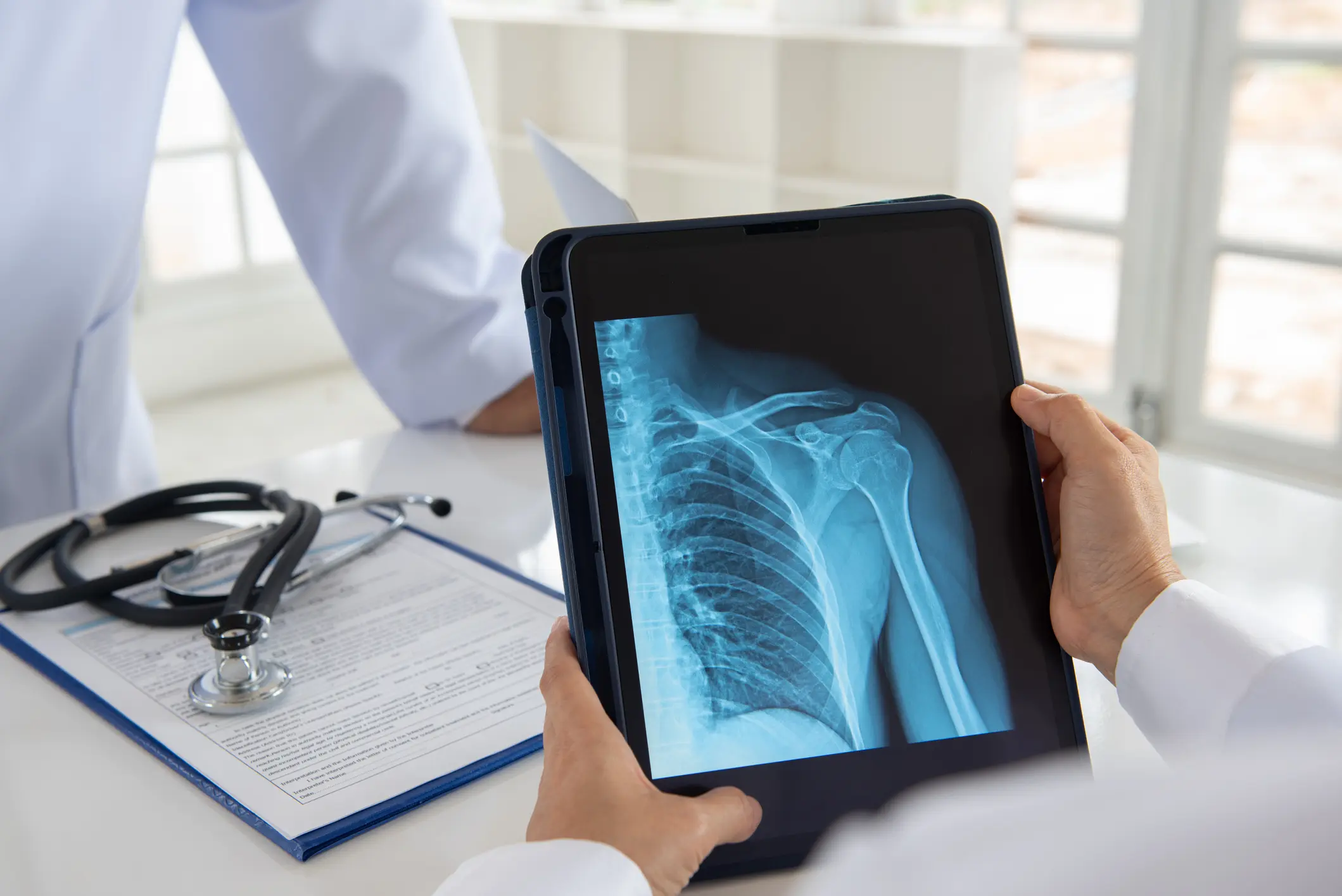
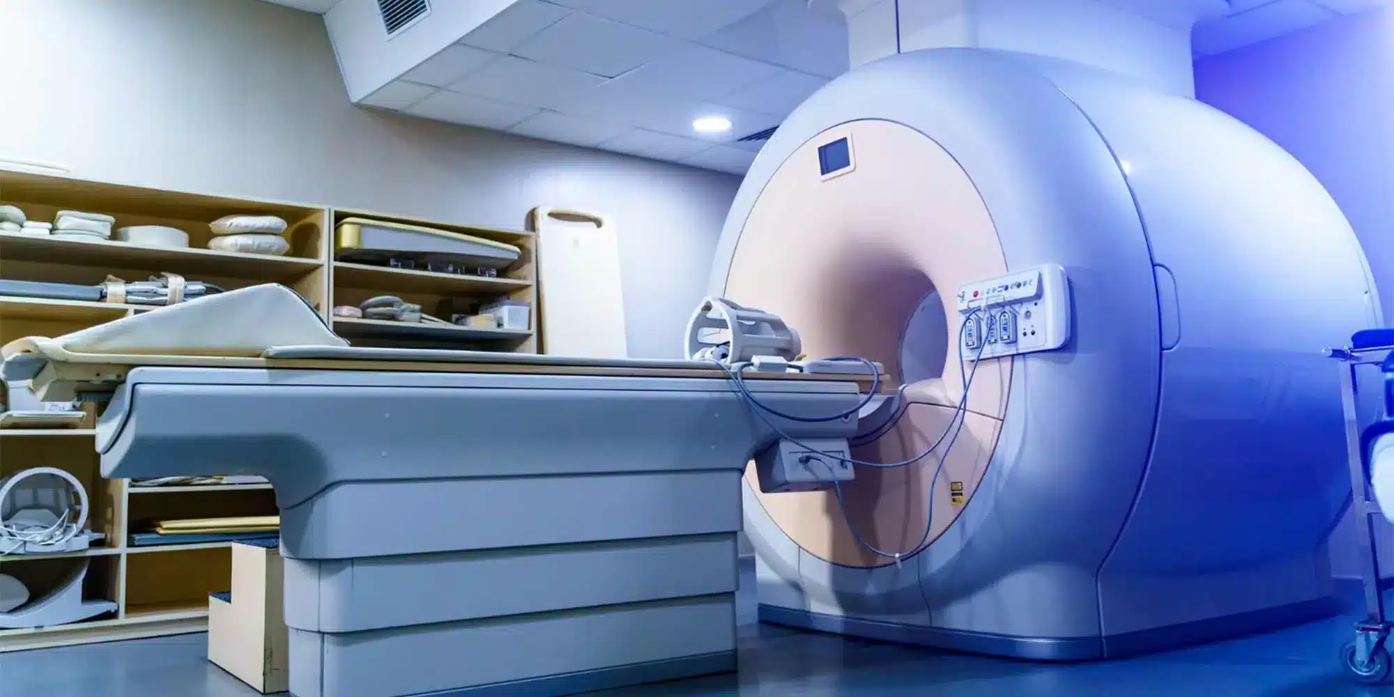
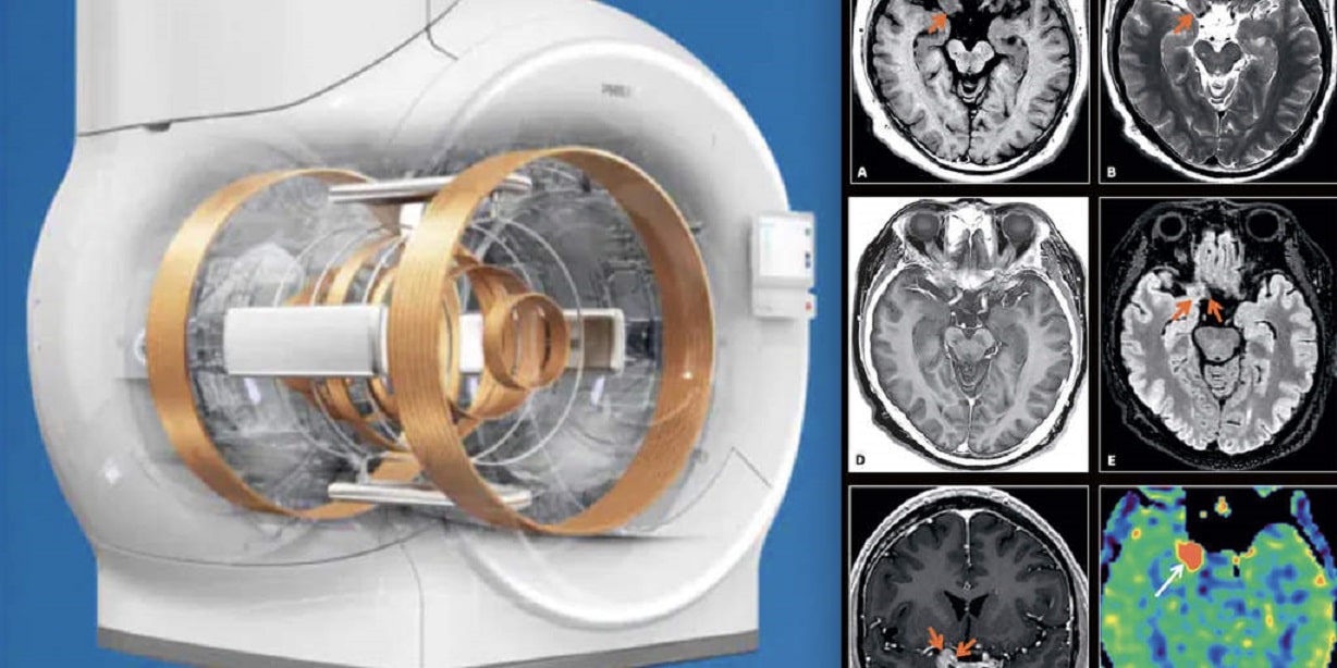
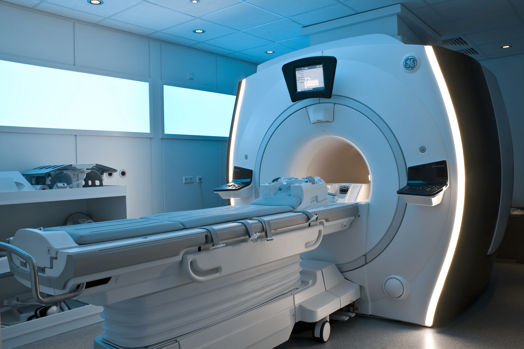
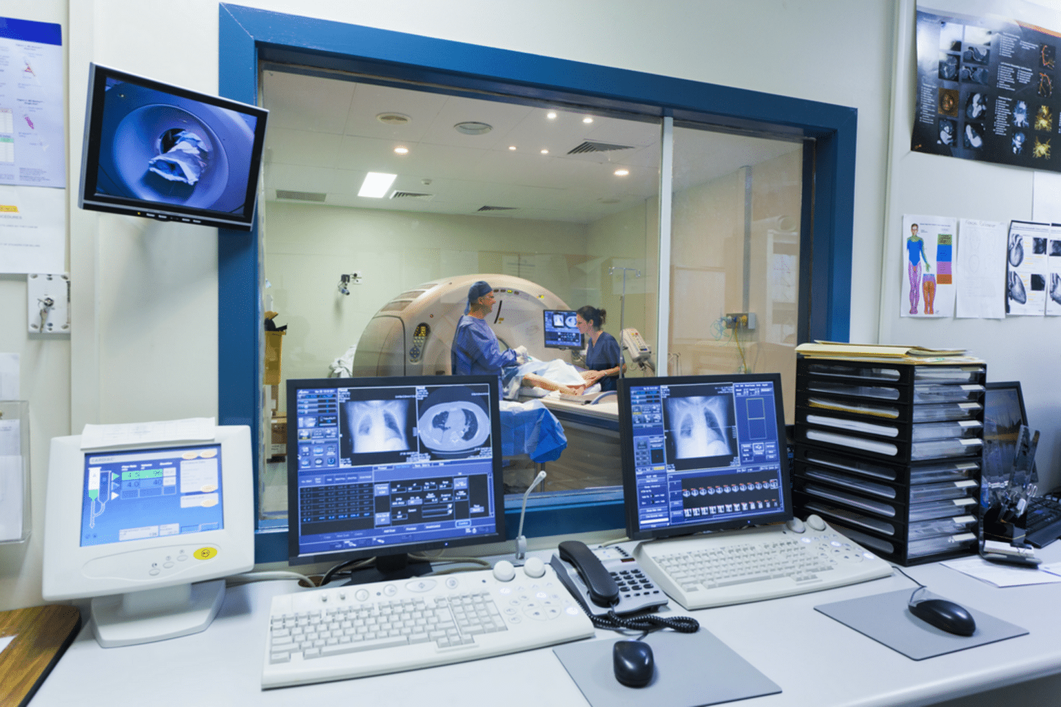
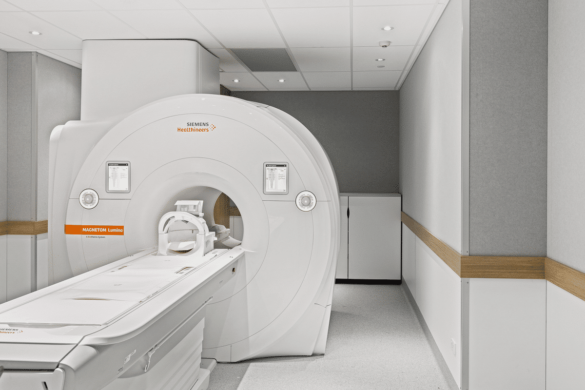
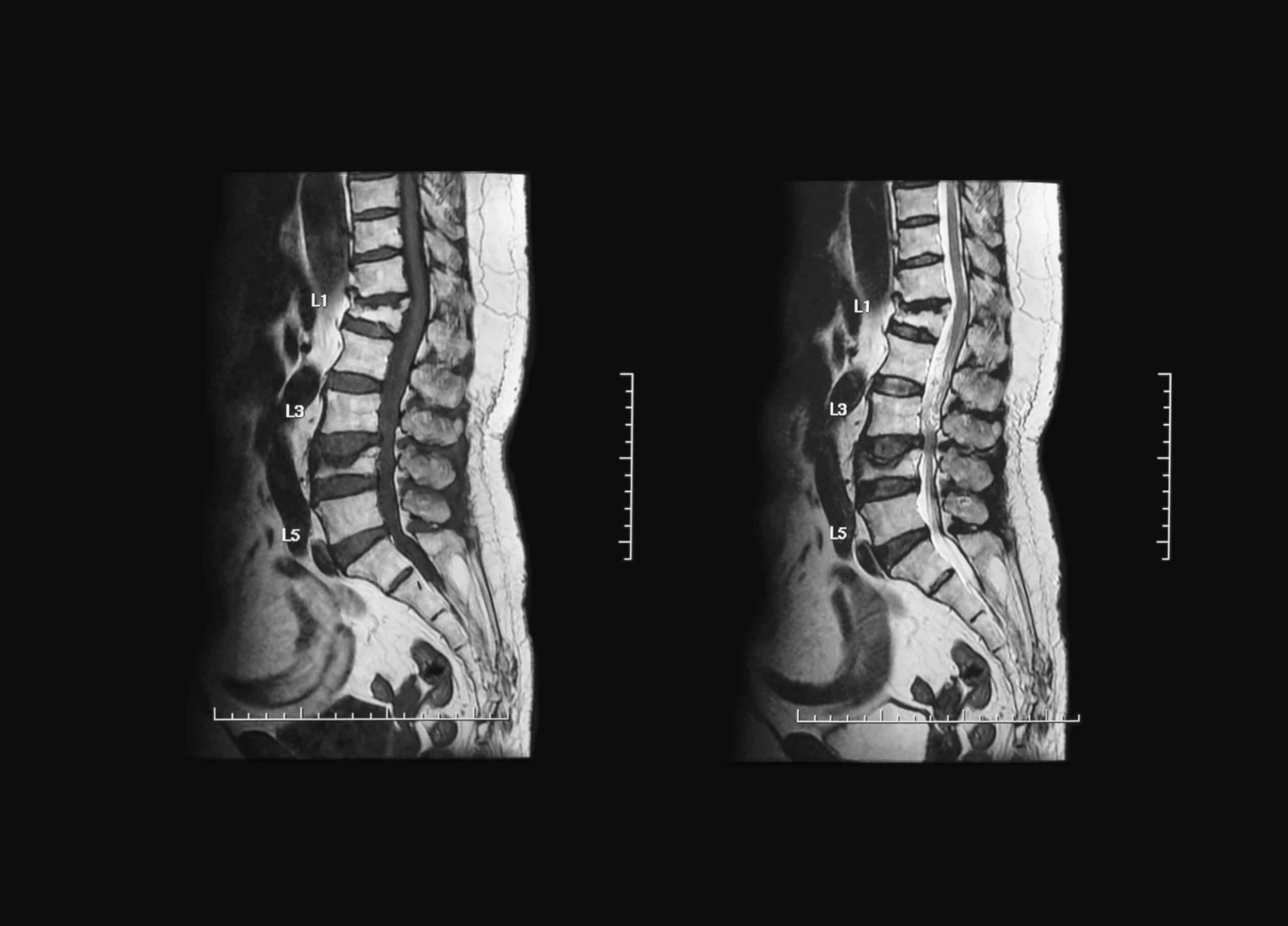
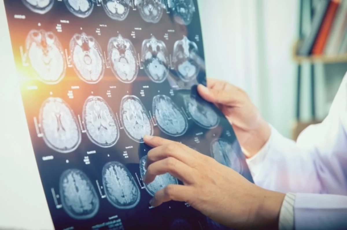
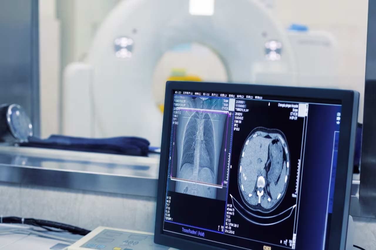
In the field of radiology, for example, artificial intelligence is primarily used to identify abnormalities in imaging scans. AI algorithms applied to computed tomography (CT) and magnetic resonance imaging (MRI) can quickly detect and segment areas of interest in scans of the lungs, liver, and brain.

2. Cancer Tumor Detection

Particular attention should be given to AI’s role in tumor imaging. It assists in detecting and monitoring cancerous tumors, improving the accuracy of cancer screenings. For instance, deep learning AI techniques have been used to analyze mammograms and accurately distinguish between malignant and benign tumors.
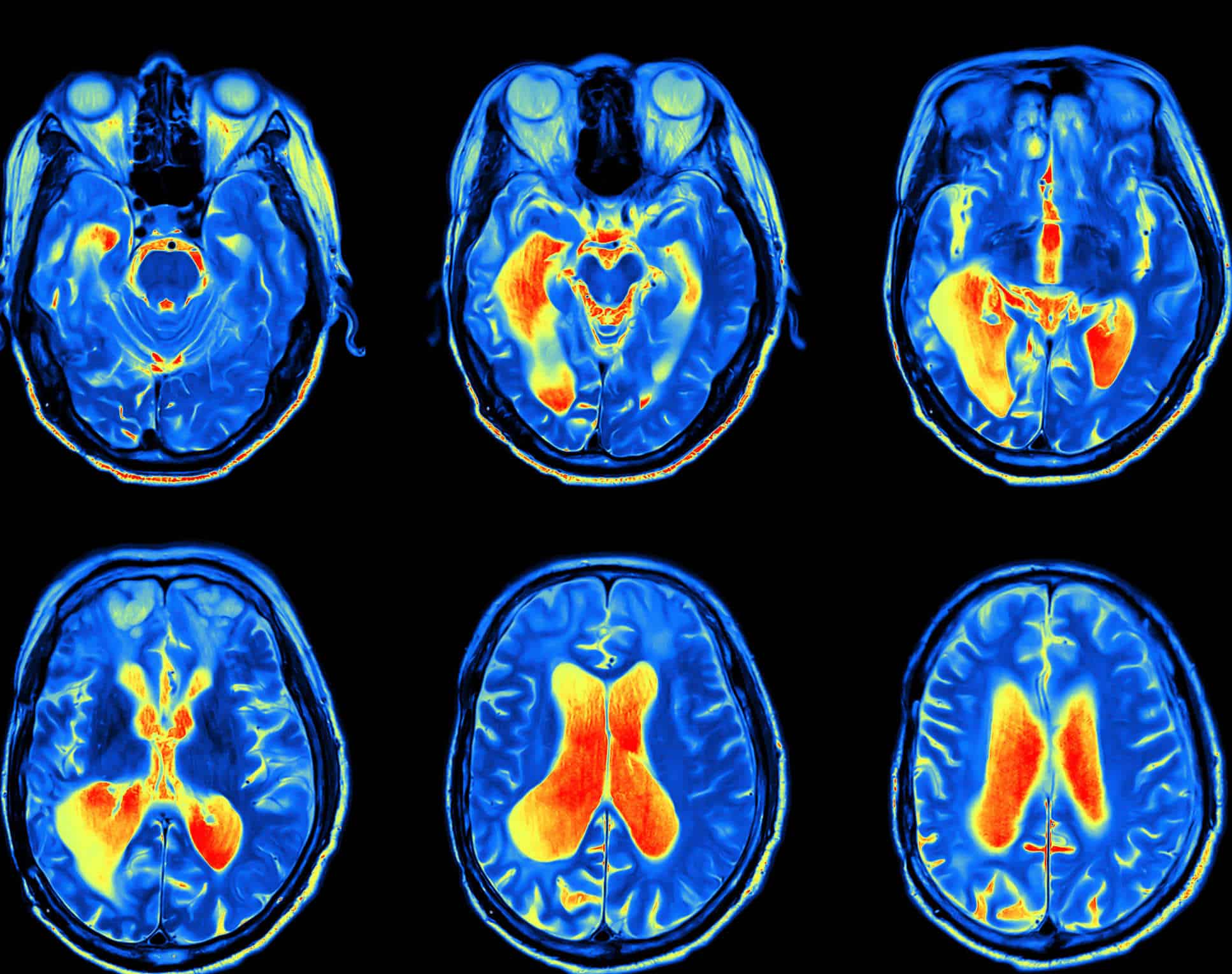
Early Detection of Diseases

3. Neurology

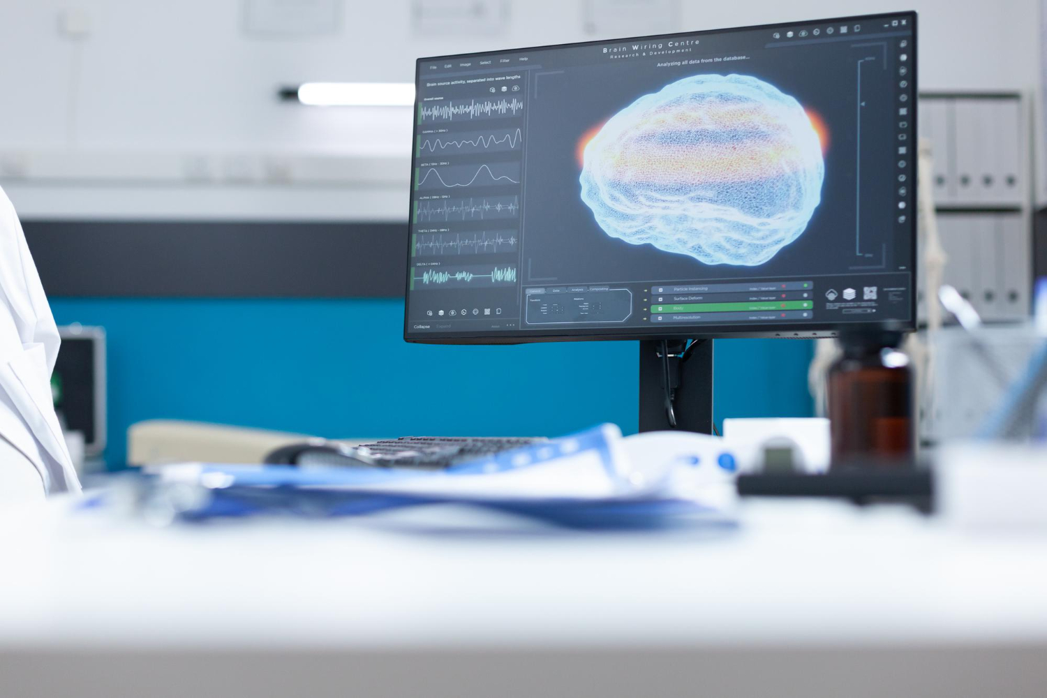
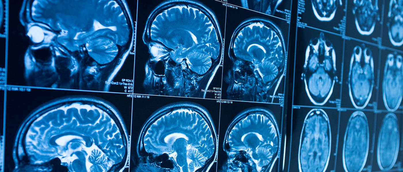
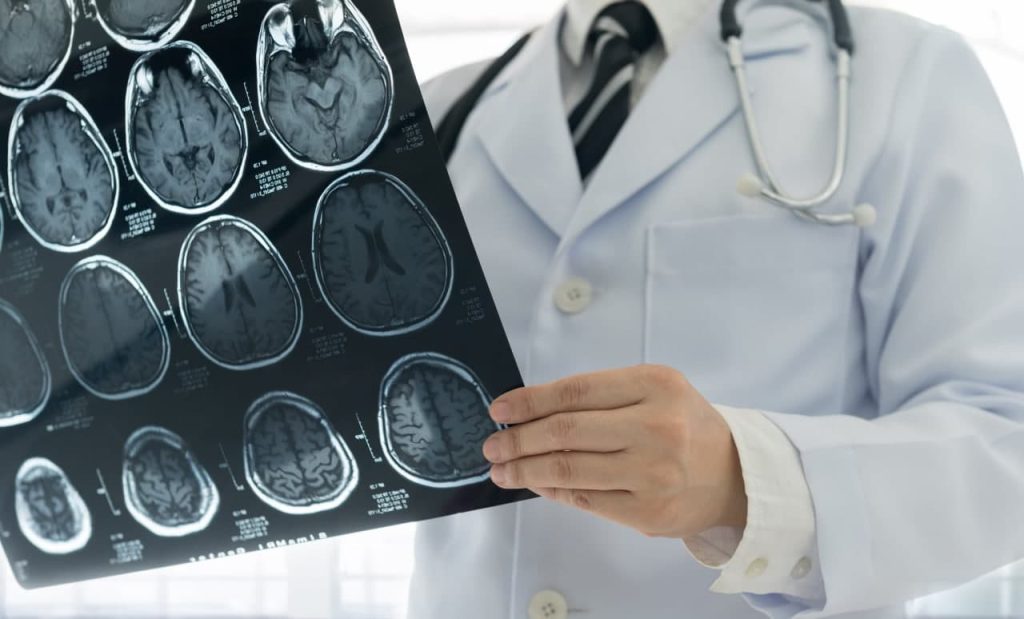
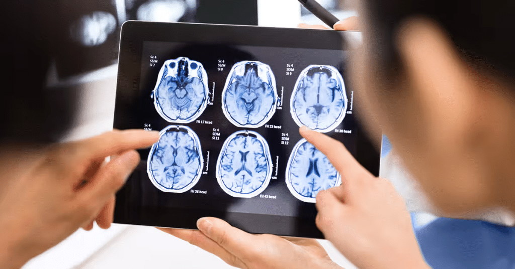
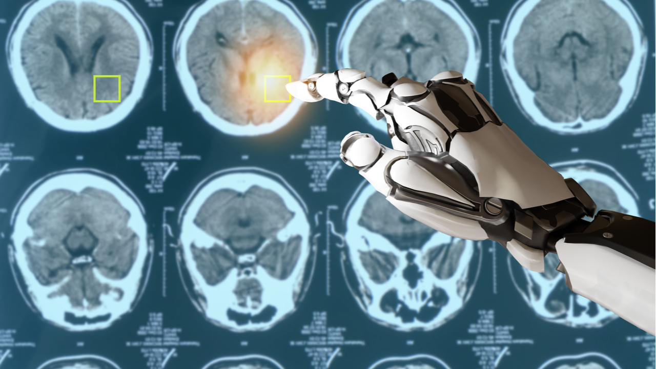
In neurology, AI enhances the analysis of brain images for conditions such as stroke, Alzheimer’s disease, and brain tumors. AI algorithms can detect subtle changes in brain tissue, allowing for early intervention and treatment. A notable example is the use of AI in diffusion-weighted imaging, a type of MRI, to quickly identify stroke-affected areas in the brain.

AI’s Impact on Diagnostic Accuracy

The integration of AI into medical imaging analysis has significantly improved diagnostic accuracy. It reduces human error and enhances early disease detection. This shift toward AI-enhanced diagnostic and imaging methods is reshaping the landscape of medical imaging technology and precision medicine. Below are the key impacts:

1. Improved Diagnosis of Various Medical Conditions

Thanks to its ability to learn from large datasets, AI algorithms have shown remarkable improvement in identifying and diagnosing medical conditions. For example, AI can match—or even surpass—the accuracy of human radiologists in detecting diseases like pneumonia in chest X-rays. This level of precision is especially critical in cases where early detection greatly influences treatment outcomes.

2. Reduction of Human Error

The second major advantage of AI in medical imaging is its consistency, which significantly reduces human error. Unlike human radiologists who may be affected by fatigue or subjective judgment, AI systems provide consistent analyses. AI support can reduce diagnostic errors in mammograms for breast cancer, thereby improving diagnostic accuracy.

Medical Imaging

3. Early Disease Detection

Early disease detection is crucial for effective treatment, and AI is playing a vital role in this area. For example, AI has been instrumental in the early detection of diabetic retinopathy—a condition that can lead to blindness if left untreated. By analyzing retinal images, AI can detect this condition with high accuracy, often before any physical symptoms appear in the patient.

Challenges and Ethical Considerations

Despite the tremendous potential of artificial intelligence in the healthcare sector and medical imaging technologies, it still faces a number of challenges and concerns that slow down its widespread adoption. These challenges include:

1. Data Privacy

The first challenge affecting AI is the concern over data privacy. AI systems require access to large datasets of patient images, which raises significant concerns about confidentiality. Ensuring the security and privacy of this sensitive data is absolutely critical. Healthcare institutions must comply with strict data protection regulations, as safeguarding this information is essential.

2. High Costs

The cost of integrating AI into existing standard imaging technologies presents a major barrier to its adoption. Acquiring AI software, along with the necessary hardware upgrades, can be extremely expensive for many healthcare institutions—especially public ones.

3. Training Medical Staff

For AI to be successfully used in medical imaging, it also requires specialized training for healthcare professionals. Radiologists and technicians must be educated on how to interpret AI results and incorporate them into clinical decision-making processes.

4. Technical Challenges

Technical issues—such as data integration and algorithmic bias—pose significant challenges. Integrating AI into current health IT systems can be complex and demands seamless compatibility. To address this, AI systems must be trained on diverse and comprehensive datasets and undergo rigorous validation processes to ensure accuracy and fairness.

It is essential to ensure the transparency of AI-supported systems in decision-making processes and to clearly define accountability for the outcomes of those decisions. Regulatory frameworks must be established to guarantee the safe and effective use of AI in the medical field. These frameworks should address key issues such as:

  • Validating algorithm reliability.
  • Official certification and approval.
  • Managing potential biases within the systems.

In conclusion, integrating AI into medical imaging is not merely a technical advancement, but a promising paradigm shift that redefines diagnosis and treatment planning. The positive impact of AI integration in imaging systems extends beyond patients, medical practitioners, and healthcare providers—it benefits anyone seeking a more efficient and error-free healthcare system.

Source: The Role of AI in Medical Imaging

Fluoroscopy: Real-Time X-ray Imaging in Medicine

Fluoroscopy is one of the most important technologies in modern medical imaging, allowing doctors and specialists to view internal body structures in real-time. This revolutionary technique has transformed diagnostic methods and interventional procedures in medicine, significantly improving diagnostic accuracy and guiding treatments more effectively. In this detailed article, we will explore the working mechanism of this technique, its applications, benefits, associated risks, and the latest advancements in this vital field of medical engineering.

What is Fluoroscopy?

Fluoroscopy is an advanced imaging technique that utilizes X-rays to generate a continuous series of images displayed as a real-time video. This technique is widely used in emergency departments, operating rooms, and diagnostic radiology clinics, enabling surgeons and physicians to monitor the movement of organs and internal structures during procedures or diagnostic examinations. The increasing reliance on fluoroscopy reflects its significance in providing accurate and immediate insights into various medical conditions.

Fluoroscopy

Fundamental Principles of Fluoroscopy

Fluoroscopy is extensively used across multiple medical specialties, enhancing the quality of healthcare. To fully understand this technique, it is essential to delve into the fundamental principles that underpin fluoroscopy, forming its scientific and technical foundation.

X-ray Mechanism

This technique operates on the same principles as conventional X-ray imaging but with the ability to produce sequential images in real time, creating a video-like sequence of still images. The process begins with generating X-rays through a specialized tube, where a high-voltage current is applied. This accelerates electrons toward a metallic target, typically tungsten—a chemical element. Upon collision, X-rays are emitted and directed toward the area of the body being examined.

Image Intensification

The image intensifier in fluoroscopy plays a crucial role in enhancing the clarity of internal details. This device amplifies the signals produced by X-rays passing through the body, resulting in a bright and clear image. Recent advancements have led to the replacement of traditional image intensifiers with flat-panel detectors, which provide high-quality images while minimizing radiation exposure.

Diverse applications of Fluoroscopy

Fluoroscopy has a wide range of medical applications, both in diagnostics and interventional procedures. This system is highly flexible and capable of providing real-time moving images, allowing doctors to monitor physiological changes instantly.

Fluoroscopy

Diagnostic Applications

Fluoroscopy is used in several essential diagnostic procedures, including:

  • Gastrointestinal Studies: Fluoroscopy is employed to assess the digestive tract, including the esophagus, stomach, and intestines. Barium studies, such as barium swallow and barium enema, help evaluate abnormalities or motility disorders.
  • Neuromuscular and Spinal Imaging: This technique is used in diagnostic imaging of the spine, particularly in myelography, which aids in assessing spinal cord conditions.
  • Joint Evaluations: For joint-related issues, fluoroscopy is also used in arthrography to evaluate abnormalities or injuries affecting the joints.

Interventional Applications

This technique is a cornerstone in medical engineering for interventional procedures, providing live imaging during operations, which contributes to:

  • Guiding Surgical Procedures: Fluoroscopy assists in catheter placement during cardiac procedures, such as coronary angiography, ensuring precise positioning and minimizing procedural risks.
  • Interventional Treatments: It is utilized in therapeutic interventions, including stent placement, bone fixation in fracture surgeries, and the insertion of delicate medical devices.
  • Minimally Invasive Procedures: This technology facilitates minimally invasive procedures requiring small or no surgical incisions, reducing patient discomfort and promoting faster recovery.

Benefits of Fluoroscopy in Medical Practice

Fluoroscopy is a pioneering technique that enhances medical care quality for several key reasons:

1) Enhanced and Accurate Diagnosis

This technology provides dynamic imaging that reveals the functional movements of internal organs, such as diaphragm motion, intestinal activity, and blood flow through vessels. This capability helps in early disease detection, as moving images can uncover functional changes that static images might not reveal.

Fluoroscopy

2) High Precision in Interventional Procedures

By offering live imaging during medical procedures, fluoroscopy enables precise manipulation of medical instruments within the body. This reduces the likelihood of surgical complications and improves success rates in procedures like stent placement and fracture fixation.

3) Reduced Surgical Invasiveness

This technique allows for numerous minimally invasive procedures, reducing the need for major surgeries. Minimally invasive techniques are less painful, promote faster recovery, and lower the risk of post-operative infections, making them a significant advancement in modern medicine.

Risks and Safety Considerations in Fluoroscopy

Despite the significant benefits of fluoroscopy, there are certain risks associated with its use that must be carefully considered.

Fluoroscopy

Radiation Exposure

Radiation exposure is one of the primary concerns when using fluoroscopy. Prolonged procedures may increase the amount of radiation a patient is exposed to. The level of exposure depends on the duration of the procedure and the examined area. To mitigate these risks, advanced techniques such as pulsed fluoroscopy and dose-reduction algorithms have been developed to minimize radiation exposure.

Contrast Media Reactions

Contrast media are often used during this technique to enhance the visibility of internal structures. However, some patients may have allergic reactions to these substances, ranging from mild skin rashes to severe anaphylactic responses. To manage these risks, pre-procedure screening and emergency medications are utilized when necessary.

Occupational Radiation Exposure

Medical professionals performing frequent fluoroscopy procedures are at risk of occupational radiation exposure. Strict protective measures, such as wearing personal protective equipment (e.g., lead aprons and thyroid shields), are essential. Additionally, regular monitoring of radiation levels ensures the safety of doctors and healthcare staff, helping to prevent potential long-term risks.

Recent Advances in Fluoroscopy Technology

The field of fluoroscopy has seen significant advancements in recent years, leading to improved image quality and reduced radiation risks. Traditional systems have been replaced with digital fluoroscopy using flat-panel detectors, which enhance image resolution while minimizing radiation exposure. This digital system provides high-definition images and allows for post-processing adjustments to refine diagnostics. Additionally, it enables seamless image sharing among medical teams for quick consultations and decision-making.

Furthermore, the integration of cone-beam computed tomography (CBCT) with fluoroscopy now offers detailed 3D imaging for complex anatomical assessments. This technology is widely used in dentistry, orthopedic surgeries, and interventional procedures requiring precise localization.

Fluoroscopy

Moreover, combining this technique with other imaging modalities such as ultrasound and magnetic resonance imaging (MRI) has led to the development of hybrid imaging systems. These systems provide both anatomical and functional insights, enhancing diagnostic accuracy and guiding interventional procedures more effectively, particularly in oncology and tumor treatments.

The Future of Fluoroscopy

The future of this technique looks promising, with continuous research and technological advancements aimed at improving procedural efficiency and reducing radiation-related risks.

Integration of Artificial Intelligence

Artificial intelligence (AI) is expected to play a pivotal role in fluoroscopy’s evolution. AI algorithms can analyze real-time images, detecting anomalies that may be overlooked by the human eye. Additionally, AI-driven techniques can optimize radiation dose reduction strategies, enhancing patient and healthcare worker safety.

Portable Fluoroscopy Systems

Research and development efforts are currently focused on creating portable fluoroscopy units that can be easily transported to emergency departments or remote areas. These mobile units will enable rapid diagnostic imaging and treatment in locations with limited medical infrastructure, thereby expanding access to essential healthcare services.

Exploring Radiation-Free Alternatives

Scientists are actively exploring non-ionizing imaging alternatives such as optical coherence tomography (OCT) and photoacoustic imaging. While these alternatives are still in experimental stages, they hold great potential for reducing radiation-related risks in this technique, potentially revolutionizing the future of medical imaging.

In conclusion, we live in an era where cutting-edge fluoroscopy technologies are transforming medical diagnostics and interventions. However, behind every advanced imaging system stands a well-trained professional who makes critical decisions. This is where HSI Healthcare Training & Consultancy comes in. Join HSI today—not just to keep up with medical advancements, but to lead them. Because in the world of healthcare, history is not just written by machines but by those who master their language. Are you ready to shape the future of medical imaging?

Source: Real-Time Imaging and Its Impact on Modern Medicine

Revolution of AI in medical imaging: A Beginner’s Guide to AI in MRI and Ultrasound

The healthcare industry is undergoing a transformative shift, thanks to AI in medical imaging, which has become a crucial tool for improving diagnostic accuracy and accelerating treatment processes. With rapid technological advancements, AI is no longer just an assistive tool but a strategic partner in medical imaging—particularly in MRI and ultrasound. This guide provides a comprehensive introduction for beginners, highlighting how AI in medical imaging is applied in MRI and ultrasound.

The Integration of AI in Medical Imaging Technologies

AI in medical imaging is one of the most significant innovations enhancing disease diagnosis methods. Through machine learning and deep learning techniques, AI-powered systems can analyze medical images with remarkable precision. This capability enables early disease detection and the development of effective treatment plans. AI’s role extends beyond expediting diagnosis—it enhances overall healthcare quality, minimizes human error, and alleviates the workload on medical professionals.

AI in medical imaging

Specifically, AI plays a vital role in MRI and ultrasound imaging, improving image quality and reducing scan time, ultimately enhancing patient comfort and lowering healthcare costs. AI in medical imaging refers to the use of machine learning and deep learning algorithms to analyze and interpret medical images. These technologies include:

  • Machine Learning – Teaching systems to recognize patterns and differences in medical images.
  • Deep Learning – Utilizing neural networks to analyze images at multiple levels, enabling accurate insights.
  • Computer Vision – Applying AI techniques to digital images for detecting fine details that may be difficult for the human eye to perceive.

By implementing these technologies, AI-driven systems can identify diseases, monitor patient progress, and provide highly accurate diagnostic recommendations. AI in medical imaging represents a revolutionary advancement in medical diagnostics, combining human expertise with computational intelligence to achieve unprecedented levels of accuracy and efficiency.

AI in medical imaging and its application in MRI

AI in medical imaging is revolutionizing MRI and ultrasound diagnostics by enhancing image analysis and improving disease detection accuracy at an unprecedented speed. This article explores the significance of MRI in medical diagnostics and how AI is optimizing this advanced imaging technology.

MRI in Medical Diagnostics

MRI (Magnetic Resonance Imaging) is a non-invasive imaging technique used to obtain detailed images of organs and tissues within the body. It relies on powerful magnetic fields and radio waves to generate high-resolution images that reveal subtle changes in biological structures. MRI plays a critical role in detecting neurological disorders, tumors, and internal injuries, making it an indispensable tool in modern medicine.

AI in medical imaging

You can check out: Understanding MRI | Principles and Applications

How AI Enhances MRI Imaging

AI in medical imaging significantly improves MRI efficiency through various innovative approaches, including:

  • Reducing Scan Time: AI-driven algorithms optimize image reconstruction, cutting scan times by up to 50%.
  • Enhancing Image Quality: Deep learning techniques reduce noise and refine image clarity, enabling more accurate diagnoses.
  • Early Disease Detection: AI-powered systems analyze MRI scans with high precision, identifying subtle abnormalities indicative of conditions such as tumors and multiple sclerosis.
  • Big Data Analysis: AI rapidly processes vast amounts of imaging data, extracting critical diagnostic insights and generating detailed reports.

Through these advancements, AI in medical imaging has become an essential partner in diagnostics, refining MRI quality and boosting early disease detection rates. This transformation in medical imaging improves healthcare efficiency and enhances patient outcomes.

AI in medical imaging and its application in Ultrasound Diagnostics

AI is also revolutionizing ultrasound imaging, refining examination quality, and improving diagnostic precision. The following sections explore the significance of ultrasound in medicine and how AI enhances this widely used imaging technique.

The Importance of Ultrasound in Medicine

Ultrasound is a fundamental diagnostic tool, particularly in obstetrics, gynecology, and internal organ assessment. This imaging method is safe, non-invasive, and uses high-frequency sound waves to produce real-time images of tissues and organs, making it an invaluable tool for monitoring fetal development and diagnosing various medical conditions.

AI in medical imaging

AI Applications in Ultrasound Imaging

AI in medical imaging enhances ultrasound accuracy through:

  • Automated Image Analysis: AI algorithms analyze ultrasound images with high precision, reducing operator dependency and minimizing diagnostic errors.
  • Anomaly Detection: Machine learning models identify abnormalities, such as fetal deformities and potential tumors, improving early diagnosis rates.
  • Guidance in Surgical Procedures: AI assists doctors during interventional procedures, increasing surgical precision and minimizing risks.
  • Real-Time Data Processing: AI-driven ultrasound systems analyze images instantly, expediting medical decision-making and treatment planning.

By integrating AI, ultrasound imaging becomes more efficient and accurate, providing high-quality diagnostic images that facilitate early disease detection and improve patient care.

You can also get acquainted with: What are The Basics of Ultrasound Technology?

The Multifaceted Benefits of AI in Medical Imaging

  1. AI-powered medical imaging systems analyze vast amounts of data with exceptional precision. These smart technologies can detect subtle abnormalities in images that may be overlooked by the human eye, significantly improving the early diagnosis of conditions such as tumors and multiple sclerosis.
  2. One of the major advantages of AI in medical imaging is the ability to expedite scanning procedures. By utilizing advanced algorithms, AI reduces scan times and enhances image reconstruction, allowing doctors to provide quicker diagnoses and shorten patient wait times.
  3. Deep learning techniques optimize image clarity by reducing noise and enhancing detail visibility. This improvement results in highly accurate medical images, which are crucial for precise diagnoses and effective treatment planning.
  4. AI-driven imaging systems generate detailed analytical reports and automatically identify disease indicators, minimizing human errors. By supporting physicians with data-backed insights, AI helps reduce the likelihood of misdiagnosis and enhances overall medical decision-making.
  5. Support for Medical Decision-Making: With its advanced analytical capabilities, AI provides detailed diagnostic data that aids in making swift and accurate treatment decisions. Doctors can rely on AI-generated insights to tailor treatment plans based on each patient’s unique condition.

AI in medical imaging

AI in MRI Imaging

Several research initiatives and practical applications have begun incorporating AI in medical imaging to enhance MRI efficiency. Notable examples include:

  • FastMRI Project: A collaboration between academic institutions and tech companies aimed at reducing scan time while improving image quality, making MRI scans more comfortable for patients.
  • Neural Image Analysis Systems: These AI-powered tools assist in the early detection of neurological disorders such as multiple sclerosis and brain tumors, offering faster and more precise diagnoses through deep learning algorithms.

AI in Ultrasound Imaging

AI is also making strides in ultrasound imaging, improving the accuracy and efficiency of diagnostic procedures:

  • Real-Time Image Analysis: AI-powered systems analyze ultrasound images instantly, enabling doctors to make immediate clinical decisions during examinations.
  • Smart Guidance Systems: These AI-driven technologies assist physicians during interventional procedures, enhancing surgical precision and minimizing risks.
  • Early Diagnosis in Obstetrics and Gynecology: AI plays a vital role in detecting fetal abnormalities and congenital defects at an early stage, allowing timely medical interventions.

Ultimately, with the rapid advancements in artificial intelligence technologies in medical imaging, it has become essential for professionals in biomedical engineering to keep up with these transformations and seize the opportunities they offer. For this reason, HSI Center provides specialized training programs that equip industry professionals with the knowledge and skills needed to apply the latest technologies, including AI, contributing to skill development and enhancing professional excellence. Get in touch with us today and discover how HSI can be your ideal partner in your career journey.

Source: Revolutionizing Radiology with Artificial Intelligence

The Use of Contrast Agents in Medical Imaging

The field of radiology has undergone remarkable transformations since Wilhelm Roentgen’s groundbreaking discovery of X-rays in 1895. Among the many advancements, the development and application of contrast agents in medical imaging stand out as pivotal contributions that have significantly enhanced the diagnostic capabilities of various imaging modalities.

The use of contrast agents in medical imaging has revolutionized diagnostics, greatly improved the visualization of internal structures and enhanced diagnostic accuracy across X-ray imaging, computed tomography (CT), magnetic resonance imaging (MRI), and ultrasound. In this article, we will explore in detail the types of contrast agents, their benefits in medical imaging, and their potential risks.

Historical Development of Contrast Agents

The historical development of contrast agents has been marked by significant milestones. Early contrast materials, such as simple salts and barium sulfate, provided initial glimpses into internal anatomical structures but were limited due to their high toxicity and poor image quality. The development of iodine-based agents in the 20th century revolutionized X-ray imaging by offering lower toxicity and improved clarity. Similarly, the introduction of gadolinium-based agents in the 1980s transformed MRI imaging, providing more precise details. Modern innovations, including nanoparticle-based imaging systems, continue to enhance specificity and safety in diagnostic imaging. Now, let’s explore what contrast agents are and their applications in medical imaging.

Contrast Agents in Medical Imaging

What Are Contrast Agents and How Are They Used?

Contrast media (MDC), or contrast agents, are substances used in diagnostic imaging to improve the clarity of high-resolution scans, such as CT and MRI. These agents enhance image quality by highlighting tissue details and potential abnormalities that would otherwise be difficult to detect. Contrast agents improve the visualization of internal structures within the body, facilitating early and accurate diagnosis of a wide range of medical conditions, including vascular diseases, tumors, and neurological disorders. They are also known as contrast media or contrast dyes, but the term “contrast agent” is the most commonly used.

As mentioned, contrast agents are an integral part of various imaging techniques, including X-rays, CT scans, MRI scans, and ultrasound. Each modality utilizes specific types of contrast media designed to improve image quality and diagnostic accuracy. For example, iodine-based contrast agents are primarily used in CT scans and angiographic studies, while gadolinium-based agents are essential for enhancing MRI scans. The introduction of microbubble contrast agents has expanded ultrasound imaging capabilities, enabling real-time, detailed assessments of blood flow and tissue perfusion.

Effects on X-ray Images: Positive, Negative, and Neutral Contrast

Contrast agents in medical imaging enhance the visibility of specific tissues or blood vessels during imaging procedures. They help improve image quality and clarity, allowing for more precise detection and diagnosis of various medical conditions. Contrast agents are categorized into three groups based on their effects on the final images:

  1. Positive Contrast Agents
  2. Negative Contrast Agents
  3. Neutral Contrast Agents

Use of contrast media in diagnostic imaging

What Are Positive Contrast Agents?

Positive contrast agents increase X-ray attenuation within the body, making tissues or structures containing these agents appear whiter or brighter on X-ray images. These agents are generally radiopaque, meaning they absorb X-rays more readily than surrounding tissues. Increased X-ray absorption leads to greater contrast between the contrast agent and surrounding tissues, making it easier to visualize specific structures or abnormalities.

Examples of Positive Contrast Agents

Common examples of positive contrast agents include iodine-based contrast media and barium sulfate. These agents are widely used in various diagnostic imaging procedures, such as angiography, CT scans, and gastrointestinal tract examinations.

What Are Negative Contrast Agents?

In contrast to positive contrast agents, negative contrast agents decrease X-ray attenuation within the body, making tissues or structures containing these agents appear darker on X-ray images. These agents are radiolucent, meaning they allow X-rays to pass through more easily than surrounding tissues. Reduced X-ray absorption, caused by contrast agents like air or carbon dioxide, creates a distinct contrast between the agent and surrounding tissues, helping highlight specific structures or abnormalities.

Examples of Negative Contrast Agents

Air and carbon dioxide are common examples of negative contrast agents used in imaging. These agents are often utilized for imaging specific areas, such as the gastrointestinal tract, where the presence of gas can provide valuable diagnostic information.

What Are Neutral Contrast Agents?

Water is considered a neutral contrast agent because it is readily available, inexpensive, and harmless to the human body. When used in abdominal imaging, water can be ingested or administered through an enema to aid in visualizing the digestive tract and other structures in the abdomen.

One of the primary benefits of using water as a contrast agent is its natural properties. Water has a density similar to many tissues and fluids in the body, meaning it does not significantly alter the overall appearance of organs and structures being imaged. This natural compatibility helps provide a clear and accurate representation of the area being examined.

Contrast Agents in X-ray Imaging

All radiographic contrast agents used in X-ray imaging can be classified based on their effects on the resulting images and the type of physical media they utilize. Some also refer to these agents as contrast dyes. In radiology, understanding these classifications is crucial in determining the appropriate iodinated contrast agent for a specific imaging procedure.

Contrast Agents in X-ray Imaging

In X-ray imaging, contrast plays a critical role in distinguishing between different tissues and structures within the body. The inherent contrast in these images results from the differential attenuation of X-rays and the density variations in anatomical structures. As X-rays pass through the body, they are absorbed or scattered depending on the density of the tissues they encounter. Dense tissues with a high atomic number, such as bones, absorb more X-rays and appear white on the image, whereas less dense tissues, such as muscles or fat, allow more X-rays to pass through and appear darker.

Contrast Agents in Computed Tomography (CT)

One of the imaging modalities that use radiographic contrast fluids is computed tomography (CT). In this case, organic iodine contrast agents are used. Today, advancements in contrast agents have led to the development of substances that are better tolerated by the body and are generally well-accepted, even by patients with severe allergies.

It is important to note that contrast agents are large molecules and are not routinely used medications. They are known for their potential intolerance and may cause unpredictable adverse reactions upon administration.


Contrast Agents in Magnetic Resonance Imaging (MRI)

In magnetic resonance imaging (MRI), contrast agents are used, but iodinated organic substances (as in CT scans) are not utilized. Instead, MRI contrast agents are primarily based on gadolinium, an element in the periodic table that belongs to the rare earth metals. Gadolinium is well tolerated by the body and has minimal side effects.

Contrast Agents in (MRI)

The contrast medium used in MRI is excreted through the kidneys, and thus, the same precautions applied to iodinated contrast agents are considered, including assessing kidney function via plasma creatinine levels before the examination.

Uses of Gadolinium Contrast Agents

Gadolinium-based contrast agents are used for various purposes, including:

  • Evaluating suspected abdominal masses to determine whether they are malignant tumors (sarcomas) or benign lipomas.
  • Studying the brain and central nervous system, including degenerative diseases such as multiple sclerosis.
  • Performing MR angiography (angio-MRI) to analyze arterial and venous vascular structures, where contrast agents are injected to enhance visualization.

Nanoparticle Contrast Agents

Nanoparticle contrast agents have the potential to revolutionize medical imaging, offering a powerful tool for disease visualization and diagnosis. These unique agents consist of tiny particles ranging from 1 to 100 nanometers, allowing them to interact with specific tissues and organs in the body, enhancing contrast in imaging techniques such as MRI, CT, and ultrasound.

Nanoparticle Contrast Agents

One of the key advantages of nanoparticle contrast agents is their ability to target specific cells or tissues, significantly improving imaging accuracy. This targeted approach not only enhances the visualization of diseased tissues but also reduces the required contrast agent dosage, minimizing potential side effects for patients.

Additionally, the small size of nanoparticles allows them to penetrate biological barriers and accumulate in specific areas, making them ideal for detecting and monitoring diseases such as cancer, cardiovascular diseases, and neurological disorders.

Elimination of Contrast Fluids

With normal kidney function, both gadolinium-based and iodinated organic contrast agents are typically eliminated within a few hours to a maximum of one day.

For contrast agents used in liver imaging, elimination occurs via the liver and bile, followed by excretion through the digestive system.

Side Effects, Considerations, and Safety

Despite their undeniable benefits, contrast agents are not without risks. Adverse reactions to contrast media can range from mild hypersensitivity reactions to severe kidney toxicity, posing significant challenges for physicians.

Understanding the safety profiles of different contrast agents and implementing proper management strategies is essential to minimizing these risks. Therefore, staying updated with the latest literature on contrast protocols is crucial to ensuring optimal patient outcomes.

Some contrast media have specific contraindications, such as:

  • Barium contrast: contraindicated in cases of colonic perforation.
  • Iodine contrast: contraindicated in severe kidney disease.
  • Contrast-enhanced studies are widely used in both X-ray and CT imaging, making them an essential component of modern diagnostic radiology.

Source: Contrast Agents (Radiographic Contrast Agents and Iodinated Contrast Media)

Radiology 101: The Key Imaging Techniques

Radiology is a field that has revolutionized the way medical conditions are diagnosed and treated. In this comprehensive guide, we will dive into the fundamentals of Radiology 101, explore the various imaging methods, the science behind them, and their crucial role in modern healthcare. Whether you are a medical student, a patient seeking complete knowledge of radiology to feel reassured during imaging procedures, or simply someone interested in medical technology, this article will provide you with a solid foundation in medical imaging techniques.

Overview of Medical Imaging Techniques

Radiology 101 includes a wide range of medical imaging techniques essential for diagnosing and monitoring various medical conditions. Understanding these techniques and their applications can help you appreciate the role of this type of rays in healthcare.

What is Radiology 101?

Radiology is the medical specialty that uses medical imaging techniques to diagnose and treat diseases within the human body. It involves the use of different imaging techniques to visualize the internal structures and functions of organs, tissues, and bones. This field has made tremendous progress over the years, enabling healthcare professionals to make more accurate diagnoses and develop precise treatment plans.

أساسيات الأشعة 101 تقنيات التصوير الطبي

What are the Basic Principles of Radiology?

Radiology relies on the principles of ionizing and non-ionizing radiation, along with advanced computer technology. These principles have led to the development of various imaging techniques, each serving its purpose in diagnosing diseases. In the following sections, we will explore these imaging methods in more detail.

X-rays

X-ray imaging, also known as radiography, is one of the oldest and most widely used techniques in radiology. It involves using X-ray beams to create images of the inside of the body. X-rays are an essential tool for examining the skeleton, detecting fractures, and identifying abnormalities in the chest and abdomen.

How Do X-rays Work?

  • Ionizing radiation is used to create images of bones and other dense structures.
  • They are commonly used to detect fractures, infections, and lung diseases.
  • When X-rays pass through the body, they are absorbed differently by various tissues.
  • Dense tissues, such as bones, absorb more X-rays, which appears white on the X-ray film.
  • On the other hand, soft tissues allow X-rays to pass through, making them appear darker on the film.
  • This contrast helps radiologists determine the condition and make an accurate diagnosis.

X-rays

Computed Tomography (CT)

Computed Tomography (CT), often referred to as CT scanning or axial CT scanning, is a type of medical imaging technique that allows for detailed three-dimensional images of the body, providing valuable insights into both anatomy and pathology.

CT combines X-ray images taken from different angles to produce detailed cross-sectional images. It is particularly useful for diagnosing complex conditions such as head injuries, tumors, and internal bleeding.

How does CT produce a three-dimensional view?

  • CT scanners work by capturing a series of X-ray images from different angles around the body.
  • These images are then processed by a computer to create cross-sectional slices, similar to cutting a loaf of bread.
  • The result is a three-dimensional representation of the scanned area, offering a more comprehensive view of the internal structures.

Computed Tomography (CT)

Magnetic Resonance Imaging (MRI)

Magnetic Resonance Imaging (MRI) is a non-invasive imaging technique that provides detailed images of soft tissues, such as the brain, spinal cord, joints, muscles, and organs. Unlike X-rays and CT, which use ionizing radiation, MRI relies on magnetic fields and radio waves. Since MRI does not use radiation, it is considered a safer option for certain patients, including pregnant women and children.

How does MRI work without radiation?

MRI machines use powerful magnets to align hydrogen nuclei in the body. Then, radio waves are applied, causing these nuclei to emit signals. These signals are captured and converted into detailed images by a computer.

Ultrasound

Ultrasound, commonly associated with pregnancy monitoring, is a versatile imaging technique that uses high-frequency sound waves to create images of different parts of the body. It is particularly useful for early detection of cancer, heart diseases, internal medicine, vascular conditions, and a variety of other medical conditions.

Ultrasound can be used for other purposes and on various body areas, including breast ultrasound, pelvic ultrasound, vascular ultrasound, and musculoskeletal ultrasound. It is effective in detecting masses and types of abnormalities within the body. Ultrasound is an important diagnostic tool, often used complementarily alongside other medical imaging techniques.

How does ultrasound produce images?

  • Ultrasound relies on the principle of sound wave reflection.
  • A transducer sends high-frequency sound waves into the body, which bounce back from different tissues and return as echoes.
  • The time it takes for these echoes to return is processed by a computer to create dynamic images.

Ultrasound

Nuclear Medicine

Nuclear medicine is a unique branch of radiology that focuses on the functional aspects of the body at the cellular level. Unlike other medical imaging techniques, it involves injecting radioactive materials known as radiopharmaceuticals, which are then tracked within the body. Nuclear medicine techniques, such as PET and SPECT scans, provide functional information about organs and tissues, making them powerful tools for diagnosing conditions such as cancer, heart disease, and thyroid disorders.

How do radiopharmaceuticals reveal cellular activity?

  • Radiopharmaceuticals emit gamma rays, which can be detected by special cameras called gamma cameras.
  • By monitoring the distribution of these radioactive substances,
  • nuclear medicine specialists gain insights into cellular functions.

The Role of Radiology in Healthcare

Radiology plays a pivotal role in modern healthcare. It not only helps diagnose diseases but also guides surgical interventions, monitors treatment effectiveness, and assists in surgical procedures.

Radiologists work closely with doctors, surgeons, and other specialists to provide accurate diagnoses and clear treatment plans. Their expertise contributes to improving patient outcomes, making radiology an indispensable component of the medical field.

Applications of Radiology

Radiology is integral to diagnosing a wide range of medical conditions, from acute injuries to chronic diseases. Below are some key applications:

  1. Trauma and Emergency Care: X-rays and CT scans quickly identify fractures, internal injuries, and bleeding, enabling rapid treatment.
  2. Neurology: MRI and CT scans are used to diagnose brain tumors, strokes, and spinal cord abnormalities.
  3. Cardiology: Echocardiograms (ultrasound of the heart) assess heart function and detect heart conditions.
  4. Oncology: CT scans, MRI, and PET scans help detect, monitor, and assess tumor growth, evaluate treatment responses, and guide biopsy procedures.
  5. Obstetrics and Gynecology: Ultrasound monitors fetal development and identifies gynecological conditions like ovarian cysts and fibroids.
  6. Gastroenterology: Ultrasound and CT scans evaluate abdominal pain and detect conditions such as gallstones, liver disease, and pancreatitis.

Radiology 101

Preparation for Radiological Exams

Preparation for radiological exams varies depending on the imaging technique used. For example, you may need to fast before a CT scan or fill your bladder for a pelvic ultrasound. The healthcare team will provide specific instructions to ensure accurate results. During the exam, you will be positioned appropriately, and the technician will operate the imaging equipment. It is essential to stay still to obtain clear images.

Benefits and Risks of Radiology

Radiology offers numerous benefits, including precise, non-invasive diagnostic capabilities that aid in the early detection and treatment of various conditions. However, it is important to be aware of potential risks, such as exposure to ionizing radiation in X-rays and CT scans. To minimize these risks and ensure patient safety, the healthcare team follows strict safety protocols.

HSI offers an opportunity for students interested in learning and studying medical imaging techniques and their applications. Through these training courses, participants gain both practical and theoretical knowledge to contribute to the advancement and development of this leading field.

X-ray and MRI: Choosing the Right Imaging Tool

When it comes to diagnostic imaging techniques, X-ray and MRI are two of the most commonly used tools to help doctors evaluate and diagnose a wide range of medical conditions. While both provide vital diagnostic information, each technology has its own uses and distinct advantages.

Differences Between X-ray and MRI

This guide will help you understand the processes, benefits, limitations, and key differences between X-rays and MRI. With all the necessary information, you’ll feel reassured and well-informed about the diagnostic option most suitable for your health condition.

X-ray and MRI- Choosing the Right Imaging Tool

X-ray

X-rays are an imaging test that uses electromagnetic waves (radiation) to create two-dimensional (2D) images of the body’s tissues and skeletal structures. The radiation used in X-rays is similar to ultraviolet (UV) radiation from the sun but has much higher energy. However, it is used in very low doses that vary depending on the area being imaged.

X-rays rely on electromagnetic radiation to create internal images of the body and are particularly effective at imaging bones and diagnosing fractures, tumors, or infections. X-rays are fast, relatively inexpensive, and well-suited for initial diagnosis, especially in emergency situations. However, they lack the ability to provide detailed views of soft tissues, such as muscles and tendons.

How Do X-rays Work?

The imaging process involves lying on a motorized table (or standing, for chest or mammogram X-rays) between an X-ray source and a detector (such as photographic plates or fluoroscopic screens).

When the X-ray source directs a beam of radiation through your body toward the detector, an image is created based on the shadows cast by different tissues, depending on how much radiation they absorb.

X-ray and MRI Choosing the Right Imaging Tool

Quick Guide to Understanding Shadows in a Typical X-ray Image:

  • Bones: Being very dense, bones absorb most of the X-rays, casting shadows that appear white.
  • Fluids, fat, and muscles: These tissues absorb moderate amounts of X-rays, appearing in varying shades of gray.
  • Lungs: Filled with air that does not block X-rays, they allow the radiation to pass through and appear black in the image.

Magnetic Resonance Imaging (MRI)

Magnetic Resonance Imaging (MRI) is a painless, non-invasive imaging technique that uses a strong magnetic field and radio waves to create detailed three-dimensional (3D) images of internal structures in the body, including organs, bones, joints, and soft tissues (such as nerves, muscles, and blood vessels).

This technique is ideal for diagnosing disorders of the brain, spinal cord, joints, or internal organs. Unlike X-rays, MRI does not use ionizing radiation, making it a safer option, especially for pregnant women and children. However, MRI is more expensive, takes longer, and may be uncomfortable for patients with claustrophobia.

How Does MRI Work?

A traditional MRI machine consists of a large cylindrical scanner that acts as a powerful magnet, with a flat motorized table that moves the patient into the device. Depending on the area being examined, you may enter the machine head-first or feet-first.

MRI works by using the magnetic field to align hydrogen atoms, particularly protons (positively charged particles at the center of atoms). Hydrogen is abundant in both water and fat.

Water makes up about 60% of the human body, flowing freely in the blood and connecting with every cell, tissue, and organ. Fat is distributed throughout the body, around the heart and blood vessels, inside the brain, bones, and nerves, and behind the eyes. This explains MRI’s high sensitivity to conditions involving abnormal fat accumulation (e.g., tumors) or increased fluid presence (e.g., cysts).

MRI

Quick Comparison Between X-rays and MRI

X-rays and MRI are both essential diagnostic tools for evaluating various medical conditions. While each technology has unique advantages, the choice of the appropriate method depends on the condition being diagnosed. Below is a quick comparison between the two:

Diagnostic Capability:

  • X-rays: Best for imaging bones; highly accurate for detecting fractures, dental issues, and spine problems.
  • MRI: Offers a broader range, providing detailed images of bones, soft tissues, and organs. Used to diagnose tumors, neurological disorders, and joint or muscle injuries.

Availability:

  • X-rays: Widely available and affordable.
  • MRI: Less common and typically reserved for cases requiring detailed imaging.

Risks:

  • X-rays: Uses ionizing radiation (which may increase cancer risk with high doses). Doses used are typically very low, but caution is advised during pregnancy.
  • MRI: Generally safe since it does not use radiation. However, the magnetic field poses risks for individuals with metal implants. Contrast agents may cause mild side effects, and the procedure can be uncomfortable for those with claustrophobia or limited mobility.

Cost:

  • X-rays: Relatively low-cost.
  • MRI: More expensive compared to X-rays.

Speed:

  • X-rays: Very fast, taking about 5 to 15 minutes.
  • MRI: Slower, taking 15 to 90 minutes depending on the area being examined.

X-ray

What Can MRI Show That X-rays Cannot?

Magnetic Resonance Imaging (MRI) is more accurate in diagnosing musculoskeletal pain caused by soft tissue injuries that X-rays cannot detect. These conditions include:

  • Ligament and tendon injuries
  • Degenerative Disc Diseases (DDD)
  • Muscle tears or strains
  • Nerve compression and damage
  • Cartilage injuries (such as meniscal tears in the knee)
    Joint disorders (such as osteoarthritis or rheumatoid arthritis)

In cases where X-rays are used as an initial imaging tool to rule out suspected conditions, an MRI or CT scan may be recommended as a complementary test to provide an accurate diagnosis and comprehensive assessment of the problem.

Uses of MRI

MRI can be used to detect abnormalities, infections, degeneration, inflammation, and diseases in soft, dense, and fluid-filled tissues in various parts of the body, including:

  • Heart
  • Liver
  • Adrenal glands
  • Kidneys
  • Hand, shoulder, elbow, and wrist
  • Foot and ankle
  • All parts of the spine, including cervical (neck), lumbar
  • (back), thoracic (mid-back), and sacral regions
  • Brain and head

There are also special types of MRI, such as:

  • Magnetic Resonance Cholangiopancreatography (MRCP): Used to detect stones, infections, and diseases in the pancreas, gallbladder, and bile and pancreatic ducts.
  • Magnetic Resonance Angiography (MRA): Used to assess the health of blood vessels.

MRI

What Can X-rays Detect?

X-rays are used to detect the following conditions:

  • Bone injuries such as fractures and dislocations
  • Bone tumors, whether cancerous or non-cancerous (additional tests may be required for a definitive diagnosis)
  • Osteoporosis (loss of bone density)
  • Scoliosis (abnormal curvature of the spine)
  • Dental issues, such as cavities, tooth decay, and abscesses
  • Lung diseases, including pneumonia and pulmonary nodules (tumors)
  • Calcifications (solid calcium deposits) in soft tissues, which may indicate specific medical conditions
  • Foreign objects lodged inside the body

Factors Influencing the Choice of Diagnostic Tool

When determining the appropriate choice between X-rays and MRI, the following factors should be considered:

Nature of the Medical Condition

  • In cases like bone fractures, X-rays are often sufficient.
  • When there are unexplained joint pains or neurological symptoms, MRI may be necessary to provide detailed images of soft tissues.

Patient Condition

  • Patients with implanted devices, such as pacemakers or metal implants, may not be suitable candidates for MRI due to the risks associated with the magnetic field.
  • Age, medical history, and previous imaging studies play an important role in selecting the appropriate tool.

Comprehensive Diagnosis

In some cases, a comprehensive diagnosis may require both tests to provide a clear and thorough picture.

Conclusion

Both X-rays and MRI are valuable diagnostic tools, each serving a specific role in identifying various medical conditions. The choice of the most suitable tool depends on the nature of the condition, the patient’s history, and diagnostic requirements. Therefore, it is essential to consult with a healthcare provider to determine the best option for your case, ensuring an accurate diagnosis and effective treatment.

Source: MRI vs X-ray: The Difference Between MRI and X-ray and Which to Choose

What are The Basics of Ultrasound Technology?

In the modern medical world, ultrasound technology stands out as one of the most groundbreaking innovations, revolutionizing both diagnosis and treatment. What began as a pioneering method to visualize the human body has evolved into a versatile tool, utilized for everything from routine pregnancy monitoring to advanced therapeutic interventions. But what exactly is ultrasound? How does it work? And why is it considered a medical revolution? Let’s take a closer look at the world of this technology.

What Is Ultrasound?

Ultrasound refers to sound waves with frequencies higher than the upper audible limit of human hearing, typically around 20,000 Hertz. In medicine, these waves are used for both diagnostic and therapeutic purposes. Diagnostic ultrasound generates images of internal organs and tissues, while therapeutic ultrasound modifies or treats tissues.

Ultrasound Technology

This technology is safe, non-invasive, and highly versatile, making it an ideal choice for various patient groups, including pregnant women and children. Unlike ionizing radiation, which can pose health risks if overused, it does not rely on ionizing radiation, thereby avoiding potential harm associated with its exposure.

How Does Ultrasound Work?

The technology relies on a device called a transducer, which emits and receives sound waves. The transducer contains piezoelectric crystals that produce sound waves when exposed to an electric current. These waves travel through the body, bouncing off internal structures and creating echoes. The transducer captures these echoes, which are then processed by sophisticated software to generate detailed images.

To ensure optimal results, a special gel is applied between the transducer and the patient’s skin. This gel eliminates air pockets that could obstruct the sound waves’ transmission, enabling accurate imaging. The technique’s precision allows for two-dimensional imaging of organs and tissues by analyzing the time and intensity of returning echoes.

Types of Ultrasounds

As a form of sound wave technology surpassing the human hearing range, ultrasounds have numerous applications thanks to its ability to penetrate tissues without causing harm. In medicine, two primary types of ultrasound are widely used: diagnostic and therapeutic. Let’s explore each in detail.

Ultrasound Technology

1) Diagnostic Ultrasound

Diagnostic ultrasound serves as the cornerstone of medical imaging. While often associated with pregnancy care—allowing parents a first glimpse of their baby—its applications extend far beyond obstetrics. It is used to image the heart, blood vessels, and various organs such as the liver, kidneys, and thyroid gland.

One significant advancement in this field is functional, which combines anatomical imaging with tissue motion and blood flow analysis. For example, Doppler ultrasounds evaluate blood flow in arteries and veins, providing crucial insights into cardiovascular health. Similarly, elastography measures tissue stiffness, aiding in early detection of conditions such as tumors and liver fibrosis.

2) Therapeutic Ultrasound

Technology’s role isn’t confined to diagnostics; it’s also a powerful therapeutic tool. High-intensity focused ultrasounds (HIFU) is a prominent example, focusing sound waves on specific areas to destroy tumors, dissolve clots, or alleviate pain without invasive procedures.

Therapeutic ultrasounds also facilitates targeted drug delivery and accelerates wound healing. Its non-invasive nature reduces recovery time and minimizes potential complications, making it a valuable option for modern medical treatments.

Ultrasound Technology

This technology has undeniably transformed the medical landscape. With its diverse applications and safety advantages, it continues to be a cornerstone of innovation in healthcare, offering diagnostic clarity and therapeutic precision.

Innovations in This Technology

The field of this technology is witnessing continuous advancements driven by research and engineering. Among the latest innovations are wireless wearable devices that enable continuous monitoring of vital signs such as heart rate and blood pressure. These devices represent a significant leap forward in telemedicine, allowing patients to receive high-quality care from the comfort of their homes.

Another notable innovation is the use of ultrasounds in precise surgical procedures. For instance, technologies like ultrasound-guided biopsies allow doctors to accurately target suspicious tissues, reducing the need for exploratory surgeries.

What Are the Everyday Applications of Ultrasound?

Ultrasound has found its way into nearly every medical specialty. In cardiology, it is used to evaluate heart function and detect conditions such as valve disorders or fluid accumulation around the heart. In emergency medicine, it is utilized to assess internal injuries or bleeding, making it an indispensable tool in critical situations.

In musculoskeletal medicine, ultrasound helps diagnose conditions like tendon tears and joint inflammations. It is also used in dermatology to evaluate skin layers and in ophthalmology to measure internal structures of the eye.

Ultrasound Technology

Addressing Safety Concerns of Ultrasound

Although it is considered safe, it must be used with care. The U.S. Food and Drug Administration (FDA) has established strict guidelines to ensure devices operate within safe limits. However, it is recommended not to use this technology for non-medical purposes, such as creating souvenir videos during pregnancy. it should only be used when there is a genuine medical need.

The Future of Ultrasound: What Lies Ahead?

The future of this technology holds immense potential. Researchers are exploring its applications in 3D bioprinting, where it is used to shape special inks within tissues to repair bones or soft tissues.

Scientists are also investigating its potential to induce hibernation-like states in patients by targeting specific brain regions. They have successfully reduced metabolic rates in animal models, paving the way for possible applications in critical care.

Ultrasound Technology

Why Does Ultrasound Matter?

Ultrasound is a cornerstone of modern medicine due to its accessibility, affordability, and versatility. Unlike many other imaging technologies, it is portable and does not require complex infrastructure, making it invaluable in rural and underserved areas.

Additionally, it provides real-time imaging, enabling rapid decision-making in critical situations. Whether guiding surgeons during procedures or monitoring a fetus’s heartbeat, it continues to save lives and improve quality of life.

In conclusion, the technology represents a significant advancement in the medical field, enabling safer and more effective diagnosis and treatment. If you work in healthcare and aim to enhance your skills and knowledge in this area, HSI offers the perfect solutions.

Through comprehensive training programs in healthcare, you can learn modern strategies and techniques to enhance your practical skills in applying this technology and other medical innovations. HSI also provides consulting and mentorship programs for professionals, fostering career development and offering innovative solutions to improve patient care and operational efficiency.

Source: Ultrasounds – National Institute of Biomedical Imaging and Bioengineering

X-Ray Imaging and Disease Diagnosis

Before the late 19th century, doctors relied on stethoscopes to diagnose lung diseases and physical touch to identify bone fractures. However, the German physicist Wilhelm Röntgen discovered X-rays and their use in imaging. Initially, the use of X-ray imaging was merely a side demonstration, but it quickly spread worldwide and became a fundamental aspect of medical diagnosis.

What is X-ray imaging?

X-ray imaging uses invisible electromagnetic energy beams to produce images of internal tissues, bones, and organs on film or digital media. Standard X-rays are performed for various reasons, including diagnosing tumors, bone injuries, dental examinations, and aiding in detecting a wide range of injuries, disorders, and diseases.

Historical_X-ray

X-Ray Imaging are performed using external radiation to produce images of the body, its organs, and other internal structures for diagnostic purposes. X-rays pass through the body’s structures onto specially treated plates, similar to camera films or digital media. The denser the structure, the whiter it appears on the film.

How do X-rays work?

Most people refer to X-ray imaging as a regular radiographic image (a single film or photo). However, X-rays are also a type of radiation used by imaging devices to create images. X-rays are also used in other types of medical imaging, such as computed tomography (CT) scans, to obtain multiple images that computers interpret to create three-dimensional pictures.

When the body is exposed to X-rays, different parts of the body allow varying amounts of X-rays to pass through. Soft tissues like blood, skin, fat, and muscles allow most X-rays to pass through, appearing dark gray on film or digital media. On the other hand, bones or tumors, which are denser than soft tissues, allow fewer X-rays to pass through and appear white on X-rays. When a bone fracture occurs, the X-ray beam passes through the broken area, which appears as a dark line on the white bone in the X-ray.

X-Ray Imaging with contrast material

Some X-Ray Imaging use contrast material (also called contrast agents or dyes). These make certain structures in the body, such as blood vessels, easier to see.

Contrast material can be in liquid, powder, or tablet form, consumed before the X-ray depending on the type of examination. Patients may receive contrast material via:

  1. Oral ingestion.
  2. Intravenous (IV) injection or spinal fluid injection.
  3. Enema.

X-Ray Imaging

How is an X-ray performed?

X-ray examinations can be performed in outpatient clinics or as part of inpatient care in hospitals. Although each facility may have specific protocols, the general steps for performing an X-ray are as follows:

1. Preparation before imaging

The patient is asked to remove any clothing or jewelry that may interfere with imaging the area being examined. If necessary, the patient is provided with a gown. The use of lotions, creams, or perfumes is avoided, as these substances can create shadows on X-rays, leading to inaccurate results. Patients may also need to refrain from eating or drinking several hours before the procedure.

2. Body positioning

The area being examined is carefully placed between the X-ray machine and a plate containing the X-ray film or a specialized image plate. Depending on the type of examination, the patient may need to lie down, sit, or stand.

3. Radiation protection

The parts of the body not being examined are covered with a lead shield to reduce radiation exposure.

4. Directing the X-ray beam

The beam is directed at the area being examined. The patient must remain still to avoid blurry images. Maintaining stillness during the imaging process ensures high-quality results.

5. Capturing images

The radiology technician stands behind a protective window to capture the image. Multiple images may be taken from different angles depending on the area being examined. For example, when imaging the chest, both front and side views are usually taken.

x-ray imaging

What are the types of X-rays?

Several types of X-rays are used to image different areas of the body. Some of the most common types include:

1- Abdominal X-rays

These X-rays help healthcare providers evaluate parts of the digestive system and diagnose conditions such as kidney or bladder stones.

2- Bone X-rays

Patients may undergo bone X-rays if a doctor suspects fractures, joint dislocations, or arthritis. Bone X-rays can also show signs of bone cancer or infections.

3- Chest X-ray

A doctor may request chest X-rays if a patient experiences symptoms such as chest pain, shortness of breath, or a persistent cough.

4- Dental X-rays

Dentists regularly take X-rays of the mouth to check for issues in the teeth, gums, or jaw.

5- Head X-rays

These images help identify skull fractures from head injuries or conditions affecting how skull bones form, such as craniosynostosis.

6- Spinal X-rays

Spinal X-rays can help diagnose spinal curvatures, herniated discs, or other spinal issues.

Other advanced medical imaging techniques using X-rays interpreted by computers include:

  • Bone density scans (DXA)
  • Computed tomography (CT scans)
  • Fluoroscopy
  • Mammography

x ray imaging

When does a patient need X-rays?

  1. Diagnosing tumors.
  2. Detecting bone injuries.
  3. Imaging arteries to examine blood vessels.
  4. CT scans for three-dimensional imaging.
  5. Fluoroscopy to monitor internal movements.
  6. Identifying the causes of symptoms like pain or swelling.
  7. Detecting signs of lung infections.
  8. Locating foreign objects inside the body.
  9. Identifying structural problems in bones, joints, or soft tissues.
  10. Planning and evaluating treatments.

What can X-Ray Imaging show?

X-rays can reveal issues such as:

  • Arthritis.
  • Bone fractures.
  • Changes or abnormalities in bone structure.
  • Spinal disc herniation.
  • Infections.
  • Kidney stones.
  • Spinal curvatures.
  • Dental cavities.
  • Tumors.

Can X-Ray Imaging show cancerous tumors?

X-Ray Imaging can reveal cancerous tumors, but they are not the primary method for detecting or diagnosing cancer. This is because tumors in certain organs may be small, hidden behind other structures (e.g., ribs in chest X-rays), or blend with normal tissues.

In conclusion

X-ray imaging has revolutionized the diagnosis of medical conditions, enabling healthcare professionals to visualize the internal structures of the human body, which has facilitated the detection of various diseases. For healthcare professionals seeking to deepen their understanding and proficiency in the field of medical imaging, the HSI Center for Health Training and Consulting offers specialized courses and consulting services that keep them up to date with the latest advancements in the medical field, helping them provide the highest level of service and care to patients.

Source: Diagnostic X-Ray Procedures

The Future of MRI Technology | Trends and Innovation

The development of MRI technology has relied on numerous contributions from scientists over the 20th and 21st centuries. Physicists such as Sir Peter Mansfield, Edward Purcell, and Felix Bloch played a prominent role in advancing this technology, alongside chemists like Paul Lauterbur and Eric Audeblad. Today, this technology continues to evolve, becoming a fundamental tool in medicine, especially in the fields of disease prevention and early detection of serious conditions such as cancer.

In addition to diagnosing many other medical conditions, MRI allows doctors to differentiate between healthy tissues and cancerous cells, greatly improving diagnostic accuracy and guiding appropriate treatments. In this article, we will explore the history of MRI technology, its applications, its significance in the medical field, its future prospects, and the role of artificial intelligence in its advancement.

A Brief Overview of MRI

Magnetic Resonance Imaging (MRI) is a non-invasive imaging technique that produces detailed three-dimensional anatomical images, often used for detecting diseases, diagnosing them, and monitoring a patient’s treatment journey. It relies on an advanced technique that excites and detects changes in the orientation of the proton spins found in water, which forms the tissues of the body.

The first device used in magnetic resonance imaging

MRI machines use powerful magnets to create a magnetic field that reorients the protons in the body to align with it. When radiofrequency pulses are applied, the protons are stimulated, causing them to fall out of alignment. When the pulses are stopped, the machine measures the energy emitted as the protons realign with the magnetic field. The time it takes for the protons to realign, and the amount of energy emitted depend on the tissue characteristics and the chemical nature of the compounds. These differences help doctors distinguish between different tissues and diagnose medical conditions with greater accuracy.

The History of MRI and Its Development Over Time

The history of MRI technology dates back to the study of magnetic resonance and how the nuclei of electrons and atoms respond to magnetism. In the 1930s, physicist I.I. Rabi developed a technique for measuring the magnetic properties and movement of atoms, which laid the foundation for what is now known as Nuclear Magnetic Resonance (NMR), which became the basis for medical MRI.

In the 1940s, physicists Felix Bloch and Edward Purcell studied the magnetic resonance properties of materials, paving the way for the use of water content in the body to develop MRI images. In 1952, Purcell and Bloch were awarded the Nobel Prize in Physics for their work.

In 1969, Dr. Raymond Damadian proposed the possibility of using MRI to distinguish cancer cells from healthy cells, and he successfully demonstrated his hypothesis in mice. Damadian discovered that MRI could differentiate between tissues based on differences in relaxation times, leading him to develop a full-body MRI scanner. In 1972, Damadian filed the first patent for this technology, and in 1977, he captured the first MRI image of the human body, a cross-sectional image of his assistant’s chest, marking the beginning of MRI imaging as we know it today.

Applications of MRI Technology

The development of MRI technology has been a significant achievement in the medical field, allowing doctors and scientists to examine the inside of the human body in fine detail using a non-invasive tool. MRI is used to diagnose a wide range of conditions, including:

  • Brain and spinal cord abnormalities
  • Tumors, cysts, and lesions in different parts of the body
  • Early screening for breast cancer in women at high risk
  • Injuries or deformities of joints such as the back and knee
  • Certain types of heart problems
  • Liver diseases and other abdominal organ conditions
  • Assessment of pelvic pain in women, such as fibroids and endometriosis
  • Suspected uterine abnormalities in women undergoing infertility evaluation

The Importance of MRI in Medicine

MRI stands out for its ability to capture detailed images of any part of the body from any imaging direction, making it an essential tool for providing high contrast of soft tissues compared to other imaging techniques like CT scans. This ability to distinguish between fat, water, muscles, and soft tissues helps doctors diagnose a variety of medical conditions more effectively.

MRI

Among the key benefits of MRI is its ability to provide detailed three-dimensional images of the targeted area, which enhances doctors’ ability to diagnose diseases accurately and efficiently. It also does not use radiation, as in X-rays or CT scans, making it a safer option for radiation-sensitive individuals such as pregnant women and children.

In addition, MRI scans have become faster and more comfortable thanks to recent technological advancements. Scans that typically take between 15 to 30 minutes provide accurate results quickly. Moreover, modern MRI machines with wide openings help reduce patient anxiety, making the scan more comfortable and secure.

Functional MRI (fMRI) also measures what the tissues are doing rather than just their appearance, helping doctors assess brain activity. By identifying regions involved in vital functions like speaking, movement, sensation, or planning, fMRI can be used to accurately assess the risks of brain surgery.

Uses of Functional MRI

  • Determining the impact of tumors on the brain
  • Assessing the effects of strokes
  • Studying head and brain injuries
  • Monitoring neurodegenerative diseases like Alzheimer’s

Technological Advancements in MRI

The field of MRI has seen remarkable progress with the emergence of new techniques that improve image accuracy and scan speed. High-field MRI systems such as 7T and 10T have been developed, offering higher image resolution and more detailed visuals than traditional systems with 1.5T or 3T. Additionally, fast imaging techniques have been developed that reduce scan time without compromising image quality. Functional MRI (fMRI) now enables real-time brain activity monitoring. There has also been significant advancement in imaging techniques for white matter in the brain, as well as advanced methods for examining brain tumors and other brain conditions. These developments help improve medical diagnoses and provide more accurate and faster results.

The future of MRI technology

Recent Innovations in MRI Technology

  1. Advancements in both device technologies and image sequencing have led to faster scan times. Techniques such as parallel imaging, simultaneous multi-slice imaging, and compressed sensing have contributed to reducing scan times while improving image quality.
  2. Functional Magnetic Resonance Imaging (fMRI) allows for direct monitoring of brain activity, providing new insights into blood flow and oxygen levels in the brain. This enhances our understanding of brain functions and disorders.
  3. Diffusion Tensor Imaging (DTI) visualizes the movement of water molecules within tissues, offering a clear view of connectivity within the brain’s white matter. This is particularly useful for detecting changes in conditions like multiple sclerosis and brain injuries.
  4. Magnetic Resonance Spectroscopy (MRS) is an advanced technique used to study brain tumors, strokes, neurological disorders, and other diseases affecting the brain.
  5. Advanced Coil Technology has led to improved designs such as array and surface coils, enhancing image quality and sensitivity. This, in turn, reduces scan times and increases diagnostic accuracy.
  6. Intraoperative MRI (iMRI) integrates MRI imaging with surgical procedures, providing real-time, live images during surgery to guide surgeons and ensure precision.
  7. Weight-Bearing MRI is performed when the patient is in a weight-bearing position, such as standing or sitting. This type of MRI is helpful for diagnosing disorders resulting from changes in posture or load bearing, such as spinal and joint diseases.

How Artificial Intelligence Impacts the Future of MRI Imaging

The integration of Artificial Intelligence (AI) into MRI technology represents a significant turning point in medical diagnostics. AI provides substantial improvements in image accuracy and quality, enhancing early and precise diagnosis. Algorithms such as machine learning and deep learning improve scan speed and efficiency, while also automating image analysis and reducing human errors. These advancements open up new possibilities for applying MRI in areas like oncology, neurology, and cardiology, allowing for more personalized and effective medical care.

How Artificial Intelligence Impacts the Future of MRI Imaging

Key Contributions of Artificial Intelligence in MRI

  • Improved Diagnostic Accuracy: AI enhances the precision of MRI image analysis, allowing for more accurate diagnoses.
  • Increased Efficiency: Through the automation of image analysis, AI reduces the time required for interpretation, enabling faster decision-making.
  • Personalized Medicine: AI offers tailored insights into patient health, aiding in the development of customized treatment plans.
  • In Oncology: AI assists in detecting tumors and distinguishing between benign and malignant lesions, improving cancer diagnosis and treatment.
  • In Neurology: AI helps in identifying early signs of neurological diseases such as Alzheimer’s and Parkinson’s, facilitating earlier intervention.
  • In Cardiology: AI is used to analyze heart images and guide treatment decisions regarding heart function and blood flow.
  • Patient Comfort: By reducing scan times and enhancing image quality without compromising accuracy, AI contributes to a more comfortable MRI experience for patients.

Future Directions in MRI Technology

Diagnostic imaging technologies are advancing rapidly, offering more precise, non-invasive methods for early disease detection. Notable developments in this field include artificial intelligence and machine learning, where algorithms are used to analyze medical images with a speed and accuracy that sometimes surpasses that of radiologists. These advancements allow for earlier detection of tumors and other diseases and help in tailoring treatments for individual patients based on their specific needs.

On the other hand, 3D and 4D Imaging provides more detailed views of the body compared to traditional 2D images, facilitating the diagnosis of complex conditions such as bone diseases and heart diseases. Additionally, Molecular Imaging allows physicians to visualize biological processes at the cellular level, aiding in the early detection of cancer.

Portable Point-of-Care Devices are also an emerging technology, enabling physicians to perform real-time scans close to patients, whether in hospitals or remote areas. In conclusion, the future of diagnostic imaging holds great promise for improving diagnostic accuracy, personalizing patient care, reducing costs, and expanding access to healthcare.

In conclusion, with the rapid advancements in the field of Magnetic Resonance Imaging (MRI), it has become essential to keep up with the latest technologies and modern practices to ensure accurate diagnoses and advanced healthcare. Therefore, HSI offers specialized courses in MRI techniques aimed at equipping medical professionals with the practical and scientific knowledge necessary to utilize this technology in the best possible way.

Source: The Future of Magnetic Resonance Imaging (MRI)

Understanding MRI | Principles and Applications

In today’s technological era, Magnetic Resonance Imaging (MRI) stands as one of the most groundbreaking innovations in medical imaging. This advanced technique allows doctors to visualize intricate details of internal tissues without requiring any surgical intervention. While MRI might seem complex to many, it is fundamentally based on straightforward scientific principles that have revolutionized modern medicine.

What Is Magnetic Resonance Imaging (MRI)?

Magnetic Resonance Imaging (MRI) is a non-invasive medical imaging technique that utilizes strong magnetic fields and radio waves to generate highly detailed images of the body’s organs and tissues. Unlike X-rays or CT scans, MRI does not use ionizing radiation, making it a safer option, especially for patients requiring frequent monitoring.

This technique is based on a physical phenomenon known as nuclear magnetic resonance (NMR). It works by stimulating hydrogen atoms in the body using a powerful magnetic field. When exposed to radio waves, these atoms emit signals, which are then captured and transformed into high-resolution, three-dimensional images.

Magnetic resonance imaging (MRI)

Key Principles of MRI

To understand how MRI functions, it is essential to grasp some fundamental principles, Firstly, strong magnetic field, one of the core components of this technology is the use of a powerful magnetic field. When a patient enters the MRI machine, their body is subjected to an extremely strong magnetic force, which alters the orientation of hydrogen atoms in their tissues. The scanner detects signals emitted by these atoms after exposure to the magnetic field.

In addition to radio frequency pulses, after the hydrogen atoms align with the magnetic field, the system of this technology emits radio frequency pulses. These pulses excite the hydrogen atoms, causing them to release energy, which is detected and processed by the scanner. Adding to signal detection and image processing that specialized sensors capture the energy signals emitted by hydrogen atoms. These signals are then processed by a computer to generate highly detailed, multi-dimensional images of the body’s internal structures.

Finally, tissue differentiation that one of the most remarkable features of this technology is its ability to distinguish between different tissue types. The rate at which hydrogen atoms return to their normal state varies depending on the tissue type, enabling the scanner to differentiate between muscles, fat, and organs with remarkable precision.

Key Advantages of MRI in Medical Imaging

Magnetic Resonance Imaging is a powerful and non-invasive medical imaging technique that offers numerous benefits, making it an essential tool for diagnosing various medical conditions. Its unique capabilities help doctors detect and assess health issues that may not be visible through other imaging methods. Below are the main advantages of MRI:

  1. High-Resolution Imaging: One of the standout benefits of MRI is its ability to produce highly detailed and clear images. This precision allows doctors to examine the body’s internal structures with exceptional accuracy, leading to improved diagnosis and more effective treatment plans.
  2. A Non-Invasive and Safe Technique: the technology is a completely non-invasive procedure, meaning it does not require surgical intervention. This reduces the risk of infection and pain, making it a safer alternative for patients. Additionally, since MRI does not use ionizing radiation, it is considered safe even for pregnant women in certain cases.
  3. Suitable for Children and Elderly Patients: Because MRI does not involve radiation exposure or invasive procedures, it is an excellent option for both children and elderly patients. This makes it a preferred imaging technique for diagnosing a wide range of conditions in these vulnerable groups.
  4. Versatile Medical Applications: MRI is capable of imaging a wide variety of tissues and organs, making it useful across numerous medical specialties, including oncology, cardiology, neurology, and orthopedics.

Magnetic resonance imaging (MRI)

Applications of MRI in Medicine

Magnetic resonance imaging is widely used in the diagnosis and treatment of various medical conditions. Here are some of its primary applications:

1) Brain and Nervous System Imaging

Magnetic resonance imaging plays a crucial role in diagnosing neurological disorders such as brain tumors, multiple sclerosis, strokes, and traumatic brain injuries. It also helps detect changes in the brain associated with neurodegenerative diseases like Alzheimer’s disease.

2) Spine and Nerve Disorders

Magnetic resonance imaging is commonly used to assess conditions affecting the spine and nervous system, including herniated discs and nerve inflammations. The high-resolution images help doctors evaluate the vertebral discs and spinal cord, facilitating precise treatment planning.

3) Cardiac and Vascular Imaging

Magnetic resonance imaging provides detailed insights into heart health by detecting weakened heart muscles and identifying vascular issues such as aneurysms or blocked arteries. This is particularly valuable for diagnosing cardiovascular diseases without the need for invasive procedures.

4) Internal Organ Examination

Magnetic resonance imaging is a vital tool for examining soft tissues such as the liver and kidneys. It aids in diagnosing conditions like tumors, hepatitis, and chronic kidney diseases with high accuracy.

5) Joint and Soft Tissue Assessment

Magnetic resonance imaging is frequently used in orthopedic and sports medicine to evaluate joint injuries, ligament tears, and tendon damage. It is also effective in diagnosing conditions like arthritis and soft tissue disorders, ensuring optimal treatment strategies.

Why Is MRI Considered a Non-Invasive Medical Imaging Technique?

Magnetic resonance imaging (MRI) is widely recognized as a non-invasive medical imaging technique. But what makes it non-invasive? The primary advantage of MRI lies in its ability to generate highly detailed images of internal organs without the need for surgical procedures. Unlike other imaging techniques such as X-rays or computed tomography (CT) scans, MRI does not rely on harmful radiation. This makes it a safer option, allowing for repeated use without concerns about radiation exposure.

Magnetic resonance imaging (MRI)

Preparation for MRI Scans

Although MRI is a non-invasive and generally safe procedure, patients need to follow specific preparatory steps to ensure accurate imaging and safety:

  1. Removing Metal Objects: Due to the strong magnetic field used in MRI, patients must remove all metallic items, including jewelry, watches, and glasses.
  2. Disclosing Medical History: It is crucial to inform the doctor about any implanted medical devices such as pacemakers or any known allergies to contrast materials that might be used in some scans.
  3. Fasting Requirements: In certain cases, fasting may be required before the scan, especially when contrast agents are involved.

Limitations and Challenges of MRI

Despite its numerous benefits, Magnetic resonance imaging does come with some limitations and challenges that should be considered:

  • Time-Consuming Process: MRI scans generally take longer compared to X-rays or CT scans.
  • High Cost: MRI is relatively expensive compared to other diagnostic imaging techniques.
  • Suitability Concerns: Individuals with metallic implants, such as pacemakers or artificial joints, may not be suitable candidates for MRI scans due to potential safety risks.

In conclusion, as medical imaging technology continues to evolve, MRI remains a crucial tool in the field. Keeping up with advancements in MRI and biomedical engineering is essential for professionals seeking excellence. HSI provides specialized training programs and innovative consulting solutions to empower healthcare professionals and institutions. Whether you’re looking to enhance your skills in medical device design or healthcare system management, HSI is your ideal partner in achieving success. Join us in shaping a brighter future in the world of biomedical engineering!

Source: National Institute of Biomedical Imaging and Bioengineering at USA

How Ultrasound Imaging Technology Works | A Beginner’s Guide

Ultrasound imaging technology is one of the most significant advancements in biomedical engineering, combining cutting-edge technology with precise hospital design to create a safe and comfortable treatment environment. This imaging method is a fundamental diagnostic tool in various medical fields, enhancing healthcare quality and facilitating non-invasive and direct medical examinations.

How High-Frequency Ultrasound Waves Work

This technology relies on high-frequency sound waves that are inaudible to the human ear. These sound waves travel through tissues and reflect off internal surfaces, creating a detailed image of internal structures. Ultrasound is widely used for diagnosing various medical conditions, making it an ideal option for emergency medicine applications and significantly contributing to the advancement of medical treatment techniques.

Ultrasound Waves

The Principle of Ultrasound Imaging

This imaging technology operates on a simple physical principle that involves generating high-frequency sound waves. A specialized device, known as a transducer, emits sound pulses and receives the reflected signals after they interact with tissues and organs. These signals are then converted into a digital image displayed on the screen. Ultrasound plays a crucial role in accurately detecting tumors and masses within the body, making it a valuable tool for early disease diagnosis.

Diverse Diagnostic Applications of Ultrasound

This technology plays a vital role in diagnosing numerous medical conditions. It is commonly used to examine vital organs such as the heart, liver, and kidneys. Additionally, it is indispensable in monitoring pregnancy and assessing fetal health. Ultrasound is also employed in precise interventional procedures, such as biopsy sample collection and guided interventions, enhancing medical accuracy while minimizing the risks associated with surgical procedures.

Ultrasound Technology and Medical Devices

This imaging process requires advanced devices known for their high accuracy and efficiency in converting sound signals into detailed images. These devices use sophisticated computational techniques to enhance image quality and precision. Operational parameters are carefully adjusted to balance signal strength and accuracy. Ultrasound provides fast, non-invasive results, making it a preferred choice among physicians and medical professionals.

Applications of Ultrasound in Medicine

One of the key advantages of this imaging technique is its versatility across different medical fields. It is used in cardiology to assess valve function and detect cardiac disorders. It is also essential in obstetrics for monitoring fetal health during pregnancy. Furthermore, it is used to examine internal organs such as the liver, kidneys, and thyroid gland. Unlike other imaging techniques, such as X-rays and CT scans, Ultrasound does not use ionizing radiation, making it a safer diagnostic alternative.

تقنية الأشعة السينية

X-ray Technology

Recent Advances in Ultrasound Imaging TechnologyModern technology has led to significant advancements in imaging techniques, including the development of 3D and 4D imaging methods. These innovations allow doctors to view more detailed anatomical structures, improving diagnostic accuracy. Additionally, artificial intelligence (AI) and machine learning enhance Ultrasound imaging efficiency by optimizing image processing and improving pattern recognition for faster and more precise diagnoses.

Portable Ultrasound Imaging Devices and Their Impact on Healthcare

In recent years, the development of portable Ultrasound devices has enabled medical professionals to conduct examinations in various settings outside hospitals, such as mobile clinics, emergency rooms, and even remote areas lacking advanced medical infrastructure.This technological evolution significantly improves healthcare quality and increases accessibility to early diagnosis.

Imaging technology is a cornerstone of modern medical advancements, integrating physical principles with digital innovations to enhance diagnostics and treatment. The design of modern hospitals also plays a crucial role in supporting this technology, providing an optimal environment for efficient and accurate diagnoses.By combining Ultrasound imaging techniques with the latest engineering innovations, healthcare facilities ensure a safe and effective setting for patient care.

This diagnostic method remains a cutting-edge tool that offers both accuracy and speed, continually evolving to meet the future demands of healthcare. As such, investing in research and development is essential to achieve the best outcomes in medicine and biomedical engineering.

in conclusion Enhance Your Biomedical Engineering Career If you aspire to advance your career as a biomedical engineer, visit HSI today to explore our training courses. Our programs will equip you with the necessary skills to compete in the job market and elevate your engineering expertise to remarkable levels.

Source: Ultrasound – Special Subjects – MSD Manual Consumer Version

Revolutionizing Early Disease Detection: The Role of MRI in Medical Diagnostics

Magnetic Resonance Imaging (MRI) is a non-invasive diagnostic tool that uses a magnetic field and radio waves to produce detailed images of the inside of the human body. It plays a crucial role in the medical field and is considered a revolution in medical diagnostics. The future of MRI technology will see significant advancements in the clarity of the images it produces, and it is expected to develop further alongside the rise of artificial intelligence. In the following sections, we will explore this technology, its role in early disease detection, and how it will continue to evolve in the future.

الرنين المغناطيسي

MRI Technology
MRI works by exposing the body to a specific magnetic field. This magnetic field exploits the physics of protons, which are abundant in the human body due to the high water content in tissues. When the body is subjected to a strong magnetic field, the hydrogen protons in the tissues align with the field. Then, radio pulses are sent through the body, temporarily altering the orientation of these protons.

After a brief period, the hydrogen protons return to their original state and release signals that are captured by the MRI machine. These signals are used to create detailed images of the internal body structures, clearly distinguishing different tissues and organs and enabling precise identification of diseases.

How MRI Can Be Used for Early Disease Detection

MRI is an effective tool for early disease detection due to the high accuracy of its images of body tissues and organs. It can be used in the early stages of tumor development, increasing the chances of successful treatment. Additionally, MRI is valuable for diagnosing various neurological conditions such as strokes and multiple sclerosis. One of its key roles in neurology is identifying changes in brain tissue even before clinical symptoms appear, significantly aiding early detection and effective treatment.

الرنين المغناطيسي

One of the important uses of MRI is in the early detection of diabetic retinopathy, a complication of diabetes. This is done by creating a digital image of the retina and examining it for early signs of the condition.

MRI is also used for early detection of breast cancer, as traditional imaging methods may not detect the disease in its early stages. Furthermore, MRI can help detect possible artery blockages or narrowing that could lead to heart muscle problems. When it comes to bones, MRI can detect early signs of arthritis and changes in surrounding bone tissue.

Innovative Solutions in MRI Technology

While MRI itself is a revolutionary tool, it is continuously evolving to offer innovative solutions for disease detection and treatment. Some of the key innovations include:

Improved Image Accuracy

MRI imaging has been enhanced using mathematical equations, particularly by applying statistical science to improve the visibility of tumors. One of the methods used is calculating the probability mass function for each image in a given time sequence. This process involves analyzing the distribution of different values within the image, helping to identify changes in tissues and internal organs.

Another technique called Kullback-Leibler divergence is used to determine the best images for detecting tumors. This mathematical measure compares two different values, helping to identify the contrast between healthy tissue and a tumor, making the tumor more visible.

Advanced Image Processing Techniques

To improve image processing, advanced algorithms like GLCM (Gray Level Co-occurrence Matrix) and PNN (Probabilistic Neural Networks) are used to detect different brain tumors. These technologies have greatly advanced medical image processing, making it easier and more accurate for doctors to identify various tumor patterns for accurate diagnosis.

Use of Artificial Intelligence

The rapid growth of artificial intelligence tools in recent years will significantly enhance MRI technology. AI assists in accurately analyzing images and handling large data sets (Big Data) to recognize complex patterns, thereby improving doctors’ ability to diagnose diseases more accurately and quickly.

Challenges Facing MRI Technology

Despite the tremendous advancements in MRI technology and its applications, there are some challenges:

  • High Cost: MRI machines are expensive to purchase and maintain, and operational costs increase when advanced techniques are used.
  • Complexity: Some modern MRI techniques can be complex, requiring high skill levels and a deep understanding of how to operate the equipment.
  • Special Needs Patients: It can be difficult for patients with special needs to undergo MRI scans, as they may struggle to stay still during the examination.
  • Patient Anxiety: Some patients may be fearful of being confined in small spaces for extended periods, requiring doctors to take extra time to reassure them.
  • Long Exam Duration: Some MRI scans take 30 to 90 minutes, which can be uncomfortable, especially for elderly patients.

The Role of Contrast Agents in MRI

Contrast agents are chemicals injected into the body before an MRI scan to enhance image clarity and improve the accuracy of details around tissues and internal organs. Contrast agents play a crucial role in MRI:

  • Increasing contrast to highlight differences between tissues, helping to distinguish between healthy and diseased tissue.
  • Certain liquid agents, such as gadolinium, are used to improve visibility of tissues.
  • Gaseous contrast agents are used in specialized cases, such as lung imaging, to interact with oxygen or carbon dioxide for clearer images.
  • They also clarify blood vessels and examine blood flow for conditions like aneurysms.
  • They assist in detecting tumors.
  • They help examine internal organs like the liver and kidneys for any pathological changes.

Innovative Contrast Agents in MRI

Over time, the contrast agents used in MRI have been developed to provide the best possible image quality and diagnostic accuracy. Some of the innovative contrast agents include:

Gadolinium-based agents, which are the most commonly used in central nervous system imaging.
Iron-based agents, typically used for liver and vascular imaging.
Manganese-based agents, which are highly effective at targeting specific organs for precise imaging.
These agents are essential components of MRI techniques, and specialists choose the appropriate agent for each specific case.

With the tremendous advancements in the medical field over recent years, it is crucial for biomedical engineers to continuously enhance their skills and expertise. This is where platforms like HSI come into play, offering courses and consultations in the medical field to help you advance in your academic journey.

on our HSI website, we offer specialized training courses and consultations in the field of biomedical engineering, led by a team of certified experts and specialists to ensure you receive the best scientific content that combines theory and practical application. For inquiries, please get in touch with us, and we will respond with all the information you need to deepen your knowledge in the field of biomedical engineering.

Source: How Diagnostic Imaging is Revolutionizing Early Disease Detection

All copyrights reserved to © HSI