How Ultrasound Imaging Technology Works | A Beginner’s Guide

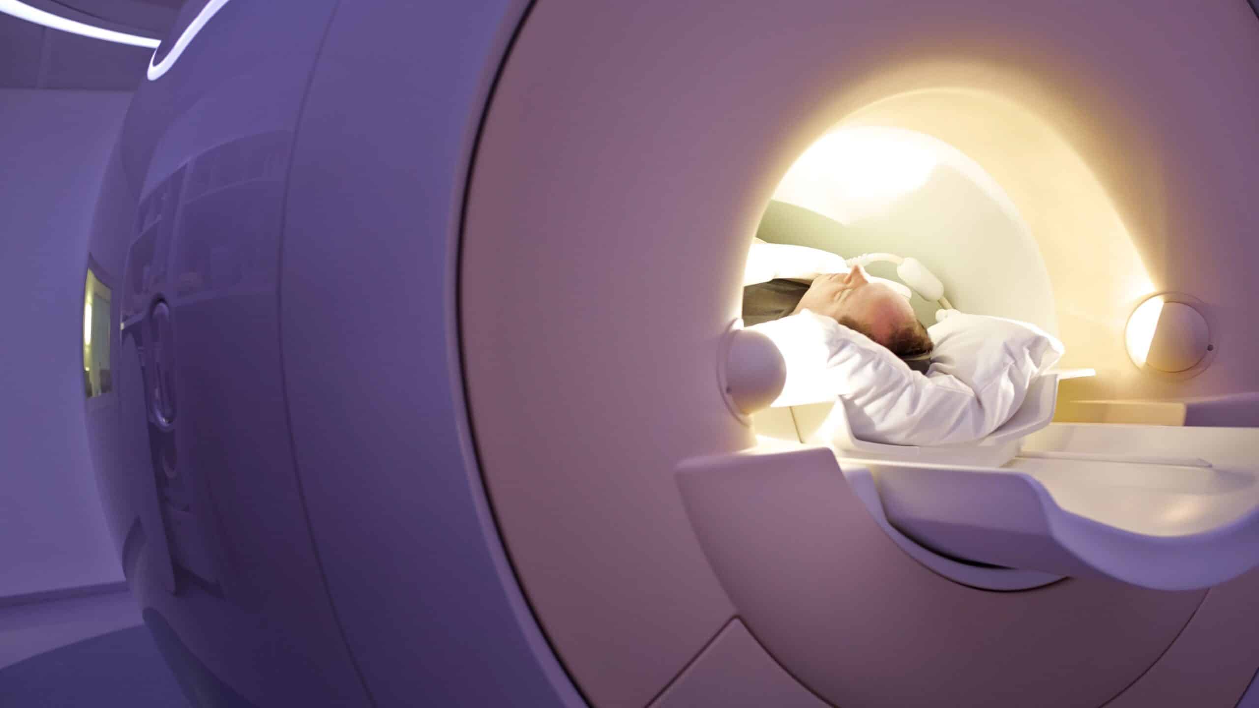
Ultrasound imaging technology is one of the most significant advancements in biomedical engineering, combining cutting-edge technology with precise hospital design to create a safe and comfortable treatment environment. This imaging method is a fundamental diagnostic tool in various medical fields, enhancing healthcare quality and facilitating non-invasive and direct medical examinations.

How High-Frequency Ultrasound Waves Work

This technology relies on high-frequency sound waves that are inaudible to the human ear. These sound waves travel through tissues and reflect off internal surfaces, creating a detailed image of internal structures. Ultrasound is widely used for diagnosing various medical conditions, making it an ideal option for emergency medicine applications and significantly contributing to the advancement of medical treatment techniques.

Ultrasound Waves

The Principle of Ultrasound Imaging

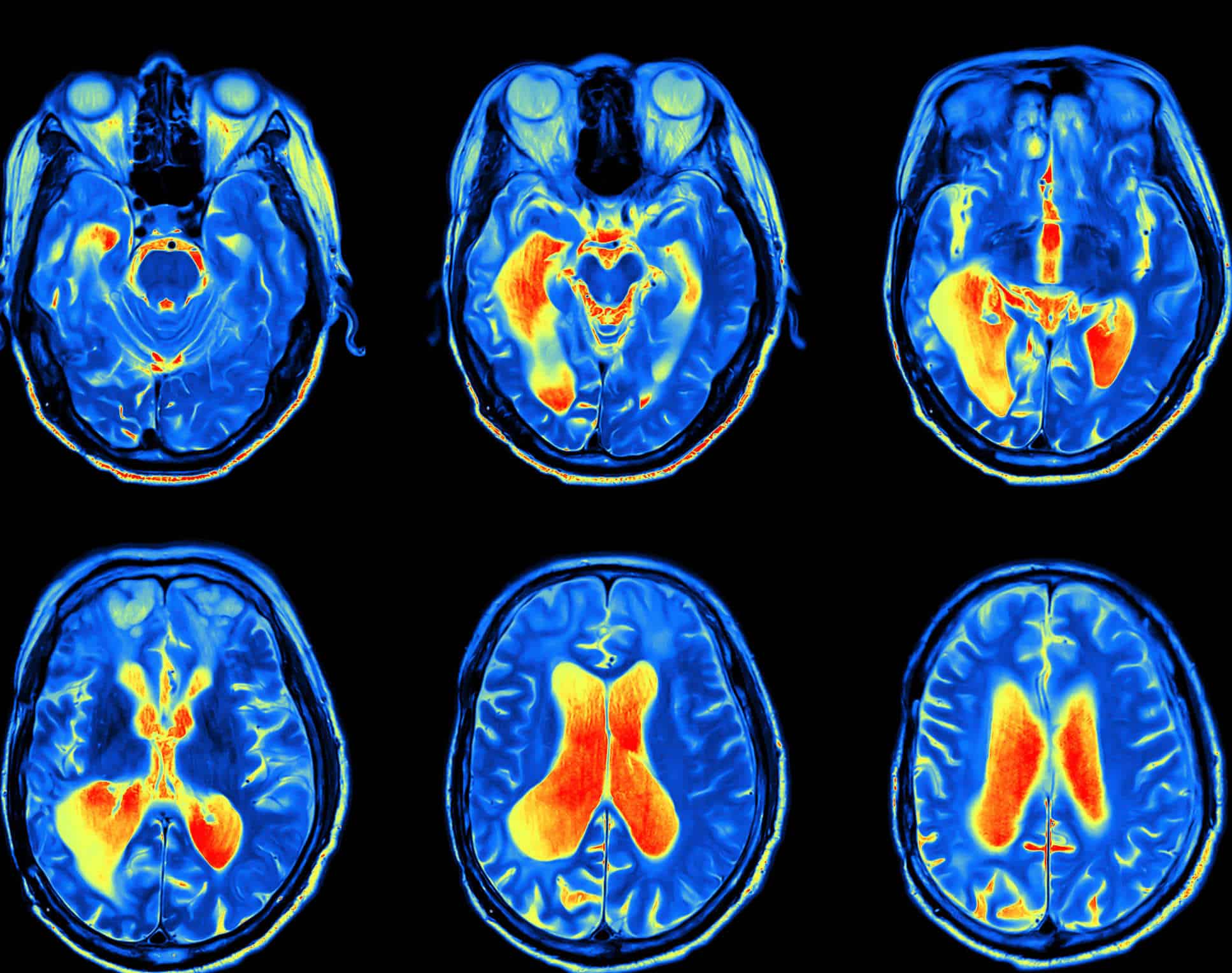
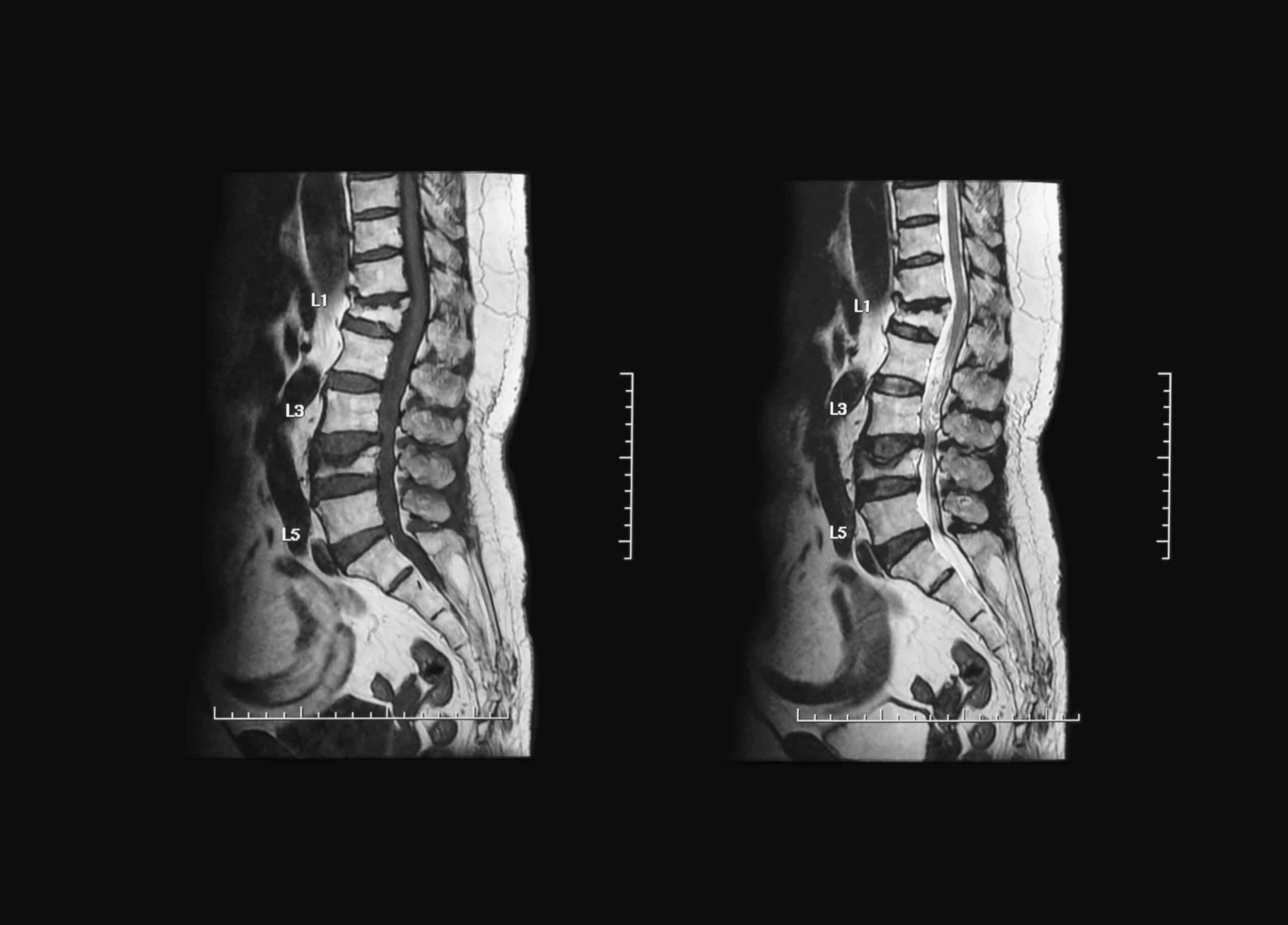
This imaging technology operates on a simple physical principle that involves generating high-frequency sound waves. A specialized device, known as a transducer, emits sound pulses and receives the reflected signals after they interact with tissues and organs. These signals are then converted into a digital image displayed on the screen. Ultrasound plays a crucial role in accurately detecting tumors and masses within the body, making it a valuable tool for early disease diagnosis.

Diverse Diagnostic Applications of Ultrasound

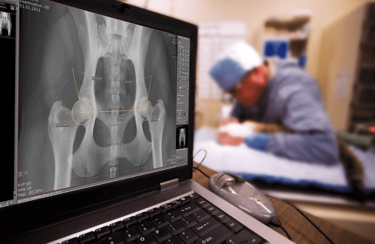
This technology plays a vital role in diagnosing numerous medical conditions. It is commonly used to examine vital organs such as the heart, liver, and kidneys. Additionally, it is indispensable in monitoring pregnancy and assessing fetal health. Ultrasound is also employed in precise interventional procedures, such as biopsy sample collection and guided interventions, enhancing medical accuracy while minimizing the risks associated with surgical procedures.

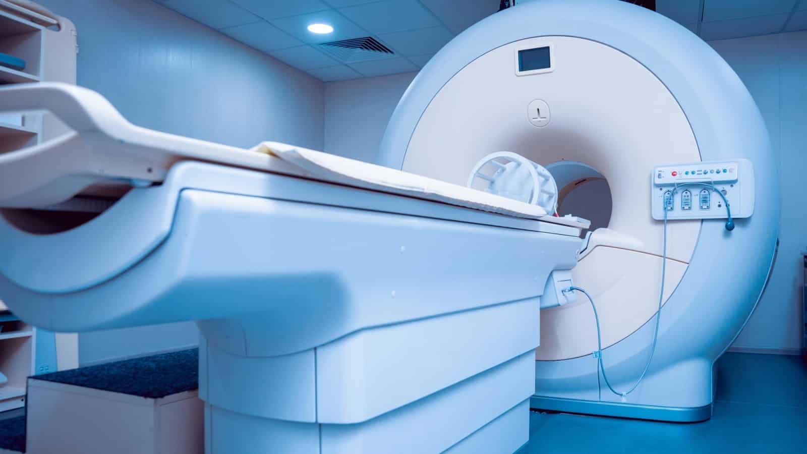
Ultrasound Technology and Medical Devices

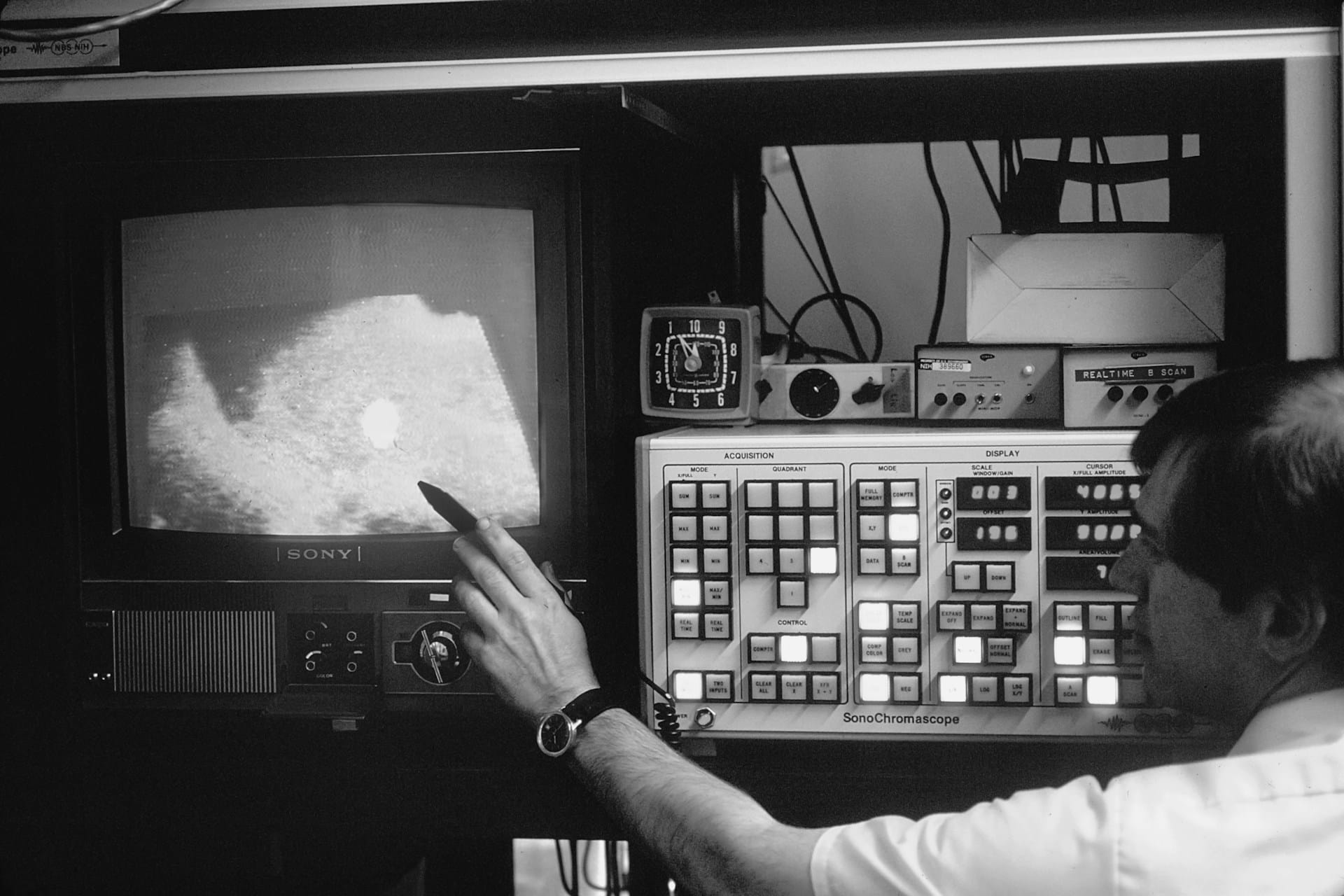
This imaging process requires advanced devices known for their high accuracy and efficiency in converting sound signals into detailed images. These devices use sophisticated computational techniques to enhance image quality and precision. Operational parameters are carefully adjusted to balance signal strength and accuracy. Ultrasound provides fast, non-invasive results, making it a preferred choice among physicians and medical professionals.

Applications of Ultrasound in Medicine

One of the key advantages of this imaging technique is its versatility across different medical fields. It is used in cardiology to assess valve function and detect cardiac disorders. It is also essential in obstetrics for monitoring fetal health during pregnancy. Furthermore, it is used to examine internal organs such as the liver, kidneys, and thyroid gland. Unlike other imaging techniques, such as X-rays and CT scans, Ultrasound does not use ionizing radiation, making it a safer diagnostic alternative.

تقنية الأشعة السينية

X-ray Technology

Recent Advances in Ultrasound Imaging TechnologyModern technology has led to significant advancements in imaging techniques, including the development of 3D and 4D imaging methods. These innovations allow doctors to view more detailed anatomical structures, improving diagnostic accuracy. Additionally, artificial intelligence (AI) and machine learning enhance Ultrasound imaging efficiency by optimizing image processing and improving pattern recognition for faster and more precise diagnoses.

Portable Ultrasound Imaging Devices and Their Impact on Healthcare

In recent years, the development of portable Ultrasound devices has enabled medical professionals to conduct examinations in various settings outside hospitals, such as mobile clinics, emergency rooms, and even remote areas lacking advanced medical infrastructure.This technological evolution significantly improves healthcare quality and increases accessibility to early diagnosis.

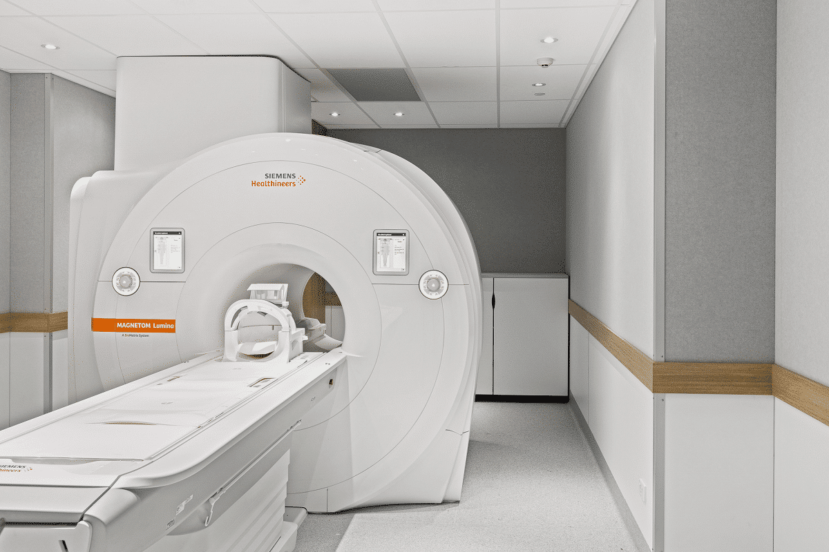
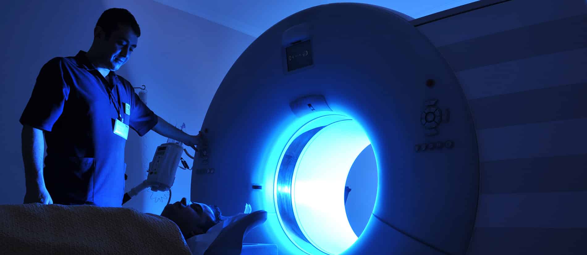
Imaging technology is a cornerstone of modern medical advancements, integrating physical principles with digital innovations to enhance diagnostics and treatment. The design of modern hospitals also plays a crucial role in supporting this technology, providing an optimal environment for efficient and accurate diagnoses.By combining Ultrasound imaging techniques with the latest engineering innovations, healthcare facilities ensure a safe and effective setting for patient care.

This diagnostic method remains a cutting-edge tool that offers both accuracy and speed, continually evolving to meet the future demands of healthcare. As such, investing in research and development is essential to achieve the best outcomes in medicine and biomedical engineering.

in conclusion Enhance Your Biomedical Engineering Career If you aspire to advance your career as a biomedical engineer, visit HSI today to explore our training courses. Our programs will equip you with the necessary skills to compete in the job market and elevate your engineering expertise to remarkable levels.

Source: Ultrasound – Special Subjects – MSD Manual Consumer Version

Revolutionizing Early Disease Detection: The Role of MRI in Medical Diagnostics

Magnetic Resonance Imaging (MRI) is a non-invasive diagnostic tool that uses a magnetic field and radio waves to produce detailed images of the inside of the human body. It plays a crucial role in the medical field and is considered a revolution in medical diagnostics. The future of MRI technology will see significant advancements in the clarity of the images it produces, and it is expected to develop further alongside the rise of artificial intelligence. In the following sections, we will explore this technology, its role in early disease detection, and how it will continue to evolve in the future.

الرنين المغناطيسي

MRI Technology
MRI works by exposing the body to a specific magnetic field. This magnetic field exploits the physics of protons, which are abundant in the human body due to the high water content in tissues. When the body is subjected to a strong magnetic field, the hydrogen protons in the tissues align with the field. Then, radio pulses are sent through the body, temporarily altering the orientation of these protons.

After a brief period, the hydrogen protons return to their original state and release signals that are captured by the MRI machine. These signals are used to create detailed images of the internal body structures, clearly distinguishing different tissues and organs and enabling precise identification of diseases.

How MRI Can Be Used for Early Disease Detection

MRI is an effective tool for early disease detection due to the high accuracy of its images of body tissues and organs. It can be used in the early stages of tumor development, increasing the chances of successful treatment. Additionally, MRI is valuable for diagnosing various neurological conditions such as strokes and multiple sclerosis. One of its key roles in neurology is identifying changes in brain tissue even before clinical symptoms appear, significantly aiding early detection and effective treatment.

الرنين المغناطيسي

One of the important uses of MRI is in the early detection of diabetic retinopathy, a complication of diabetes. This is done by creating a digital image of the retina and examining it for early signs of the condition.

MRI is also used for early detection of breast cancer, as traditional imaging methods may not detect the disease in its early stages. Furthermore, MRI can help detect possible artery blockages or narrowing that could lead to heart muscle problems. When it comes to bones, MRI can detect early signs of arthritis and changes in surrounding bone tissue.

Innovative Solutions in MRI Technology

While MRI itself is a revolutionary tool, it is continuously evolving to offer innovative solutions for disease detection and treatment. Some of the key innovations include:

Improved Image Accuracy

MRI imaging has been enhanced using mathematical equations, particularly by applying statistical science to improve the visibility of tumors. One of the methods used is calculating the probability mass function for each image in a given time sequence. This process involves analyzing the distribution of different values within the image, helping to identify changes in tissues and internal organs.

Another technique called Kullback-Leibler divergence is used to determine the best images for detecting tumors. This mathematical measure compares two different values, helping to identify the contrast between healthy tissue and a tumor, making the tumor more visible.

Advanced Image Processing Techniques

To improve image processing, advanced algorithms like GLCM (Gray Level Co-occurrence Matrix) and PNN (Probabilistic Neural Networks) are used to detect different brain tumors. These technologies have greatly advanced medical image processing, making it easier and more accurate for doctors to identify various tumor patterns for accurate diagnosis.

Use of Artificial Intelligence

The rapid growth of artificial intelligence tools in recent years will significantly enhance MRI technology. AI assists in accurately analyzing images and handling large data sets (Big Data) to recognize complex patterns, thereby improving doctors’ ability to diagnose diseases more accurately and quickly.

Challenges Facing MRI Technology

Despite the tremendous advancements in MRI technology and its applications, there are some challenges:

  • High Cost: MRI machines are expensive to purchase and maintain, and operational costs increase when advanced techniques are used.
  • Complexity: Some modern MRI techniques can be complex, requiring high skill levels and a deep understanding of how to operate the equipment.
  • Special Needs Patients: It can be difficult for patients with special needs to undergo MRI scans, as they may struggle to stay still during the examination.
  • Patient Anxiety: Some patients may be fearful of being confined in small spaces for extended periods, requiring doctors to take extra time to reassure them.
  • Long Exam Duration: Some MRI scans take 30 to 90 minutes, which can be uncomfortable, especially for elderly patients.

The Role of Contrast Agents in MRI

Contrast agents are chemicals injected into the body before an MRI scan to enhance image clarity and improve the accuracy of details around tissues and internal organs. Contrast agents play a crucial role in MRI:

  • Increasing contrast to highlight differences between tissues, helping to distinguish between healthy and diseased tissue.
  • Certain liquid agents, such as gadolinium, are used to improve visibility of tissues.
  • Gaseous contrast agents are used in specialized cases, such as lung imaging, to interact with oxygen or carbon dioxide for clearer images.
  • They also clarify blood vessels and examine blood flow for conditions like aneurysms.
  • They assist in detecting tumors.
  • They help examine internal organs like the liver and kidneys for any pathological changes.

Innovative Contrast Agents in MRI

Over time, the contrast agents used in MRI have been developed to provide the best possible image quality and diagnostic accuracy. Some of the innovative contrast agents include:

Gadolinium-based agents, which are the most commonly used in central nervous system imaging.
Iron-based agents, typically used for liver and vascular imaging.
Manganese-based agents, which are highly effective at targeting specific organs for precise imaging.
These agents are essential components of MRI techniques, and specialists choose the appropriate agent for each specific case.

With the tremendous advancements in the medical field over recent years, it is crucial for biomedical engineers to continuously enhance their skills and expertise. This is where platforms like HSI come into play, offering courses and consultations in the medical field to help you advance in your academic journey.

on our HSI website, we offer specialized training courses and consultations in the field of biomedical engineering, led by a team of certified experts and specialists to ensure you receive the best scientific content that combines theory and practical application. For inquiries, please get in touch with us, and we will respond with all the information you need to deepen your knowledge in the field of biomedical engineering.

Source: How Diagnostic Imaging is Revolutionizing Early Disease Detection

All copyrights reserved to © HSI