The Future of Wearable ECG Monitoring Devices

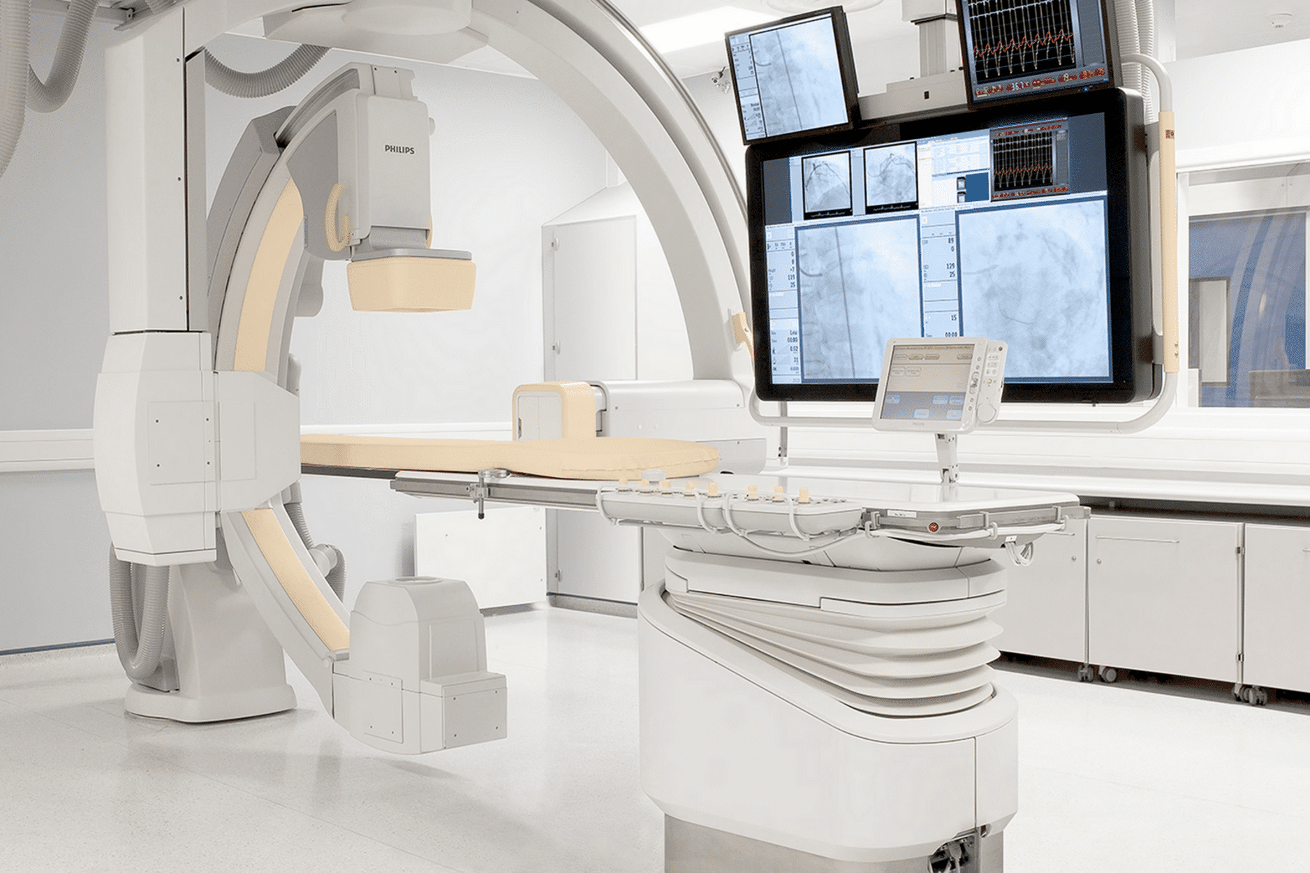
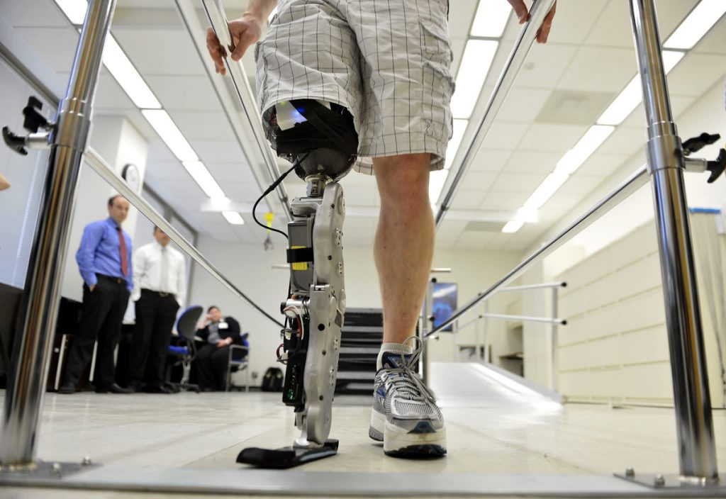
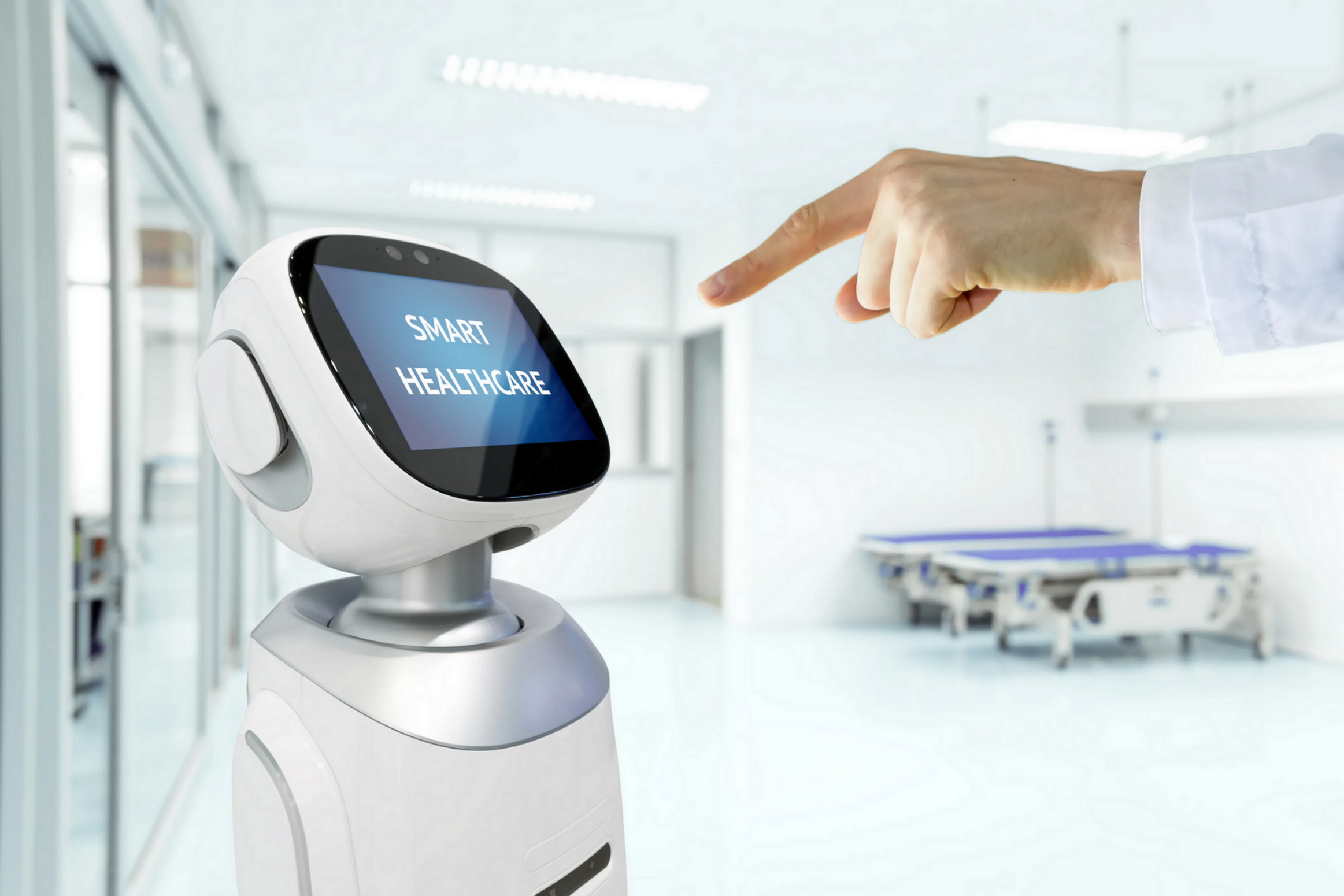
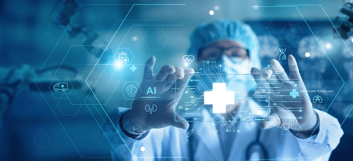
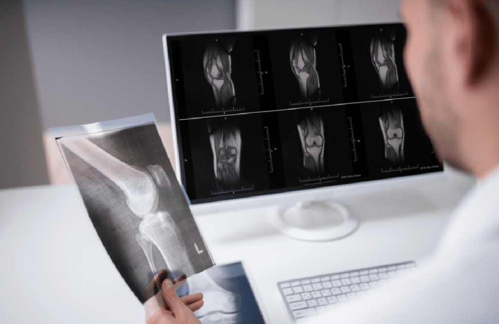
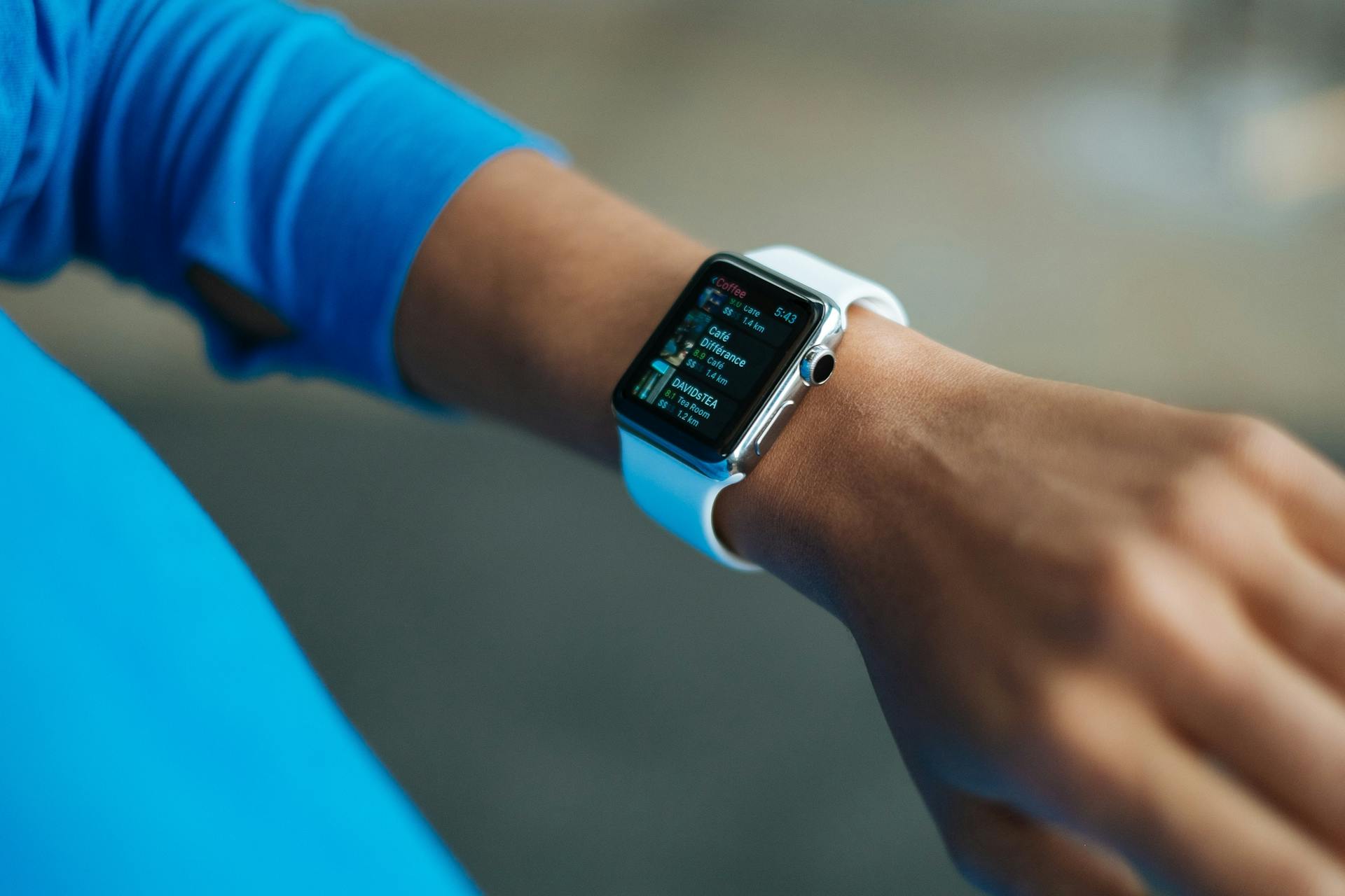
The healthcare field has witnessed significant developments thanks to technological advancements that have enabled the use of AI-powered wearable ECG monitoring devices. Since the early days of using various monitoring and diagnostic techniques, patient conditions have been observed more accurately than before. This has led to improved treatment quality and more efficient medical procedures. The process is no longer limited to visits to hospitals and traditional medical centers; patients can now monitor their health at home using these smart devices.

Combining health monitoring technologies with artificial intelligence

The idea of integrating wearable monitoring technologies with artificial intelligence began several decades ago, with scientists and innovators developing early prototypes for early diagnosis. Today, with noticeable improvements in computing capabilities and the availability of vast amounts of data, AI plays a pivotal role in analyzing medical information and providing accurate recommendations, thereby enhancing the performance of wearable ECG monitoring devices.

Wearable ECG

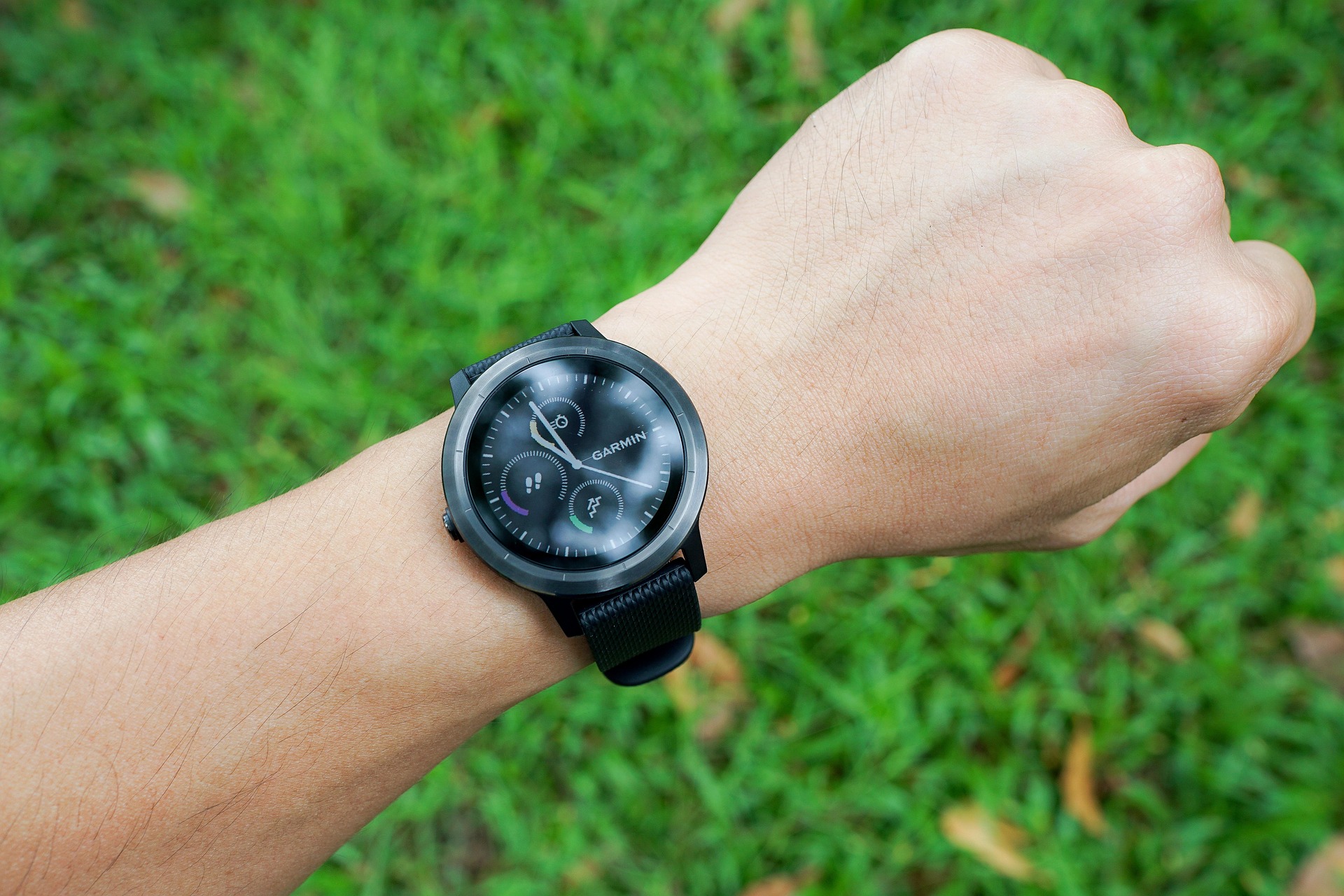
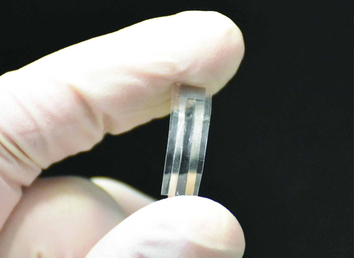
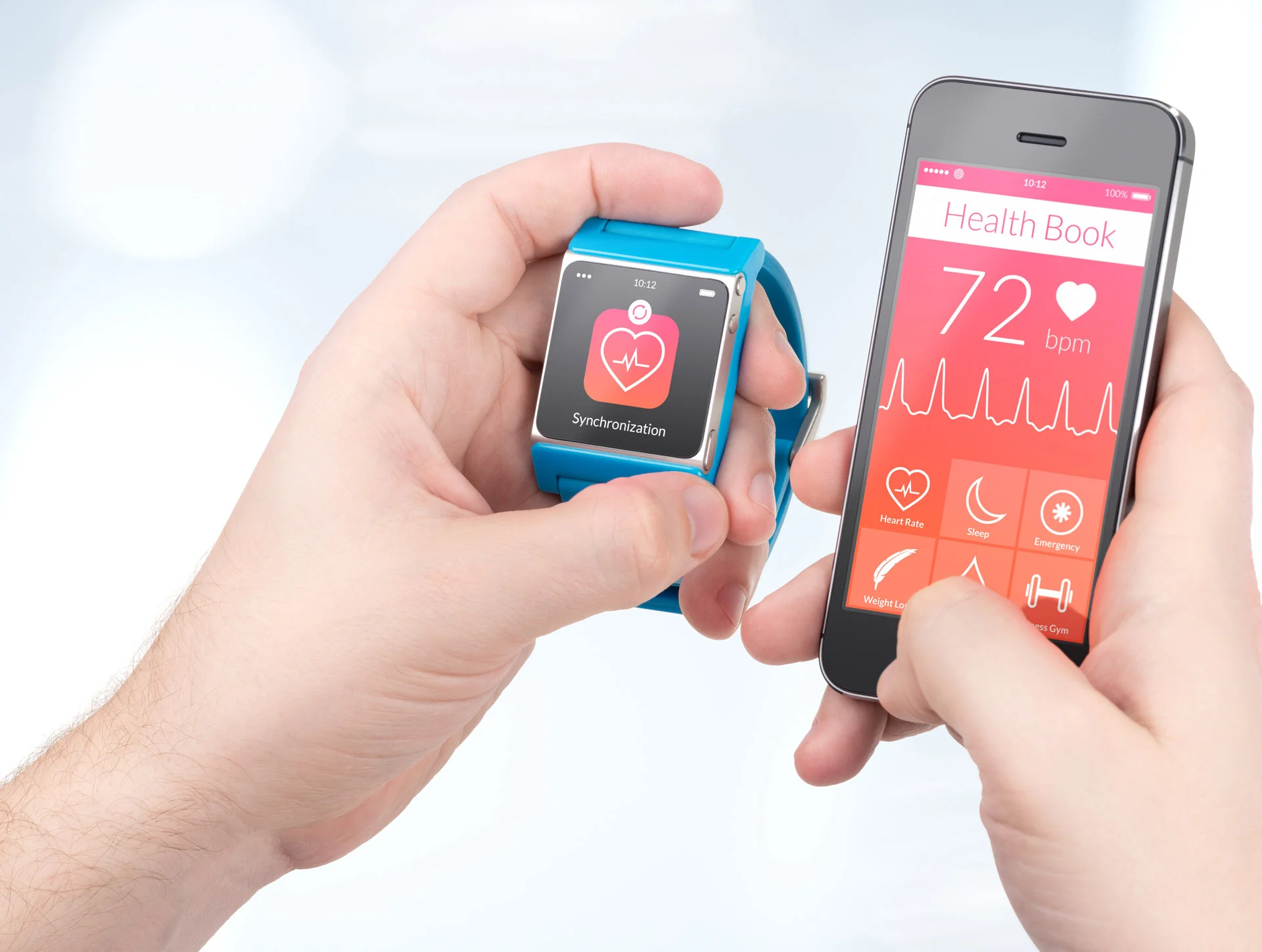
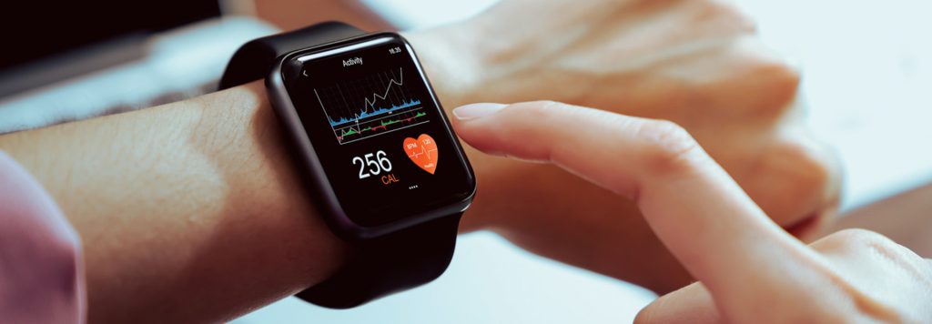
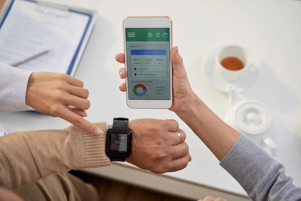
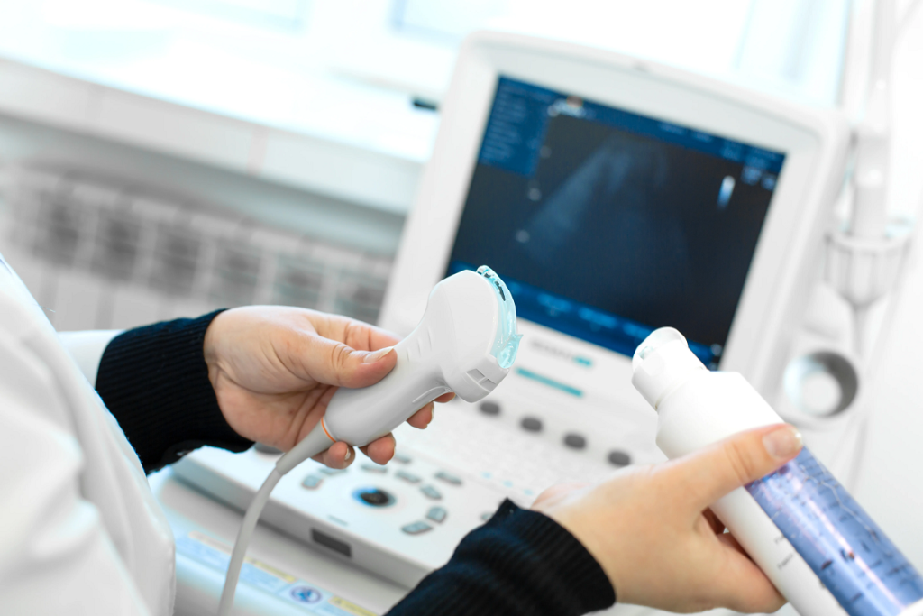
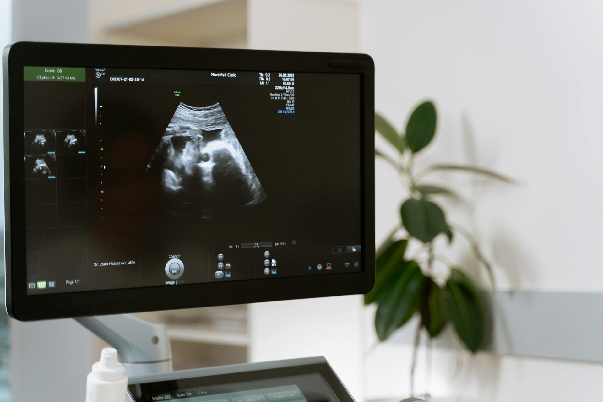
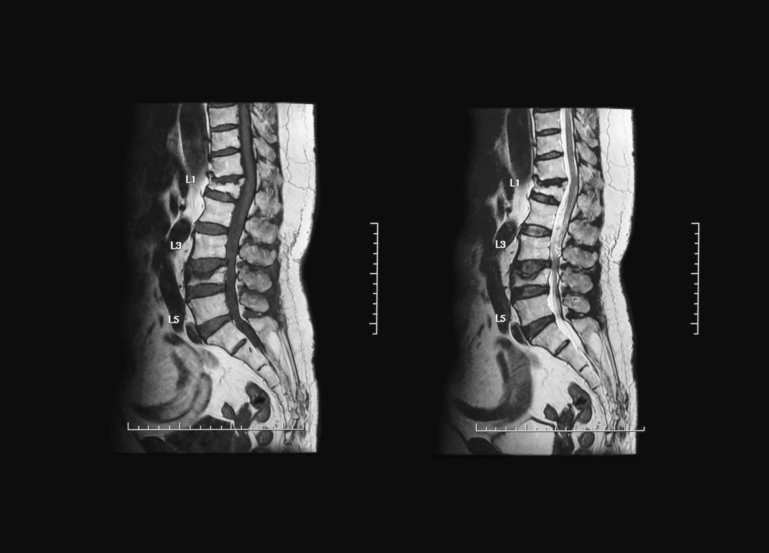
These devices are used to measure a wide range of vital signs, such as heart rate, blood pressure, blood oxygen levels, and electrocardiogram (ECG) activity. These measurements have narrowed the gap between patients and doctors, allowing for early medical intervention and preventing the development of chronic diseases.

Research and Evaluation Methodology

Modern studies rely on a comprehensive analysis of published research in the field of wearable ECG monitoring devices and artificial intelligence. The references and data are classified through several stages:

  • Identifying Sources and Data: The process began by gathering sources from global databases such as Google Scholar, resulting in thousands of articles discussing AI and its applications in healthcare.
  • Reviewing Titles and Abstracts: A meticulous filtering process was carried out to select the most relevant articles, focusing on studies that addressed the use of wearable ECG monitoring devices in disease diagnosis.
  • Technical Evaluation: The review included an assessment of the research methodologies and techniques used in each study, highlighting the technological innovations and the AI used in analyzing patient data.
  • Data Integration: The data was merged to create a comprehensive view of how wearable ECG monitoring devices impact workflow in medical institutions and enhance the quality of healthcare.

This methodology illustrates that progress in wearable ECG monitoring devices is not solely dependent on technical advancements but also on how they integrate with existing electronic medical record systems and established AI systems.

Research Findings and Key Innovations

There are several remarkable outcomes from research in this field, including:

1- Improved Early Diagnosis and Continuous Monitoring

Numerous studies have demonstrated that wearable ECG monitoring devices contribute to the early detection of arrhythmias, such as atrial fibrillation. These devices offer continuous monitoring of vital heart indicators, enabling doctors to detect any abnormal changes immediately. One study indicated that using these devices could reduce diagnostic errors by up to 30%, thereby improving patient treatment outcomes.

2- Reduced Need for Frequent Medical Visits

Thanks to wearable ECG monitoring devices, patients can now monitor their health remotely, reducing the need for repeated hospital visits. This approach not only eases the burden on healthcare systems but also saves time and effort for patients, especially those with chronic conditions that require constant monitoring.

3- Enhanced Quality of Medical Data

Continuous and effective data collection is one of the key advantages of wearable ECG monitoring devices. These devices provide accurate data regarding the patient’s health status, which helps train AI models to analyze patterns and predict potential health risks. Moreover, this data can be integrated with electronic medical records, offering a comprehensive system for monitoring and documenting medical cases.

4- Enhanced Interaction Between Patients and Healthcare Providers

The use of wearable ECG monitoring devices fosters an interactive environment between patients and doctors, as physicians can monitor data in real time and provide immediate medical consultations. This direct interaction helps build trust between both parties and enables the delivery of more personalized and effective healthcare.

Opportunities for Innovation in Healthcare

Integrating wearable ECG monitoring devices with AI technologies marks a transformative shift in healthcare. These innovations are expected to improve the efficiency of diagnosis and treatment, as AI can analyze data faster and more accurately than traditional methods while reducing healthcare costs. By minimizing medical errors and optimizing time and resource management, personalized medicine is enabled, where continuous data provides an accurate picture of each patient’s condition. This allows for the customization of treatment plans to meet individual needs, enhances preventive care, and ensures continuous monitoring by identifying health risks before they escalate and enabling early intervention.

Challenges Facing Device Implementation

Despite the clear benefits, wearable ECG monitoring devices face some core challenges. One of the main issues is managing the vast amounts of data generated by these devices, which require effective and secure storage and analysis. Integrating the generated data with existing electronic medical records demands the development of new protocols and updates to technical infrastructure, privacy, and security measures. Protecting patients’ health information from cyberattacks and breaches is of utmost priority. Additionally, doctors and nurses need to trust and adopt the new technology as an aid in providing healthcare without undermining their traditional skills and expertise.

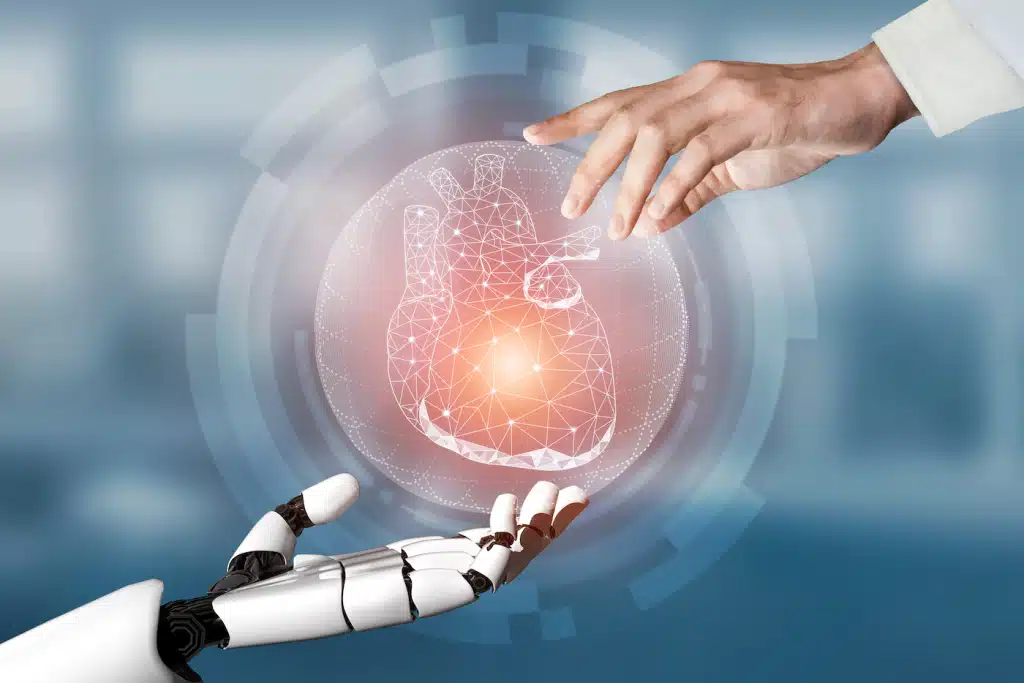
The Role of Artificial Intelligence in Device Development

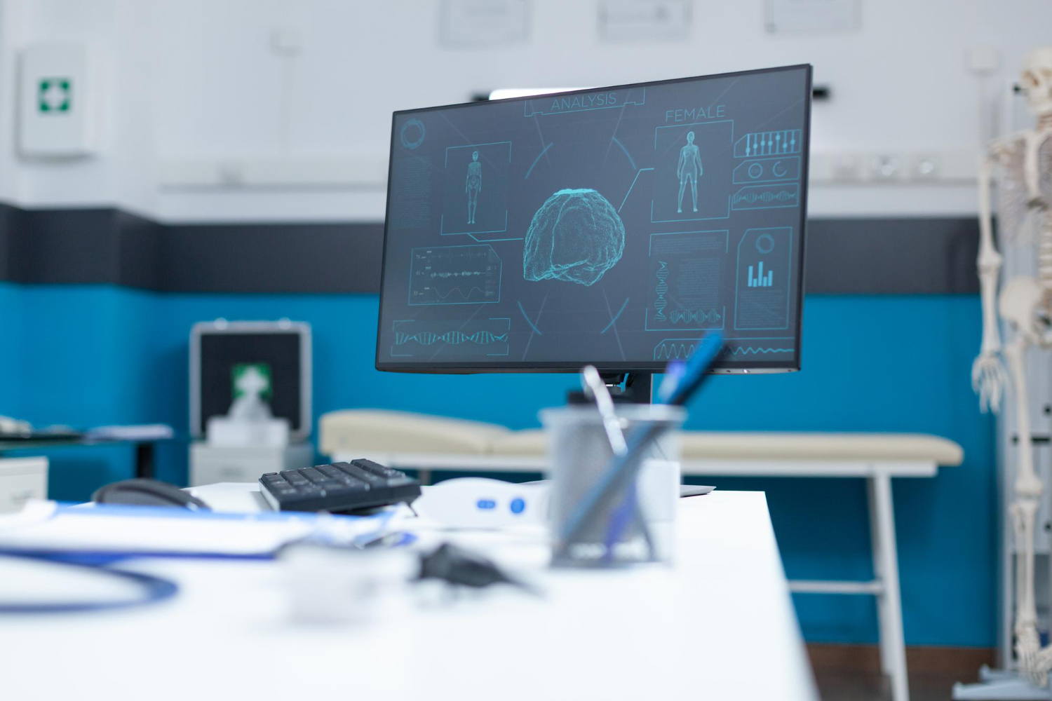
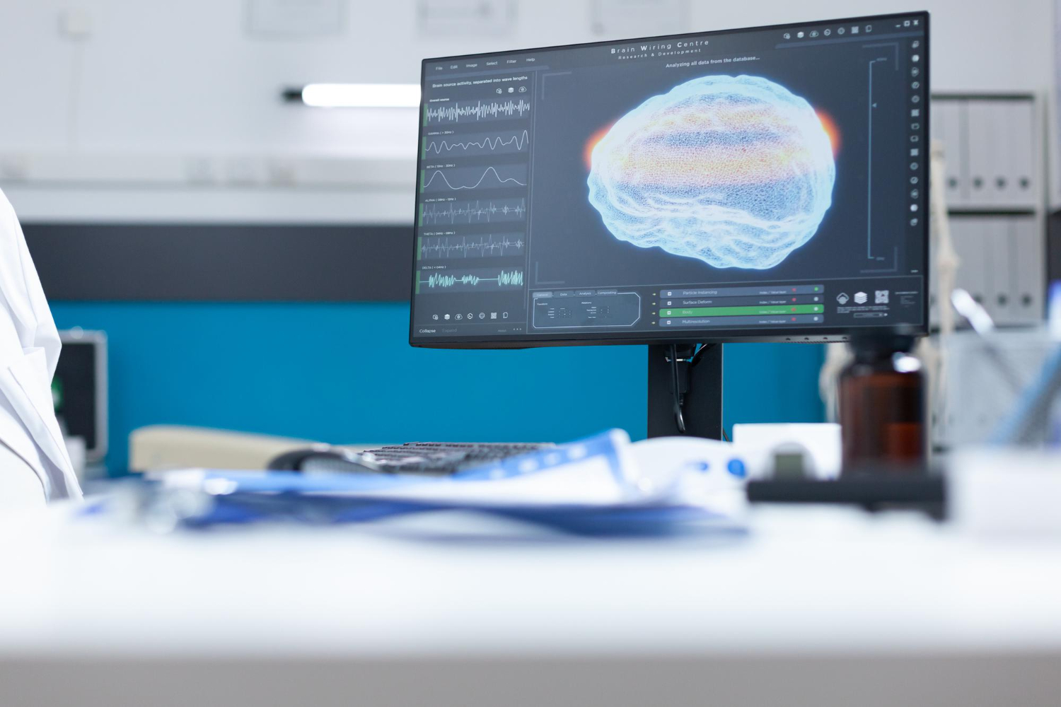
Artificial intelligence is considered the pivotal element that will transform wearable ECG monitoring devices from mere measuring tools into integrated diagnostic systems. AI relies on machine learning and deep learning techniques to analyze data patterns and deliver accurate predictions regarding a patient’s health status. Through these analyses, diagnostic accuracy can be improved and medical error rates reduced. Moreover, AI enables the development of interpretative models that explain the decision-making process, thereby increasing users’ confidence in the system.

Current Applications in Healthcare

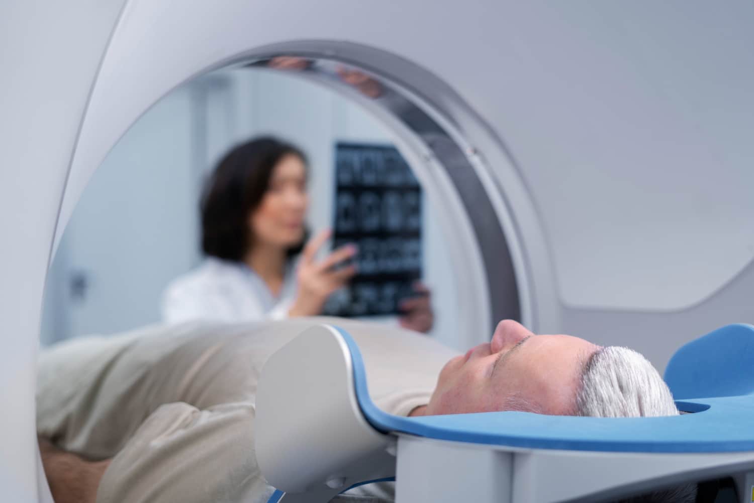
Wearable ECG monitoring devices are currently used in many medical institutions, where they are integrated with smartphone applications and remote monitoring systems. For example, they are employed for the early detection of arrhythmias, continuously monitoring heartbeats to detect any irregularities. They also facilitate remote patient monitoring, especially for those suffering from chronic diseases, allowing doctors to track their condition without the need for frequent visits. Additionally, data analysis through AI algorithms provides precise treatment recommendations based on the collected data.

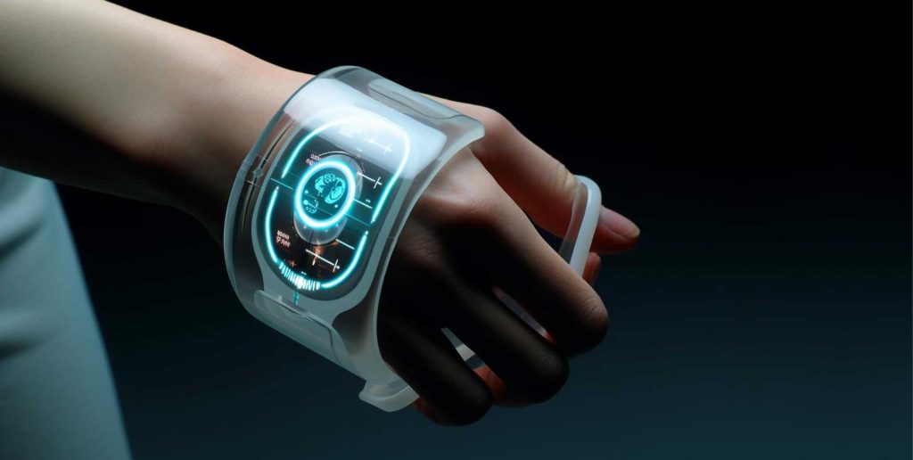
It is expected that the future will bring unprecedented developments in wearable ECG monitoring devices, particularly with advancements in AI and wireless communications. Some future trends include increased integration with advanced AI systems, which will lead to continuously improved diagnostic accuracy and analysis. Moreover, the development of more comfortable and wearable devices, with improved manufacturing materials for enhanced flexibility and lightness, will allow for extended use without discomfort. The scope of usage is also expected to expand beyond just ECG monitoring to include other health indicators, creating a comprehensive health system based on continuous monitoring and early prevention. Additionally, advanced protocols for data protection and cybersecurity will be developed to ensure safety and privacy.

At HSI, we believe that empowering medical engineers with modern knowledge and skills is key to the future. Therefore, we invite you to join our specialized training courses as we delve into these technologies and prepare you to play an active role in shaping the future of smart healthcare.

The development of biodegradable implants through AI revolution

In recent years, the field of biodegradable implants has undergone a transformative shift, thanks to the integration of AI in biomaterials and medical engineering. This evolution has moved us away from traditional trial and error methods toward a more precise, efficient, and intelligent process. Advanced algorithms now accelerate every stage from design and manufacturing to post implant monitoring. As artificial intelligence continues to influence global innovation, it’s crucial to dive deeper into this revolutionary fusion and understand its impact on the future of medicine. Let’s explore!

What Are Biodegradable Implants?

According to leading global medical institutions, biodegradable implants are medical devices designed to support or replace damaged tissues or bones inside the body. Over time, these implants naturally degrade into harmless components that are either absorbed or expelled by the body. This innovation presents a safer and more adaptable alternative to traditional permanent implants such as metal or plastic which often require additional surgeries for removal after healing.

The degradation rate of these implants depends on the type and structural composition of the material, allowing healthcare professionals to customize the duration of biological support based on the patient’s specific needs and the nature of the injury.

Biodegradable Implants

The Importance of Biodegradable Implants in Regenerative Medicine

Biodegradable implants play a critical role in the advancement of regenerative medicine. Their ability to support tissue healing without the need for follow-up surgeries significantly enhances patient recovery. These implants gradually dissolve after fulfilling their biological function, minimizing post-operative complications and surgical burdens.

By offering a smart and safe alternative to permanent implants, biodegradable devices improve clinical outcomes, especially for complex injuries or patients with chronic conditions. They also reduce emotional and physical stress, making treatments more efficient and patient friendly.

How AI Is Transforming Biodegradable Implant Design

As medical technology rapidly advances, artificial intelligence (AI) has emerged as a transformative force in the design of biodegradable implants. Traditional biomedical engineering standards are now being redefined, with AI in biomaterials enabling researchers to achieve unprecedented levels of precision and innovation. Let’s explore how this cutting-edge synergy is reshaping implant development.

1) Microscopic Structural Design with Machine Learning

  • Machine learning algorithms play a vital role in analyzing thousands of biological engineering samples to extract the optimal porous structures. These designs aim to strike the perfect balance between mechanical strength and degradation rate.
  • Artificial Neural Networks (ANNs) are trained on real-world mechanical data, generating precise implant structures that support fluid flow and nourish bone cells without compromising durability. This level of microstructural control would be nearly impossible without the capabilities of AI.

Biodegradable Implants

2) Numerical Simulation and Advanced Modeling Strategies

  • The design process of biodegradable implants greatly benefits from finite element methods (FEM), which simulate real-world body forces acting on the implant. These simulations help predict deformation and stress distribution before the manufacturing stage begins.
  • By integrating AI in biomaterials, researchers can refine these models further. Continuous training using laboratory and clinical test data allows AI systems to enhance simulation accuracy and bridge the gap between virtual predictions and real-life performance. This dramatically improves both the safety and efficiency of the final implant.

Also get acquainted with: The Role of AI in the Early Detection of Diseases through Medical Imaging

Smart Material Selection and Manufacturing

The success of biodegradable implants relies heavily on the careful selection and advanced manufacturing of smart materials. This phase is crucial for achieving the ideal balance between biocompatibility, durability, and controlled degradation.

1) AI-Powered Evaluation of Chemical Compositions

  • Both natural polymeric membranes (such as collagen and gelatin) and synthetic ones (like PLA and PGA) are commonly used in implant procedures. Through AI in biomaterials, thousands of material compositions can be virtually screened to identify the most suitable options based on biological, mechanical, and degradation criteria.
  • Molecular interaction modeling techniques—such as pharmacophore modeling—are applied to replace highly inflammatory components with atoms or functional groups that minimize immune responses. This process significantly enhances the safety and biocompatibility of the implant.

2) Additive Manufacturing and 3D Bioprinting

  • 3D bioprinting technology allows for the creation of patient-specific implants using CT and MRI scan data. These customized implants ensure uniform pore distribution, which is essential for effective tissue integration.
  • AI-driven control systems optimize the printing path and improve print efficiency, reducing production time, material waste, and overall costs. This not only enhances sustainability but also ensures a more accessible and scalable implant solution.

Biodegradable Implants

Intelligent Post-Implant Monitoring and Automation

After implantation, smart monitoring systems play a vital role in tracking the performance of biodegradable implants. These systems enable early interventions and tailored adjustments to improve healing outcomes.

1) Ultrasound and MRI-Based Monitoring

Computer vision algorithms are used to analyze imaging data—such as ultrasound and MRI scans—to monitor implant degradation and bone integration with high precision. These tools alert physicians to any delays in tissue fusion or abnormal implant morphology, allowing for timely medical decisions.

2) IoMT and Real-Time Healing Tracking

Next-generation implants are being equipped with micro-sensors that measure localized temperature, pH levels, and internal bone pressure. This real-time data is processed by AI algorithms to build predictive models of healing progress, offering personalized treatment recommendations for each patient.

Biodegradable Implants

Personalized Patient-Centered Care

Deep personalization in medical implant design is revolutionizing recovery experiences and treatment outcomes. With the integration of AI in biomaterials, it is now possible to create biodegradable implants tailored to each patient’s unique physiological profile.

1) Comprehensive Patient Data Analysis

  • Advanced AI systems collect and process extensive patient data—including age, weight, bone density, and medical history—to generate a precise digital model of the skeletal structure.
  • This model becomes the blueprint for designing a custom biodegradable implant that meets specific strength, flexibility, and degradation rate requirements. This high level of personalization reduces the risk of implant rejection and speeds up healing.

2) Continuous Learning and Smart Drug Delivery

  • Some innovative biodegradable implants now include embedded drug-releasing molecules—such as bone growth factors or localized pain relievers—that are gradually released as the implant degrades.
  • Using deep learning algorithms, AI in biomaterials dynamically adjusts drug release rates based on the patient’s healing stage and biological feedback, creating a more responsive and effective treatment.

Biodegradable Implants

Ethical Challenges and Technical Considerations

As AI in biomaterials continues to evolve, the field faces several ethical, technical, and regulatory hurdles that must be addressed to ensure safety, transparency, and trust.

  • Data Availability and Diversity: Machine learning models require vast and diverse datasets, but current clinical implant trials are still limited, particularly for rare or complex cases. This data scarcity can hinder the accuracy and generalization of AI predictions.
  • Explainability and Trust: Medical professionals demand transparent AI models that clearly explain how decisions are made. Without interpretability, physicians may hesitate to fully adopt AI-driven implant systems.
  • Regulatory Approval and Global Standards: Medical device regulations vary from country to country, often leading to extended approval timelines for AI-based implants. Aligning these frameworks is key to accelerating innovation without compromising safety.

The Future of Biodegradable Implants and AI Integration

Looking ahead, the integration of AI in biomaterials is set to expand beyond orthopedic applications, driving innovation across the broader landscape of regenerative medicine.

  1. Nano-Tech Integration: Future biodegradable implants are expected to incorporate smart nanomaterials like nanoparticles that deliver antibiotics or growth factors in response to cellular signals enhancing healing and infection prevention.
  2. Smart Self-Diagnostics: Research is also exploring micro-sensors embedded within implants that detect inflammation markers and autonomously trigger alerts or therapeutic adjustments, improving real time care and patient safety.
  3. Expanding Beyond Bone: The benefits of biodegradable implants will extend to vascular systems, neural tissues, and soft organ reconstruction, unlocking new potential in personalized, less-invasive healthcare solutions.

In the end, in a world rapidly advancing toward digital transformation and medical innovation, biodegradable implants powered by AI in biomaterials represent a breakthrough in precision, performance, and personalization. At HSI Healthcare Simulation and Innovation Center, we’re proud to be at the forefront of this change. Our comprehensive training programs, led by expert professionals and enriched with hands-on learning and specialized consulting, equip you with the tools to lead the future of medical care. Be part of the transformation.

Source: Application of Artificial Intelligence at All Stages of Bone Tissue Engineering

The Role of AI in the Development of Smart Prosthetics and the Challenges They Face

Over the past few years, artificial intelligence (AI) and smart prosthetics have begun transforming early prosthetic solutions. These technologies are not merely futuristic concepts but practical tools that are already enhancing how individuals embark on their prosthetic journeys.​

Understanding Early Smart Prosthetics and Their Challenges

Before delving into the specifics of this technology, it’s crucial to understand what early smart prosthetics are and their significant importance. These are the initial prosthetic devices provided to patients shortly after surgery, following wound healing and before the residual limb has fully stabilized.​

The Role of AI in the Development of Smart Prosthetics

The Role of Artificial Intelligence in Enhancing Prosthetic Movement

Imagine a prosthetic limb that not only mimics movement but also learns, adapts, and responds as if it were a natural extension of the body. This is what AI technologies offer in the field of prosthetics, revolutionizing their functionality by enabling more intelligent and fluid movements that adapt to the user’s needs. From predictive algorithms to machine learning, AI is fundamentally transforming prosthetics, making them more efficient and their functions more realistic and human-like.​

1- Processing and Adaptation

AI enhances prosthetic movements by processing vast amounts of data collected from sensors embedded in the device. These sensors monitor muscle signals, joint angles, and external conditions such as surface type or temperature. The AI system analyzes this data in real-time, allowing for precise adjustments to ensure smooth movement and immediate responsiveness.

2- Learning and User Adaptation

Machine learning, a branch of AI, drives smart prosthetics capable of adapting to individual user needs. By analyzing repetitive movements, the prosthetic can learn user patterns and understand their movement preferences. Over time, the device becomes more intelligent, anticipating the user’s intentions even before they are executed.

For example, an AI-powered prosthetic arm can distinguish between muscle signals associated with grasping a water cup and those for holding a pen, allowing for seamless transitions between tasks, making the device a natural extension of the body rather than just an assistive tool.

3- Enhancing Balance and Improving Gait

In lower-limb smart prosthetics, AI plays a crucial role in maintaining balance and improving gait patterns. By analyzing how the user distributes their weight and moves across different surfaces, the prosthetic can make precise adjustments to ensure stability.

For instance, walking on a sandy beach requires a different pattern than walking on a flat sidewalk. A smart prosthetic leg can detect these changes and adjust its stiffness, angle, or stride length, accordingly, reducing fatigue and ensuring the user’s safety.

Smart Prosthetics

The Importance of Early Prosthetic Fitting

Fitting a prosthetic limb at an early stage plays a crucial role in the recovery process, as it helps to:​

  • Reduce swelling​
  • Improve blood circulation​
  • Prevent joint stiffness​
  • Allow the patient to begin standing and moving​
  • Preserve muscle strength and balance​

Difficulties in Early Device Fitting

One of the biggest challenges facing early smart prosthetic fittings is the continuous change in the shape of the residual limb. Swelling may increase and decrease, the skin may be sensitive, and the shape of the residual limb is not yet final. Therefore, prosthetic sockets need to be adjusted as healing continues, making it difficult to design a socket that is both snug and comfortable.​

A socket that is too tight may cause pain or injury, while one that is too loose may lead to instability and increase the risk of imbalance. Thus, achieving the right fit requires a delicate balance and continuous care, where smart technology can make a real difference in prosthetic design.​

The technology behind smart prosthetics

​The technology behind smart prosthetics relies on a combination of hardware components and software to function efficiently. Sensors, actuators, and processors work together to collect data, analyze it, and execute precise movements.​

Advanced Algorithms

AI algorithms transform raw sensor data into actionable insights. For instance, a machine learning model can analyze thousands of steps to determine how a user’s gait changes when moving uphill versus downhill. The algorithm uses this knowledge to adjust the prosthetic’s movement automatically, ensuring a more seamless experience.​

Companies developing smart prosthetics should prioritize the accuracy and efficiency of these algorithms. Collaborating with data scientists and software engineers can help refine these systems, ensuring reliable performance under various conditions.​

Artificial foot

Sensor Integration

Sensors are the cornerstone of AI-powered prosthetics, gathering the necessary data to enable the system to learn and interact. These sensors include:​

  • Myoelectric Sensors: Capture electrical signals emitted by muscles.​
  • Gyroscopes: Measure stability and assist in adjusting movement.​
  • Pressure Sensors: Monitor weight distribution to ensure natural walking patterns.​

The data collected by these sensors is transmitted to an integrated processor within the prosthetic limb, where AI algorithms analyze it in real time. This allows the device to respond immediately to any changes in the user’s movement or surrounding environment, providing a smoother and more effective user experience.​

How AI is Transforming Early Prosthetic Design

AI brings speed, precision, and predictive capabilities to the prosthetic design process, facilitating a faster and more accurate match between the prosthetic design and the individual. This is achieved through:​

1- AI-Assisted Socket Design

In traditional socket design, a prosthetist uses measurements and molds to create a well-fitting socket. With smart prosthetics, it’s now possible to digitally scan the limb and input this data into advanced modeling software. This software predicts how the limb will change over time and suggests socket shapes that can adapt accordingly.​

2- Predicting Limb Volume Changes

One of the most challenging aspects of early prosthetic fitting is the continuous change in limb volume, which may shrink or shift weekly. AI can now track these changes using data from sensors and user inputs, even predicting future changes.​

3- Matching Appropriate Components to Each User

AI’s role extends beyond socket design to selecting the appropriate foot or knee for each user. Based on gait patterns, strength, age, and goals, AI can suggest prosthetic combinations that offer the best balance between support and flexibility. This enhances the success of the initial fitting, helps the user move faster with fewer adjustments, reduces trial time, and accelerates the transition to a more stable walking pattern.​

How AI is Transforming Early Prosthetic Design

The Future of Smart Prosthetics

The future of smart prosthetics extends beyond the devices themselves to building integrated systems that enhance user experience by incorporating complementary technologies.​

For example, mobile applications can serve as auxiliary tools, allowing users to monitor their prosthetics’ performance, adjust settings, and access tutorials or troubleshooting guides. These apps can also collect valuable data for companies, providing insights into how users interact with their devices and identifying areas for improvement.​

Additionally, wearable devices like smartwatches or fitness trackers can enhance functionality. By synchronizing with AI-powered prosthetics, these devices can provide real-time feedback on activity levels, body posture, and gait, helping users make informed decisions about their health and mobility.​

Expanding Access by Reducing Costs

While smart prosthetics offer advanced functionality, they often come with high production costs. To make these devices accessible to a broader range of users, companies should adopt cost-effective manufacturing techniques and explore alternative materials.​

Technologies like 3D printing can play a pivotal role in reducing production expenses. By leveraging additive manufacturing, companies can quickly produce complex prosthetic components at a lower cost. When combined with AI, these components provide the same high-level performance as traditionally manufactured parts.​

In conclusion, integrating AI into prosthetic design represents a revolutionary step toward improving users’ quality of life, offering them more natural movements and an enhanced user experience. In this context, HSI Center stands out as a leader in biomedical engineering and healthcare solutions, offering specialized training courses and professional consultations aimed at empowering professionals and organizations to make impactful contributions to the healthcare industry. ​

​The source: How AI and Smart Prosthetics Are Improving Early Fit Prosthetic Solutions.

Comprehensive Guide to Choosing Medical Equipment for Your Hospital

Medical equipment is considered the cornerstone of delivering effective and high-quality healthcare within hospitals. It plays a pivotal role in diagnosing diseases, treating patients, and monitoring their conditions. With rapid technological advancements, choosing the right medical equipment has become a complex task that requires a careful study of several factors to ensure the best patient outcomes and improve operational efficiency within the hospital. Making informed decisions regarding the selection of medical equipment directly impacts the quality of healthcare services provided, patient safety, and the economic viability of the medical institution.

With a wide range of options available, those responsible for choosing medical equipment may find themselves facing a significant challenge. The vast diversity in device types, specifications, technologies, and manufacturers necessitates the existence of a comprehensive guide that assists in making the right decisions. This article aims to serve as that guide by discussing the fundamental criteria to consider when selecting medical equipment for your hospital, with a focus on patient needs, safety, and ensuring accuracy and reliability.

Choosing Medical Equipment

Essential Criteria to Consider When Choosing Medical Equipment
The process of choosing the right medical equipment requires a careful evaluation of several essential criteria to ensure that the health needs of patients are met and the hospital’s objectives are achieved.

Patient Needs and Safety

The first and most important factor to consider when choosing medical equipment is the specific needs of patients and their safety. The hospital must accurately determine the demographic profile of the patients it serves, including their average ages, common health conditions, and the medical specialties provided. For instance, if the hospital serves a large number of children, it is crucial to select medical equipment specifically designed to meet their needs, taking into account their small sizes and unique physiological requirements. Similarly, the medical furniture should be comfortable and practical to accommodate patients of different ages and categories.

Choosing Medical Equipment

Ensuring patient safety is of utmost importance. Medical equipment should be chosen with built-in safety features, such as alarm systems that alert the medical staff in case of any malfunction or abnormal readings. The equipment should also include automatic shut-off mechanisms to reduce potential risks and infection control procedures to prevent the spread of diseases. For example, when selecting medical equipment such as vital signs monitors, features such as continuous monitoring, alerts for abnormal readings, and secure data transmission to prevent unauthorized access must be considered.

Another critical aspect is compatibility with existing healthcare systems to ensure smooth integration and efficient data management. It is essential to ensure that the new equipment can be easily integrated with the electronic health record (EHR) system and other medical devices used within the hospital. This integration helps streamline workflows, improve data accuracy, and enhance overall clinical efficiency.

Accuracy and Reliability

Accuracy and reliability are key characteristics when choosing medical equipment. Inaccurate or unreliable devices can lead to misdiagnosis and ineffective treatment, thereby endangering patient health. To ensure accuracy, it is necessary to select medical equipment that has undergone rigorous testing and obtained the necessary regulatory approvals. Look for quality certifications such as ISO 13485, which pertains to quality management systems in the medical device industry and ensures that manufacturers adhere to global safety and quality standards.

It is also important to research the reputation of the manufacturer and evaluate its performance in real-world environments. Obtaining feedback from other healthcare professionals who have used the equipment can provide valuable insights into its performance under clinical conditions. Additionally, the availability of support and maintenance services should be taken into account to ensure that any issues can be promptly resolved, minimizing downtime.

Economic Viability and Long-Term Value

While the initial cost of medical equipment is an important factor, it is equally crucial to assess the long-term value and economic viability of the selection. A cost-benefit analysis should be conducted to determine whether the benefits of the equipment outweigh its expenses in the long run. Factors such as durability, warranty coverage, and the potential for future upgrades or modifications should be considered.

Investing in high-quality equipment that offers long-term value can lead to cost savings and improved patient outcomes over time. For example, choosing medical equipment with advanced features and connectivity capabilities can enhance efficiency and data management, resulting in better clinical decision-making and a reduction in manual paperwork. Additionally, the availability of spare parts, as well as maintenance and repair costs, should be considered. Often, choosing medical equipment from reputable manufacturers ensures a reliable supply of spare parts and access to a network of authorized service centers, which minimizes downtime and extends the lifespan of the equipment.

Ensuring Quality and Safety in Choosing Medical Equipment

To guarantee that the selected medical equipment meets the highest standards of quality and safety, hospitals must follow several key steps.

Verifying Quality Certifications and Industry Standards:

Hospitals should meticulously check quality certifications and industry standards when choosing medical equipment. The ISO 13485 certification is one of the most important in this field, as it indicates that the manufacturer has a quality management system that meets the requirements for medical devices. Additionally, it is essential to ensure that the equipment has obtained the necessary regulatory approvals from both local and international authorities, such as the U.S. Food and Drug Administration (FDA) or the CE mark in Europe.

Conducting a Comprehensive Risk Assessment for the Equipment:

The process of choosing medical equipment should include a thorough risk assessment of the potential hazards associated with using the equipment. It is important to identify and evaluate the risks that patients and healthcare providers may face and to implement measures to mitigate these risks. For example, ensuring that the equipment is user-friendly can reduce the likelihood of user errors, and selecting devices made of biocompatible materials can minimize the risk of adverse reactions in patients. Furthermore, appropriate protocols for cleaning and sterilizing the equipment should be established to prevent the spread of infections.

Impact on Hospital Efficiency and Patient Outcomes

The proper selection of medical equipment has a significant impact on the efficiency of hospital operations and patient outcomes.

Accurate and Timely Diagnosis of Diseases:

Modern and advanced medical equipment contributes to the precise and prompt diagnosis of diseases. Advanced imaging devices such as Magnetic Resonance Imaging (MRI), Computed Tomography (CT) scans, and Electrocardiogram (ECG) machines provide detailed images and accurate information that help physicians detect diseases at early stages and precisely determine their characteristics. Early diagnosis can lead to more effective treatment and increase the chances of full recovery.

Improved Treatment Outcomes:

The impact of choosing the right medical equipment extends to improving the treatment outcomes of diseases. Advanced medical devices, such as ventilators, blood pumps, artificial heart devices, and minimally invasive surgical instruments, provide effective tools for delivering appropriate treatment for a variety of conditions. These technologies help accelerate the recovery process, reduce the risk of complications, and enhance the overall quality of life for patients.

Reducing the Risk of Injuries and Complications:

Selecting the appropriate medical equipment can significantly reduce the risk of injuries and complications among patients. Modern patient monitoring systems offer continuous surveillance of vital signs and provide timely alerts in case of any abnormal changes, allowing the medical staff to intervene swiftly. Moreover, the use of minimally invasive surgical tools reduces the size of incisions, shortens recovery periods, and minimizes the risk of infections and other complications associated with traditional surgery.

Enhancing Operational Efficiency in Hospitals:

The adoption of modern medical equipment leads to improved operational efficiency within hospitals. Advanced devices can streamline workflows, enhance data management, and reduce the length of hospital stays. For instance, remote patient monitoring systems can decrease the need for frequent in-person visits to the hospital or clinic, thereby reducing healthcare costs. Additionally, the implementation of automated systems minimizes human error and increases the speed and accuracy of clinical processes.

Choosing the right medical equipment for your hospital is a strategic decision that profoundly affects the quality of healthcare, patient outcomes, and operational efficiency. It requires a comprehensive and well-thought-out approach that considers patient needs, safety, accuracy, reliability, training, technical support, and a detailed analysis of economic viability and long-term value.

Collaboration among doctors, technicians, and administrators is essential during the equipment selection process to ensure that the needs of all stakeholders are met. The selection of medical equipment should be viewed as an ongoing process that necessitates regular evaluation and updates to keep pace with technological advancements and changing patient needs. By making informed decisions, hospitals can ensure that they are equipped with the best medical equipment to provide high-quality and effective healthcare to their patients.

If you wish to receive comprehensive courses in the field of biomedical engineering to develop yourself and enhance your skills, HSI provides you with the necessary tools to ensure a bright future as a biomedical engineer. All you need to do is check out our services and contact us for more information.

Wearable Medical Devices: How AI is Shaping the Future of Healthcare

In recent years, the healthcare sector has witnessed groundbreaking technological advancements, with the integration of artificial intelligence (AI) bringing a paradigm shift in patient care. One of the most notable developments is wearable medical devices, which are revolutionizing healthcare. These innovative devices not only transform how we monitor and manage our health but also unlock new possibilities in medical research and personalized medicine. In this article, we will explore these advancements in detail.

What Are Wearable Medical Devices?

Wearable medical devices are small, portable electronic devices worn on the body, capable of collecting, analyzing, and transmitting a wide range of health data. These devices can monitor various vital signs, such as heart rate, blood pressure, and temperature, providing real-time information that can be used for early disease detection or chronic condition management. With technological advancements, these devices have become more sophisticated, offering features such as ECG monitoring, continuous glucose monitoring (CGMs), and even sleep tracking.

Wearable Medical Devices - How AI is Shaping the Future of Healthcare

Benefits of AI-Powered Wearable Medical Devices

The integration of AI in wearable medical devices is transforming healthcare by providing patients and healthcare providers with continuous, data-driven insights. This personalized approach enhances patient care, particularly in managing chronic diseases and promoting preventive health measures. By tailoring interventions to individual needs, AI-powered wearables improve patient outcomes, enhance healthcare services, and empower individuals to take control of their health. Here are some key benefits of incorporating AI into these devices:

1. Enhanced Monitoring and Early Detection

AI-powered wearable devices continuously monitor vital signs and detect irregularities, enabling early detection of health risks such as arrhythmias or sudden changes in blood sugar levels—often before noticeable symptoms appear. This allows timely interventions and has shown promising results in detecting atrial fibrillation, potentially preventing strokes.

2. Personalized Treatment Plans

By analyzing individual health data, AI tailors’ treatment recommendations to meet specific patient needs, optimizing medication dosages, exercise routines, or dietary adjustments for more effective care. For instance, AI can adjust insulin doses for diabetics based on real-time glucose readings or suggest lifestyle modifications for hypertension patients.

3. Better Chronic Disease Management

Continuous monitoring enables more effective management of chronic diseases, particularly where one-size-fits-all treatments often fall short, such as diabetes, hypertension, and cardiovascular conditions. This reduces complications and hospital admissions, leading to more responsive care.

Wearable Medical Devices - Future of Healthcare

4. Empowering Patients

AI-powered wearable medical devices enhance patient engagement by providing real-time health insights, personalized health tips, and reminders, enabling individuals to manage their health more effectively.

5. Improved Patient Outcomes

By facilitating early interventions and continuous data-driven analysis, wearable devices improve long-term healthcare outcomes. Preventing severe complications, reducing hospital admissions, and minimizing emergency visits ultimately enhance patients’ quality of life.

6. A Key Role in Preventive Medicine

AI-powered medical devices play a crucial role in preventive medicine. Through continuous monitoring of vital signs and daily activities, these devices help individuals identify potential health risks and make informed decisions to prevent illnesses. For example, a wearable device tracking physical activity and sleep patterns can provide valuable insights into lifestyle habits that may contribute to obesity or sleep disorders.

7. Enabling Individuals to Take Charge of Their Health

Patients can easily track their progress and share data with their doctors, facilitating informed decision-making in diagnosis and treatment. For example, individuals with chronic conditions can use wearables to monitor symptoms and make necessary lifestyle or medication adjustments, ultimately leading to better health outcomes and quality of life.

How Wearable Medical Devices Support Medical Research

The benefits of wearable medical devices extend beyond patients; they also offer significant advantages to the medical research community. In clinical trials, these devices play a crucial role in collecting accurate and reliable data. Traditional data collection methods often rely on self-reported information or intermittent measurements, which can be prone to errors. In contrast, wearable devices provide continuous and objective data, offering a more comprehensive view of a patient’s health.

By integrating these devices into clinical trials, researchers can gather real-time data on how patients respond to treatments, identify potential side effects, and monitor adherence to prescribed therapies. This wealth of information allows for more precise assessments of the efficacy and safety of new treatments, ultimately accelerating drug development and bringing new medications to market faster.

How Do Wearable Medical Devices Work?

Wearable medical devices are equipped with sensors that continuously collect data on vital signs, physical activity, and other health metrics. This data is then transmitted to a smartphone or another connected device for analysis and interpretation. Some devices provide real-time feedback, while others store the information for healthcare professionals to review later.

Wearable Medical Devices

Types of Wearable Medical Devices

  1. Wearable medical devices come in a variety of forms, catering to different health needs and preferences. Here are some common examples:
  2. Fitness Trackers: Monitor physical activity, heart rate, sleep patterns, and calorie consumption.
  3. Smartwatches: Many smartwatches include health monitoring features, such as heart rate tracking, blood pressure monitoring, and ECG functionality.
  4. Continuous Glucose Monitors (CGMs): Essential for individuals with diabetes, these devices provide real-time blood sugar readings without the need for finger pricks.
  5. Wearable ECG Monitors: Detect irregular heart rhythms and other cardiac issues, enabling early intervention and heart attack prevention.
    Smart Rings: Compact devices offering various health monitoring features, including heart rate tracking, blood oxygen levels, and sleep analysis.
  6. Wearable Biosensors: Advanced devices capable of measuring multiple biomarkers, such as stress levels and skin temperature.
  7. AI-Powered Devices: Enhance data analysis, allowing for precise health monitoring and predictive insights.
  8. Sleep Management Devices: Track sleep patterns and provide actionable insights to improve sleep quality and overall health.
  9. Wearable Hearing Aids: Advanced hearing aids and smart earbuds that offer hearing assistance, health tracking, and seamless connectivity.
  10. Smart Glasses: Enhance vision, provide real-time data, and assist in rehabilitation or accessibility solutions.
  11. Wearable Robots: Aid mobility and rehabilitation, supporting physical therapy and improving movement.
  12. Smart Clothing: Garments embedded with sensors to track physical activity, posture, and health metrics.

The Future of Wearable Medical Devices

The field of wearable medical devices is advancing rapidly, and we can expect to see smaller, more comfortable devices with expanded capabilities. Integration with artificial intelligence (AI) will enable more sophisticated data analysis and personalized recommendations. Moreover, wearable technology will play an increasingly vital role in managing chronic diseases, monitoring mental health, and providing remote patient care.

How AI is Shaping the Future of Healthcare

Challenges Facing Wearable Devices

While wearable medical devices significantly enhance healthcare, they also face several challenges that must be addressed:

  1. Data Privacy and Security: Protecting sensitive health data is crucial to prevent unauthorized access.
  2. Regulatory Compliance: The development and commercialization of these devices must adhere to strict regulatory guidelines to ensure safety and effectiveness.
  3. User Adoption: Ensuring that individuals wear and use these devices correctly is essential for accurate data collection and effective health management.
  4. Battery Life: Limited battery life can be inconvenient and hinder continuous monitoring.
  5. Data Accuracy: These devices rely on sensors to monitor vital signs, but external factors such as body movement, environmental conditions, or sensor placement can impact data accuracy.

Conclusion

Despite these challenges, wearable medical devices have revolutionized healthcare by offering innovative solutions for monitoring, diagnosing, and treating a wide range of health conditions. With advancements in sensor technology, AI integration, and personalized healthcare, these devices are becoming more accurate, user-friendly, and accessible. As they continue to evolve, they hold great promise for improving patient outcomes, enhancing healthcare management, and reducing the burden on traditional healthcare systems.

source: Wearable Medical Devices: The Future of Healthcare

Fluoroscopy: Real-Time X-ray Imaging in Medicine

Fluoroscopy is one of the most important technologies in modern medical imaging, allowing doctors and specialists to view internal body structures in real-time. This revolutionary technique has transformed diagnostic methods and interventional procedures in medicine, significantly improving diagnostic accuracy and guiding treatments more effectively. In this detailed article, we will explore the working mechanism of this technique, its applications, benefits, associated risks, and the latest advancements in this vital field of medical engineering.

What is Fluoroscopy?

Fluoroscopy is an advanced imaging technique that utilizes X-rays to generate a continuous series of images displayed as a real-time video. This technique is widely used in emergency departments, operating rooms, and diagnostic radiology clinics, enabling surgeons and physicians to monitor the movement of organs and internal structures during procedures or diagnostic examinations. The increasing reliance on fluoroscopy reflects its significance in providing accurate and immediate insights into various medical conditions.

Fluoroscopy

Fundamental Principles of Fluoroscopy

Fluoroscopy is extensively used across multiple medical specialties, enhancing the quality of healthcare. To fully understand this technique, it is essential to delve into the fundamental principles that underpin fluoroscopy, forming its scientific and technical foundation.

X-ray Mechanism

This technique operates on the same principles as conventional X-ray imaging but with the ability to produce sequential images in real time, creating a video-like sequence of still images. The process begins with generating X-rays through a specialized tube, where a high-voltage current is applied. This accelerates electrons toward a metallic target, typically tungsten—a chemical element. Upon collision, X-rays are emitted and directed toward the area of the body being examined.

Image Intensification

The image intensifier in fluoroscopy plays a crucial role in enhancing the clarity of internal details. This device amplifies the signals produced by X-rays passing through the body, resulting in a bright and clear image. Recent advancements have led to the replacement of traditional image intensifiers with flat-panel detectors, which provide high-quality images while minimizing radiation exposure.

Diverse applications of Fluoroscopy

Fluoroscopy has a wide range of medical applications, both in diagnostics and interventional procedures. This system is highly flexible and capable of providing real-time moving images, allowing doctors to monitor physiological changes instantly.

Fluoroscopy

Diagnostic Applications

Fluoroscopy is used in several essential diagnostic procedures, including:

  • Gastrointestinal Studies: Fluoroscopy is employed to assess the digestive tract, including the esophagus, stomach, and intestines. Barium studies, such as barium swallow and barium enema, help evaluate abnormalities or motility disorders.
  • Neuromuscular and Spinal Imaging: This technique is used in diagnostic imaging of the spine, particularly in myelography, which aids in assessing spinal cord conditions.
  • Joint Evaluations: For joint-related issues, fluoroscopy is also used in arthrography to evaluate abnormalities or injuries affecting the joints.

Interventional Applications

This technique is a cornerstone in medical engineering for interventional procedures, providing live imaging during operations, which contributes to:

  • Guiding Surgical Procedures: Fluoroscopy assists in catheter placement during cardiac procedures, such as coronary angiography, ensuring precise positioning and minimizing procedural risks.
  • Interventional Treatments: It is utilized in therapeutic interventions, including stent placement, bone fixation in fracture surgeries, and the insertion of delicate medical devices.
  • Minimally Invasive Procedures: This technology facilitates minimally invasive procedures requiring small or no surgical incisions, reducing patient discomfort and promoting faster recovery.

Benefits of Fluoroscopy in Medical Practice

Fluoroscopy is a pioneering technique that enhances medical care quality for several key reasons:

1) Enhanced and Accurate Diagnosis

This technology provides dynamic imaging that reveals the functional movements of internal organs, such as diaphragm motion, intestinal activity, and blood flow through vessels. This capability helps in early disease detection, as moving images can uncover functional changes that static images might not reveal.

Fluoroscopy

2) High Precision in Interventional Procedures

By offering live imaging during medical procedures, fluoroscopy enables precise manipulation of medical instruments within the body. This reduces the likelihood of surgical complications and improves success rates in procedures like stent placement and fracture fixation.

3) Reduced Surgical Invasiveness

This technique allows for numerous minimally invasive procedures, reducing the need for major surgeries. Minimally invasive techniques are less painful, promote faster recovery, and lower the risk of post-operative infections, making them a significant advancement in modern medicine.

Risks and Safety Considerations in Fluoroscopy

Despite the significant benefits of fluoroscopy, there are certain risks associated with its use that must be carefully considered.

Fluoroscopy

Radiation Exposure

Radiation exposure is one of the primary concerns when using fluoroscopy. Prolonged procedures may increase the amount of radiation a patient is exposed to. The level of exposure depends on the duration of the procedure and the examined area. To mitigate these risks, advanced techniques such as pulsed fluoroscopy and dose-reduction algorithms have been developed to minimize radiation exposure.

Contrast Media Reactions

Contrast media are often used during this technique to enhance the visibility of internal structures. However, some patients may have allergic reactions to these substances, ranging from mild skin rashes to severe anaphylactic responses. To manage these risks, pre-procedure screening and emergency medications are utilized when necessary.

Occupational Radiation Exposure

Medical professionals performing frequent fluoroscopy procedures are at risk of occupational radiation exposure. Strict protective measures, such as wearing personal protective equipment (e.g., lead aprons and thyroid shields), are essential. Additionally, regular monitoring of radiation levels ensures the safety of doctors and healthcare staff, helping to prevent potential long-term risks.

Recent Advances in Fluoroscopy Technology

The field of fluoroscopy has seen significant advancements in recent years, leading to improved image quality and reduced radiation risks. Traditional systems have been replaced with digital fluoroscopy using flat-panel detectors, which enhance image resolution while minimizing radiation exposure. This digital system provides high-definition images and allows for post-processing adjustments to refine diagnostics. Additionally, it enables seamless image sharing among medical teams for quick consultations and decision-making.

Furthermore, the integration of cone-beam computed tomography (CBCT) with fluoroscopy now offers detailed 3D imaging for complex anatomical assessments. This technology is widely used in dentistry, orthopedic surgeries, and interventional procedures requiring precise localization.

Fluoroscopy

Moreover, combining this technique with other imaging modalities such as ultrasound and magnetic resonance imaging (MRI) has led to the development of hybrid imaging systems. These systems provide both anatomical and functional insights, enhancing diagnostic accuracy and guiding interventional procedures more effectively, particularly in oncology and tumor treatments.

The Future of Fluoroscopy

The future of this technique looks promising, with continuous research and technological advancements aimed at improving procedural efficiency and reducing radiation-related risks.

Integration of Artificial Intelligence

Artificial intelligence (AI) is expected to play a pivotal role in fluoroscopy’s evolution. AI algorithms can analyze real-time images, detecting anomalies that may be overlooked by the human eye. Additionally, AI-driven techniques can optimize radiation dose reduction strategies, enhancing patient and healthcare worker safety.

Portable Fluoroscopy Systems

Research and development efforts are currently focused on creating portable fluoroscopy units that can be easily transported to emergency departments or remote areas. These mobile units will enable rapid diagnostic imaging and treatment in locations with limited medical infrastructure, thereby expanding access to essential healthcare services.

Exploring Radiation-Free Alternatives

Scientists are actively exploring non-ionizing imaging alternatives such as optical coherence tomography (OCT) and photoacoustic imaging. While these alternatives are still in experimental stages, they hold great potential for reducing radiation-related risks in this technique, potentially revolutionizing the future of medical imaging.

In conclusion, we live in an era where cutting-edge fluoroscopy technologies are transforming medical diagnostics and interventions. However, behind every advanced imaging system stands a well-trained professional who makes critical decisions. This is where HSI Healthcare Training & Consultancy comes in. Join HSI today—not just to keep up with medical advancements, but to lead them. Because in the world of healthcare, history is not just written by machines but by those who master their language. Are you ready to shape the future of medical imaging?

Source: Real-Time Imaging and Its Impact on Modern Medicine

Biomechanics and the Role of Artificial Intelligence in Prosthetic Design

The technological integration in biomechanics and the role played by machine learning and artificial intelligence in prosthetic design have become a boon for individuals with disabilities. Leading manufacturers of rehabilitation aids have utilized the concept of neural networks to simulate various anatomical and biomechanical functions of missing human body parts. The involvement of human interaction with various factors such as electronic circuits, software, robotics, etc., has had a revolutionary impact in the field of rehabilitation, leading to the development of devices like prosthetic legs, human body-controlled prosthetics, and exoskeletons. In this article, we will discuss the role of Biomechanics and artificial intelligence in prosthetic design and rehabilitation.

Prosthetics: Innovation in Medical Compensation

Prosthetics include any artificial device that replaces a missing body part due to injury, disease, or congenital defects. Some companies have developed various types of prosthetics, ranging from purely cosmetic to advanced devices made using titanium and carbon fibers. Regarding the manufacturing of these prosthetics using biomechanics, the process is quite similar to making medical insoles. The end of the amputated limb is digitally scanned, and then a special socket is designed to which the prosthetic is attached. Using electrodes, electrical signals from the muscles are received and sent to an electronic control board inside the prosthetic, which translates these signals into actual movements.

Biomechanics

Understanding Prosthetic and Orthotic Engineering

The field of prosthetics focuses on creating artificial limbs, while orthotic specialists design devices to support or correct musculoskeletal conditions. Traditionally, this field relied on manual measurements, mechanical adjustments, and trial-and-error methods. Although these techniques were effective, they often lacked precision and adaptability to achieve optimal results.

Artificial Intelligence in Prosthetic Design is changing this landscape by integrating machine learning, robotics, and sensing technology. These tools transform static devices into dynamic systems capable of learning and evolving based on individual user needs. The result is a new level of functionality that mimics natural movement and intelligently responds to unique biomechanical challenges.

History of Artificial Intelligence in Prosthetics and Orthotics

The first intelligent prosthetic limb was developed by Charles A. Blatchford & Sons, Ltd in 1993, with an improved version in 1995 called the Intelligent Prosthesis Plus. In 1998, Blatchford developed an adaptive prosthetic limb combining three operating mechanisms—hydraulic, pneumatic, and microprocessor control. A fully microprocessor-controlled knee was developed in 1997 by Ottobock, known as the C-leg. In 2015, Blatchford introduced Linx, the world’s first fully integrated limb with seven sensors and four CPUs throughout the leg, allowing coordination and synchronization of the knee and ankle joints by sensing and analyzing data about the user’s movement and activities to make them more natural.

The first prosthetic hand was launched by Touch Bionics in 2007, featuring individually powered fingers and a thumb with gripping capabilities. The design was later integrated with a rotating thumb known as i-limb ultra and i-limb revolution designs, implemented using Biosim and the My i-limb application. Bebionic was commercially launched in the market in 2010 by RSL Steeper.

Biomechanics in Prosthetic Design

The concept of a brain-computer interface (BCI) implemented by neural prosthetics or mind-controlled prosthetics that can recognize real-time data and tools to achieve nearly natural function is a current requirement. A mind-controlled smart prosthetic arm based on electroencephalography was presented at the IEEE Conference in 2016 but has not yet been commercialized. Researchers are on the path to developing more complex devices that mimic the natural brain by implementing artificial intelligence on onboard computers that read and respond to neural signals transmitted to prosthetics and robotic orthotics that enhance the function of amputated and paralyzed body parts.

Application of Artificial Intelligence in Prosthetic Design

Artificial intelligence (AI) is driving a significant transformation in the engineering of prosthetics and orthotics, converting traditional devices into intelligent, adaptive tools. These innovations not only enhance mobility but also improve the overall quality of life for individuals with physical disabilities. By leveraging the latest advancements in AI for prosthetic design, these devices have become more responsive, intuitive, and integrated into daily routines. As AI progresses, it bridges the gap between human capability and technological support in ways previously deemed impossible.

The integration of AI with biomechanics has significantly improved prosthetic control, enabling amputees to operate prosthetic limbs more effectively. Recently, a brain-controlled prosthetic (a form of myoelectric control) was introduced as the latest advancement in AI-assisted control systems. A joint project between the Pentagon and Johns Hopkins University’s Applied Physics Laboratory (APL) led to the development of a modular prosthetic limb entirely controlled by brain-implanted sensors. This innovation even aims to restore the sense of touch by sending electrical pulses from the limb to the sensory cortex.

Types of AI Applications in Prosthetic Design

The application of biomechanics and Artificial Intelligence in Prosthetic Design can be categorized based on the affected area:

  1. Lower limb prosthetics and orthotics
  2. Upper limb prosthetics and orthotics
  3. Rehabilitation aids such as automated mobility devices

Prosthetic

Role of Biomechanics and AI in Prosthetic Design

  1. Adaptive Prosthetics: AI-powered prosthetics utilize algorithms to instantly adapt to user movements and environmental changes.
  2. Dynamic Adjustment: AI-driven prosthetic legs adjust gait and force distribution dynamically when transitioning from flat surfaces to stairs or uneven terrain.
  3. Customized Design: AI-enhanced technology leverages imaging data and user feedback to design devices that offer optimal fit and improved function.
  4. Generative Design Tools: These tools craft prosthetic sockets that precisely fit individual anatomy, enhancing comfort and reducing pressure points.
  5. Neural Integration: AI-based brain-machine interfaces allow users to control prosthetics via neural signals.
  6. Neural Signal Interpretation: AI interprets brain activity to execute precise movements in prosthetics, providing users with greater autonomy.
  7. Predictive Maintenance: AI technology tracks device usage to predict wear and tear, ensuring timely maintenance and longer lifespan.
  8. Smart Orthotic Braces: Equipped with sensors, these braces alert users to potential issues before failures occur.
  9. Biomechanical Simulation: AI simulates human movement to optimize prosthetic and orthotic designs before production.
  10. Digital Assessment: Digital tools evaluate balance, alignment, and weight distribution, allowing for precise adjustments during design.

Technological Innovations in AI

  1. Generative AI in Design: AI maximizes functionality while minimizing material usage by creating efficient, user-specific designs.
  2. Virtual Modeling: Virtual models simulate device performance, allowing for precise adjustments and reduced development cycles.
  3. Self-Learning Prosthetics: Prosthetics equipped with self-learning systems improve through use and feedback over time.
  4. Advanced Sensor Integration: AI-driven sensors analyze movement patterns and pressure, continuously enhancing device performance.

Application-of-AI-in-Prosthetic-Design

Challenges and Ethical Considerations

  1. Data Privacy: Protecting sensitive user information collected by AI systems.
  2. Accessibility: Ensuring these technologies are available to underserved populations.
  3. Bias in AI Models: Mitigating errors that could affect device functionality.
  4. Regulatory Compliance: Meeting stringent medical standards.

Future Trends

  1. Neuroprosthetics: Improved brain-device communication may restore sensory feedback.
  2. Hybrid Bio-Devices: Combining AI with genetically modified tissues could achieve seamless body integration.
  3. On-Demand Manufacturing: AI and 3D printing can streamline custom prosthetic fabrication, reducing costs and wait times.
  4. Emotionally Intelligent Devices: Future devices may adapt to user emotions, providing additional support during stress or fatigue.

In conclusion, the role of biomechanics and Artificial Intelligence in Prosthetic Design has revolutionized the engineering of prosthetics and orthotics by transforming them into intelligent tools that enhance independence and mobility. Despite significant advancements in recent years, there remains a need for more affordable and accessible prosthetics to ensure availability for individuals with disabilities, regardless of their financial means, without hindering their opportunity to live normally.

Herein lies the role of HSI Center in offering innovative solutions in the field of medical engineering. HSI is committed to advancing the sector through a comprehensive range of specialized courses and high-quality healthcare solutions, contributing to enhancing the quality of life for individuals in need of these technologies.

Source: AI in Biomechanics: From Creating Cosmetic Prosthetics to Making Metahumans

Soft skills for engineers of biomedical 2025 | 5 basic skills

In an era of rapid advancements in biomedical engineering, technical knowledge alone is no longer sufficient for success. Soft skills for engineers have become a crucial component in the career development of any biomedical engineer aiming to excel in this competitive field. In this article, we will explore five fundamental soft skills that blend personal and professional aspects, forming the cornerstone of a successful future for biomedical engineers in 2025.

Soft Skills for Biomedical Engineers

In the growing field of biomedical engineering, soft skills are essential for success and innovation. Biomedical engineers must integrate a combination of scientific knowledge, technical expertise, and interpersonal skills to excel in their roles. Below are the key soft skills that biomedical engineers need to thrive in the evolving landscape of biomedical technology and healthcare.

1) Effective Communication and Collaboration Skills

Effective communication is one of the most critical soft skills for engineers, especially in a field that requires collaboration with multidisciplinary teams. Biomedical engineering is not solely about technical expertise; it also involves the ability to explain complex concepts clearly to doctors, nurses, scientists, and other healthcare professionals.

soft skills for engineers

The Importance of Communication in the Workplace

In biomedical projects, engineers from various disciplines, such as electronics, mechanics, and biology, collaborate to develop medical devices or innovative treatment solutions. Therefore, engineers must be capable of:

  • Explaining Complex Ideas in Simple Terms: This helps facilitate knowledge transfer among professionals with different backgrounds.
  • Negotiating and Conflict Resolution: Engineers should be able to manage discussions and resolve personal conflicts that may arise during teamwork.
  • Delivering Presentations and Reports: Strong presentation skills ensure that projects meet required standards and receive necessary support from all stakeholders.

Additionally, active listening is a crucial part of soft skills for engineers. It enables them to quickly grasp team feedback and develop integrated solutions that align with workplace needs. Given the intense competition and complex challenges in the industry, continuous collaboration and effective communication significantly enhance an engineer’s ability to innovate and accelerate project execution.

2) Problem-Solving and Creative Innovation

Biomedical engineers face complex challenges that require innovative and effective solutions, making problem-solving and creative thinking fundamental pillars in medical project development.

Integrating Creativity with Problem-Solving Methodologies

Developing advanced medical devices demands a deep understanding of medical challenges and a precise analysis before proposing innovative solutions. This highlights the importance of:

  • Critical Problem Analysis: Studying all aspects of a problem and identifying strengths and weaknesses in the system.
  • Generating New Ideas: Utilizing creativity to design devices or systems that enhance treatment outcomes.
  • Experimentation and Practical Application: Testing new ideas in a simulated environment or on a small scale before full implementation.

soft skills for engineers

By refining their soft skills, engineers can systematically and creatively tackle problems, delivering solutions that exceed expectations and contribute to improving healthcare services. Furthermore, combining analytical thinking with creativity fosters an innovative work environment where technical expertise meets renewal and invention.

3) Analytical Thinking and Scientific Research

Analytical thinking and scientific research are core elements and the driving force behind innovation in biomedical engineering. These soft skills for engineers expand the boundaries of knowledge and open new horizons for medical technology advancements.

The Role of Research and Analysis in Performance Improvement

Engineers face challenges that require a deep understanding of data and precise analysis to develop practical solutions that directly aid patient treatment and assist medical professionals. This process involves:

  • Data Collection and Analysis: Utilizing advanced scientific research methods to analyze results and ensure the safety and effectiveness of medical devices.
  • Continuous Experimentation: Conducting repeated trials to refine prototypes and improve them based on analytical findings.
  • Practical Application of Research: Translating research outcomes into technological solutions applicable in hospitals and medical centers.

Enhancing engineers’ analytical and research skills enables them to gain deeper insights into medical needs, leading to the development of cutting-edge devices that improve patient quality of life. Moreover, the ability to analyze information and extract valuable conclusions is key to progress in this field, especially with the continuous advancements in information technology and big data.

You can also check out: The Importance of Sales Skills for Biomedical Engineers and How to Master Them

4) Adaptability and Continuous Learning

Another essential soft skill for engineers, particularly in biomedical engineering, is the ability to adapt and engage in continuous learning. Given the rapid technological advancements in the healthcare sector, staying updated is crucial for long-term success.

soft skills for engineers

Challenges and Changes in the Work Environment

Biomedical engineering is one of the most rapidly evolving fields, with new technologies emerging regularly and market demands constantly shifting. Engineers must:

  • Stay Updated with Innovations: Follow the latest research, technologies, and breakthroughs relevant to their field.
  • Be Open to Change: Embrace new learning methodologies and develop practical strategies to handle evolving challenges.
  • Invest in Self-Development: Participate in training courses, workshops, and scientific events to enhance their knowledge and skills.

Adaptability is a key soft skill for engineers that enhances resilience and confidence in handling industry changes. Engineers who commit to continuous learning not only keep up with current trends but also invest in their professional future, ensuring they remain competitive and innovative in their field.

5) Ethical Professionalism and Compliance with Standards

Given the critical nature of biomedical engineering, where patient safety and well-being are at stake, ethical professionalism and adherence to industry standards are among the most vital soft skills for engineers in this domain. Technical expertise alone is not enough—engineers must also uphold ethical responsibility in their work.

soft skills for engineers

The Importance of Ethics and Regulatory Standards

When designing medical devices and therapeutic applications, engineers must ensure that all processes align with strict ethical and regulatory guidelines, including:

  • Compliance with Regulatory Bodies: Adhering to standards set by organizations like the FDA and ISO to guarantee product safety and effectiveness.
  • Protecting Patient Privacy: Ensuring that all technological solutions respect patient rights and maintain data confidentiality.
  • Social Responsibility: Practicing transparency and integrity in all professional interactions.

Strengthening ethical professionalism ensures that engineers play an active role not only in technological advancements but also in patient safety and community welfare. Upholding high ethical standards and industry compliance builds trust and credibility, which are invaluable assets in a successful engineering career.

In the rapidly evolving field of biomedical engineering, success is not solely dependent on technical knowledge. Soft skills for engineers, such as adaptability, analytical thinking, problem-solving, and ethical professionalism, are critical for innovation and effective collaboration in multidisciplinary environments.

If you are looking to advance your career in biomedical engineering, HSI is your gateway to excellence. We offer a comprehensive range of specialized training programs led by industry experts to help you acquire the skills necessary to enhance your career prospects and make a real impact in the healthcare sector. Join us now and be part of the future of biomedical engineering!

Source: Top Skills You Need for a Career in Biomedical Engineering

Smart Hospital Design: Integrating IoT and AI for Better Patient Care

In an era where healthcare challenges and pressure on medical systems are increasing, Smart Hospital Design emerges as an innovative solution that leverages technology to enhance medical outcomes. This concept integrates advanced technologies such as the Internet of Things (IoT), Artificial Intelligence (AI), and other digital systems to create a seamless environment that supports early disease diagnosis, enables continuous patient monitoring, and optimizes resource management effectively.

Understanding Smart Hospital Design

Smart Hospital Design refers to the integration of modern technologies within a hospital’s infrastructure to achieve comprehensive and efficient patient care. This concept goes beyond traditional healthcare by utilizing intelligent systems that communicate in real time, providing instant data on patient conditions, medical equipment, and environmental factors within the hospital. Through this approach, smart hospital design contributes to:

  • Enhancing Patient Experience: By offering personalized healthcare services and precise monitoring of their condition.
  • Improving Operational Efficiency: Through automation of administrative and medical tasks, reducing human errors.
  • Supporting Decision-Making: By analyzing data and predicting future needs using AI-driven algorithms.

Smart Hospital Design

Essential Technologies in Smart Hospital Design

The integration of cutting-edge technologies is a driving force behind Smart Hospital Design, enabling healthcare facilities to enhance efficiency, patient care, and medical outcomes. Many countries are actively adopting these innovations to transform traditional hospitals into smart healthcare environments. Below are the key technologies shaping smart hospital design.

1) Internet of Things (IoT)

The Internet of Things (IoT) serves as the backbone of Smart Hospital Design, consisting of a network of connected smart devices that collect real-time data. Its applications include:

Continuous Monitoring: Smart sensors track vital signs such as blood pressure, heart rate, and oxygen levels, allowing medical teams to respond swiftly to any abnormalities.

  • Asset Management: Hospitals utilize RFID technology and sensors to monitor the location of medical equipment, reducing time spent searching for critical devices.
  • Environmental Control: Automated systems adjust lighting, temperature, and humidity levels to create an optimal healing environment for patients.

2) Artificial Intelligence (AI)

The integration of AI in healthcare plays a crucial role, especially in smart hospital design, as it helps in:

  • Medical Data Analysis: AI-driven algorithms analyze medical imaging and clinical data, leading to faster and more accurate disease diagnoses.
  • Risk Prediction: AI-powered predictive models help physicians anticipate potential complications, enabling early interventions.
  • Surgical Precision: AI-assisted surgical systems support surgeons in making precise decisions, reducing human error and improving surgical outcomes.

Smart Hospital Design

3) Big Data Analytics

The collection and analysis of big data are integral to Smart Hospital Design, offering benefits such as:

  • Electronic Health Record (EHR) Management: Massive amounts of patient data are stored and analyzed to provide a comprehensive view of a patient’s health across departments.
  • Health Trend Monitoring: Data analytics helps detect disease patterns and guide healthcare policies to better address community health needs.

4) Robotics and Assistive Technologies

Robotics play a significant role in Smart Hospital Design, contributing to surgical assistance, advanced robotic systems enhance precision in surgeries, leading to shorter recovery times and fewer complications. And automated routine tasks, robots perform essential functions like medication delivery and medical supply transportation, reducing the burden on healthcare staff and allowing them to focus on patient care.

5) Telemedicine Technologies

Modern telemedicine solutions expand healthcare accessibility, particularly in remote areas, by enabling remote consultations, patients can receive medical advice from specialists without the need for in-person visits. And chronic disease management by smart devices continuously monitor patients’ conditions and send regular health reports to medical teams for proactive care.

Smart Hospital Design

Benefits of Smart Hospital Design

  • Smart hospital design enhances patient care by enabling real-time monitoring of vital signs, allowing doctors to provide immediate and precise treatment, ultimately improving recovery rates.
  • It optimizes hospital resource management by tracking medical equipment locations and managing work schedules efficiently, reducing costs, streamlining operations, and improving healthcare service quality.
  • Medical errors are minimized through the automation of critical processes. Smart systems relying on data analysis provide instant alerts in case of any malfunction or safety threshold breach.
  • With data analytics and AI-driven predictions, well-informed decisions are made based on accurate insights, leading to better clinical outcomes and improved healthcare delivery.

Challenges in Implementing Smart Hospital Design

Despite these challenges, Smart Hospital Design represents the future of healthcare, offering innovative solutions to enhance patient outcomes and streamline hospital operations.

  1. Investment Cost: Integrating advanced technologies requires significant investments in infrastructure and medical equipment. Many hospitals struggle to secure the necessary funding to implement such modern systems.
  2. Data Security: With increased digital connectivity within hospitals, the risk of patient data exposure to cyberattacks rises, necessitating the implementation of advanced security solutions to ensure data confidentiality and integrity.
  3. Medical Staff Training: Transitioning to smart hospital designs requires training healthcare professionals to effectively use modern technologies, which can take additional time and effort.
  4. System Integration: Ensuring seamless communication and integration between different systems is a major challenge, as all hospital devices must operate in perfect harmony without disruptions.

Smart hospital Design - Singapore General Hospital

Models of Integrating IoT and AI in Hospital Design

The healthcare sector is undergoing a radical transformation with the adoption of IoT and AI technologies in smart hospital design. These innovations enhance medical service quality by improving diagnostic accuracy, reducing errors, and accelerating emergency response. Below are some leading examples showcasing how these technologies are revolutionizing patient experience and operational efficiency in hospitals.

1- Johns Hopkins Hospital

Johns Hopkins Hospital is a leading example of Smart Hospital Design, incorporating AI and robotics into surgical procedures. These innovations have significantly improved surgical precision and reduced patient recovery times. Additionally, its electronic health record (EHR) system allows doctors to access patient information quickly and efficiently, streamlining medical decision-making.

2- Cleveland Clinic

Cleveland Clinic has embraced telemedicine as part of its Smart Hospital Design, expanding access to healthcare services. Patient rooms are equipped with IoT devices that continuously monitor vital signs, ensuring immediate medical intervention when necessary. This approach enhances patient safety and enables real-time health tracking.

3- Singapore General Hospital

Singapore General Hospital utilizes big data analytics as a core component of its Smart Hospital Design strategy. By analyzing patient admission patterns, the hospital optimizes logistics and resource management. Additionally, automated transport systems powered by robotics efficiently deliver medical supplies, reducing operational delays and enhancing efficiency.

Finally, Smart Hospital Design represents a bold step toward the future of healthcare, where IoT and AI-driven systems create a more efficient, accurate, and patient-centered environment. These advancements improve patient experiences, enhance staff performance, and revolutionize medical service delivery.

Source: Smart Hospitals, Integrating Technology into Healthcare Design Management

Revolution of AI in medical imaging: A Beginner’s Guide to AI in MRI and Ultrasound

The healthcare industry is undergoing a transformative shift, thanks to AI in medical imaging, which has become a crucial tool for improving diagnostic accuracy and accelerating treatment processes. With rapid technological advancements, AI is no longer just an assistive tool but a strategic partner in medical imaging—particularly in MRI and ultrasound. This guide provides a comprehensive introduction for beginners, highlighting how AI in medical imaging is applied in MRI and ultrasound.

The Integration of AI in Medical Imaging Technologies

AI in medical imaging is one of the most significant innovations enhancing disease diagnosis methods. Through machine learning and deep learning techniques, AI-powered systems can analyze medical images with remarkable precision. This capability enables early disease detection and the development of effective treatment plans. AI’s role extends beyond expediting diagnosis—it enhances overall healthcare quality, minimizes human error, and alleviates the workload on medical professionals.

AI in medical imaging

Specifically, AI plays a vital role in MRI and ultrasound imaging, improving image quality and reducing scan time, ultimately enhancing patient comfort and lowering healthcare costs. AI in medical imaging refers to the use of machine learning and deep learning algorithms to analyze and interpret medical images. These technologies include:

  • Machine Learning – Teaching systems to recognize patterns and differences in medical images.
  • Deep Learning – Utilizing neural networks to analyze images at multiple levels, enabling accurate insights.
  • Computer Vision – Applying AI techniques to digital images for detecting fine details that may be difficult for the human eye to perceive.

By implementing these technologies, AI-driven systems can identify diseases, monitor patient progress, and provide highly accurate diagnostic recommendations. AI in medical imaging represents a revolutionary advancement in medical diagnostics, combining human expertise with computational intelligence to achieve unprecedented levels of accuracy and efficiency.

AI in medical imaging and its application in MRI

AI in medical imaging is revolutionizing MRI and ultrasound diagnostics by enhancing image analysis and improving disease detection accuracy at an unprecedented speed. This article explores the significance of MRI in medical diagnostics and how AI is optimizing this advanced imaging technology.

MRI in Medical Diagnostics

MRI (Magnetic Resonance Imaging) is a non-invasive imaging technique used to obtain detailed images of organs and tissues within the body. It relies on powerful magnetic fields and radio waves to generate high-resolution images that reveal subtle changes in biological structures. MRI plays a critical role in detecting neurological disorders, tumors, and internal injuries, making it an indispensable tool in modern medicine.

AI in medical imaging

You can check out: Understanding MRI | Principles and Applications

How AI Enhances MRI Imaging

AI in medical imaging significantly improves MRI efficiency through various innovative approaches, including:

  • Reducing Scan Time: AI-driven algorithms optimize image reconstruction, cutting scan times by up to 50%.
  • Enhancing Image Quality: Deep learning techniques reduce noise and refine image clarity, enabling more accurate diagnoses.
  • Early Disease Detection: AI-powered systems analyze MRI scans with high precision, identifying subtle abnormalities indicative of conditions such as tumors and multiple sclerosis.
  • Big Data Analysis: AI rapidly processes vast amounts of imaging data, extracting critical diagnostic insights and generating detailed reports.

Through these advancements, AI in medical imaging has become an essential partner in diagnostics, refining MRI quality and boosting early disease detection rates. This transformation in medical imaging improves healthcare efficiency and enhances patient outcomes.

AI in medical imaging and its application in Ultrasound Diagnostics

AI is also revolutionizing ultrasound imaging, refining examination quality, and improving diagnostic precision. The following sections explore the significance of ultrasound in medicine and how AI enhances this widely used imaging technique.

The Importance of Ultrasound in Medicine

Ultrasound is a fundamental diagnostic tool, particularly in obstetrics, gynecology, and internal organ assessment. This imaging method is safe, non-invasive, and uses high-frequency sound waves to produce real-time images of tissues and organs, making it an invaluable tool for monitoring fetal development and diagnosing various medical conditions.

AI in medical imaging

AI Applications in Ultrasound Imaging

AI in medical imaging enhances ultrasound accuracy through:

  • Automated Image Analysis: AI algorithms analyze ultrasound images with high precision, reducing operator dependency and minimizing diagnostic errors.
  • Anomaly Detection: Machine learning models identify abnormalities, such as fetal deformities and potential tumors, improving early diagnosis rates.
  • Guidance in Surgical Procedures: AI assists doctors during interventional procedures, increasing surgical precision and minimizing risks.
  • Real-Time Data Processing: AI-driven ultrasound systems analyze images instantly, expediting medical decision-making and treatment planning.

By integrating AI, ultrasound imaging becomes more efficient and accurate, providing high-quality diagnostic images that facilitate early disease detection and improve patient care.

You can also get acquainted with: What are The Basics of Ultrasound Technology?

The Multifaceted Benefits of AI in Medical Imaging

  1. AI-powered medical imaging systems analyze vast amounts of data with exceptional precision. These smart technologies can detect subtle abnormalities in images that may be overlooked by the human eye, significantly improving the early diagnosis of conditions such as tumors and multiple sclerosis.
  2. One of the major advantages of AI in medical imaging is the ability to expedite scanning procedures. By utilizing advanced algorithms, AI reduces scan times and enhances image reconstruction, allowing doctors to provide quicker diagnoses and shorten patient wait times.
  3. Deep learning techniques optimize image clarity by reducing noise and enhancing detail visibility. This improvement results in highly accurate medical images, which are crucial for precise diagnoses and effective treatment planning.
  4. AI-driven imaging systems generate detailed analytical reports and automatically identify disease indicators, minimizing human errors. By supporting physicians with data-backed insights, AI helps reduce the likelihood of misdiagnosis and enhances overall medical decision-making.
  5. Support for Medical Decision-Making: With its advanced analytical capabilities, AI provides detailed diagnostic data that aids in making swift and accurate treatment decisions. Doctors can rely on AI-generated insights to tailor treatment plans based on each patient’s unique condition.

AI in medical imaging

AI in MRI Imaging

Several research initiatives and practical applications have begun incorporating AI in medical imaging to enhance MRI efficiency. Notable examples include:

  • FastMRI Project: A collaboration between academic institutions and tech companies aimed at reducing scan time while improving image quality, making MRI scans more comfortable for patients.
  • Neural Image Analysis Systems: These AI-powered tools assist in the early detection of neurological disorders such as multiple sclerosis and brain tumors, offering faster and more precise diagnoses through deep learning algorithms.

AI in Ultrasound Imaging

AI is also making strides in ultrasound imaging, improving the accuracy and efficiency of diagnostic procedures:

  • Real-Time Image Analysis: AI-powered systems analyze ultrasound images instantly, enabling doctors to make immediate clinical decisions during examinations.
  • Smart Guidance Systems: These AI-driven technologies assist physicians during interventional procedures, enhancing surgical precision and minimizing risks.
  • Early Diagnosis in Obstetrics and Gynecology: AI plays a vital role in detecting fetal abnormalities and congenital defects at an early stage, allowing timely medical interventions.

Ultimately, with the rapid advancements in artificial intelligence technologies in medical imaging, it has become essential for professionals in biomedical engineering to keep up with these transformations and seize the opportunities they offer. For this reason, HSI Center provides specialized training programs that equip industry professionals with the knowledge and skills needed to apply the latest technologies, including AI, contributing to skill development and enhancing professional excellence. Get in touch with us today and discover how HSI can be your ideal partner in your career journey.

Source: Revolutionizing Radiology with Artificial Intelligence

3D Printing in Medicine: Transforming Patient Care

3D printing is one of the most groundbreaking technologies revolutionizing our world today. With the rapid advancement of technology reshaping various industries, the medical field has embraced this innovative tool to enhance patient care and provide customized, effective medical solutions. Among the sectors experiencing remarkable progress due to 3D printing, healthcare stands at the forefront. This article explores this advanced technology, its diverse applications, and its significant impact on medicine and healthcare.

What is 3D Printing in Medicine?

3D printing, also known as additive manufacturing in the medical field, is a process of creating three-dimensional objects from digital models specifically designed for medical purposes. This technology builds successive layers of materials such as bioplastics, metals, and even living cells to produce customized medical devices, prosthetics, and precise anatomical models, such as heart valves.

heart valves

Imagine a surgeon preparing for a complex heart surgery. Using CT scan or MRI data, a detailed 3D digital model of the patient’s heart can be created. This model serves as a guide for a 3D printer to produce an exact replica of the heart, allowing the surgeon to meticulously plan the procedure. Such applications not only improve surgical accuracy but also reduce the time patients spend in the operating room.

Current Applications of 3D Printing in Hospitals

Several significant applications of 3D printing stand out in the healthcare sector, particularly in hospitals. These applications play a crucial role in providing direct solutions and accurate diagnoses for patients with various internal and external medical conditions. Below are some of the most notable applications:

1. Personalized Medical Solutions

One of the primary advantages of 3D printing in healthcare is its ability to offer personalized medical solutions for each patient. For instance, children who require prosthetic limbs can receive customized prosthetics designed to fit their growing bodies perfectly. These tailored prosthetic limbs not only provide comfort but also enhance functionality, enabling patients to lead active and normal lives.

2. Surgical Planning with Anatomical Models

3D printing is revolutionizing surgical planning by enabling the creation of realistic anatomical models. These models, which replicate organs, bones, and complex soft tissues, allow surgeons to better understand difficult cases. By using these models, surgeons can plan procedures in advance and practice techniques before entering the operating room, reducing risks and increasing success rates.

3D printing

3. 3D-Printed Prosthetics and Implants

Medical implants and prosthetics require patients to adapt to them. However, bioprinting (3D printing of organs) has changed the game by enabling the creation of patient-specific implants designed to match the anatomy of organs.

4. Applications in Dentistry

3D printing is also making significant advancements in dentistry. Today, dentists can create crowns, bridges, and even orthodontic devices directly in their clinics using 3D printers. This eliminates the need to send orders to external laboratories, reducing patient waiting times and enhancing overall satisfaction and experience.

Benefits of 3D Printing in Healthcare

3D printing is transforming the medical field by offering faster production, cost-effective solutions, and customized medical devices. This technology is reshaping patient care, making treatments more accessible and efficient. Below, we explore the key benefits of 3D printing in healthcare.

Speed and Efficiency

One of the most significant benefits of 3D printing in healthcare is the speed at which medical devices and anatomical models can be produced. In emergency medical situations, rapid production can be a lifesaving factor. Traditional manufacturing methods may take weeks or even months to create a customized medical device, whereas 3D printing can achieve this in just hours or days.

3D printing

Cost Reduction

Although the initial investment in 3D printing technology can be high, the long-term savings are substantial. The ability to produce customized medical devices on demand reduces storage costs and minimizes unnecessary waste. Additionally, hospitals can manufacture many essential devices in-house, reducing transportation and logistical expenses.

Challenges Facing 3D Printing in Healthcare

Although there are many advantages and useful applications of three-dimensional printing technology, this technology faces some difficult challenges in the field of healthcare, these challenges are formed in the following important points:

Regulatory Barriers

One of the major challenges limiting the widespread adoption of 3D printing in healthcare is regulatory complexity. Medical devices produced through 3D printing must comply with strict safety and efficacy standards to ensure patient well-being. Regulatory bodies, such as the U.S. Food and Drug Administration (FDA), continuously develop guidelines for these devices. However, as technology evolves, regulatory frameworks must adapt accordingly.

Material Limitations

Despite the expanding range of materials suitable for medical 3D printing, significant challenges remain. One key issue is the search for biocompatible materials that can withstand medical conditions while meeting regulatory standards. Researchers are actively working on innovative materials, such as bio-ink for tissue printing and advanced polymers for long-term implants.

3D printing

Technological Barriers

Scalability and precision remain critical challenges in certain medical 3D printing applications. For example, while 3D printing can create highly intricate structures, achieving the necessary accuracy for delicate devices—such as drug delivery systems—remains a significant obstacle. Overcoming these limitations is essential for expanding the scope of 3D printing in medical applications.

The Future of 3D Printing

As we explore the vast potential of 3D printing, it becomes clear that its future in healthcare is incredibly promising. Emerging technologies such as bioprinting, which involves printing living tissues and organs, are nearing reality. Imagine a world where patients no longer need to wait years for organ transplants—organs could be printed using their own cells.

This breakthrough could significantly reduce wait times and lower the risk of organ rejection. Additionally, personalized medicine through 3D-printed drugs with tailored dosages and controlled release properties holds enormous potential for more effective treatments.

3D printing

Case Studies and Real-World Applications

Several real-world studies and applications have successfully demonstrated the impact of 3D printing in medical devices. Here are some noteworthy examples:

  1. Northwestern University: Researchers at Northwestern University are leveraging 3D-printed scaffolds for tissue engineering and regenerative medicine. They successfully created a 3D-printed ovary, which was implanted in mice, enabling them to ovulate and reproduce. This breakthrough marks a significant step toward advancements in human reproductive medicine.
  2. Veterans’ Health Administration: The Veterans Health Administration has adopted 3D printing to develop customized prosthetic limbs for veterans. This initiative has not only enhanced the fit and functionality of prosthetic devices but also reduced wait times and costs associated with providing these essential medical solutions.

In conclusion, 3D printing is undoubtedly reshaping the future of healthcare by offering precise, customized, and effective medical solutions. As advancements continue, expertise in this field remains crucial for professionals leading this technological revolution.

HSI plays a vital role in this transformation by offering advanced training programs and innovative consultancy services to equip medical and engineering professionals with the necessary skills. Whether you’re looking to enhance your expertise in designing medical devices or managing healthcare systems, join us on this journey toward a brighter future in medical engineering. Be part of the change that makes a difference in patients’ lives and society as a whole.

Source: Creality – specialized in the production of printers developed with advanced technologies

The Future of AI in Healthcare: What to Expect

The role of artificial intelligence (AI) in healthcare has emerged as a transformative force, offering unprecedented opportunities to enhance patient care and advance the medical field. By 2025, the impact of AI has become evident with numerous applications in medical imaging, diagnostics, personalized medicine, and virtual health assistance. However, the true potential of AI in healthcare has yet to fully unfold, with significant advancements and challenges expected in the coming years. In today’s article, we will explore the current and anticipated role of AI in healthcare and the long-term impact it is likely to have on the medical field.

What is the Role of AI in Healthcare?

Artificial intelligence refers to the ability to simulate human intelligence and perform complex automated tasks using computers and machine learning technologies. AI-powered devices aim to replicate human cognitive abilities but can also surpass them in many areas, particularly when it comes to quickly analyzing vast amounts of big data to identify patterns, variations, and trends. In healthcare, AI is already transforming the patient experience, how doctors practice medicine, and how the pharmaceutical industry operates.

AI in Healthcare

The Role of AI in Healthcare

AI in healthcare encompasses a wide range of technological advancements that enable machines to perform tasks such as learning, problem-solving, and decision-making—tasks that typically require human intelligence. By identifying patterns and connections, AI-powered systems analyze and interpret text, images, audio, and video. These algorithms are essential for machine learning, a branch of AI that allows healthcare systems to learn from data and continually improve across various functions. This includes recommendation systems, natural language processing, and image recognition.

Current Applications of AI in Healthcare

Artificial intelligence is rapidly being integrated by healthcare organizations across several domains, including:

1- Drug Development and Discovery

By analyzing massive datasets to identify promising drug candidates, AI accelerates the process of discovering and developing new medications. AI-powered simulations can save time and costs by modeling drug interactions and predicting drug efficacy.

2- Predictive Analytics and Risk Assessment

Through the evaluation of patient data and environmental variables, AI supports public health by predicting the likelihood of disease outbreaks. Early interventions based on predictive analytics improve patient outcomes by managing health issues and preventing hospital readmissions.

The Future of AI in Healthcare

3- Disease Detection and Diagnosis

AI-driven algorithms can accurately identify diseases such as cancer, fractures, and cardiovascular disorders by analyzing medical images like MRIs, CT scans, and X-rays. Machine learning algorithms enhance early disease detection by recognizing patterns and abnormalities in medical data.

4- Treatment Planning and Personalization

AI assists medical practitioners in creating personalized treatment plans based on patients’ clinical, genetic, and lifestyle information. Tailoring treatment recommendations to unique health profiles improves therapeutic outcomes.

5- Administrative Efficiency

AI-powered applications help healthcare organizations automate administrative tasks such as billing, scheduling, and patient inquiries. Robotic process automation (RPA) ensures more efficient claims processing and medical data management.

6- Remote Monitoring and Telemedicine

Real-time health data collection is made possible by AI-enabled sensors and smart devices, facilitating remote patient monitoring. Telemedicine systems use AI to conduct virtual consultations, enabling patients to access medical care from anywhere in the world.

The Future of AI in Healthcare What to Expect

The Future of Artificial Intelligence in Healthcare

Artificial intelligence (AI) is a broad term encompassing various interconnected processes. Below are some of the most common applications of AI in healthcare:

  • Machine Learning (ML): The process of training algorithms to perform tasks such as information classification and outcome prediction using datasets like medical records.
  • Deep Learning: A subset of machine learning that creates neural networks capable of handling more complex tasks using larger datasets, longer training periods, and multiple layers of machine learning algorithms.
  • Natural Language Processing (NLP): The application of machine learning to human spoken and written language. In healthcare, NLP is used to comprehend published research, reports, notes, and documentation.

AI is also employed in computer systems to automate administrative and clinical processes through Robotic Process Automation (RPA). Many healthcare organizations use RPA to streamline daily operations in their facilities and enhance patient experiences. AI can be applied in three key areas within healthcare:

  1. Patient-Oriented AI
  2. Physician-Oriented AI
  3. Administration and Operations-Oriented AI

Will AI Change the Future of Healthcare Delivery?

AI is reshaping healthcare delivery and diagnostics across various stages of the value chain. AI systems can accurately evaluate medical images to assist healthcare professionals in diagnosing issues. AI-powered technologies expedite the analysis of pathology slides, improving diagnostic precision. Additionally, AI models leverage patient data and medical histories to predict disease outbreaks and develop personalized treatment systems based on lifestyle, clinical history, and genetic variables.

The Future of AI in Healthcare

AI-enabled telemedicine systems offer remote consultations, while wearable technologies and sensors facilitate real-time health monitoring and early intervention. With tailored educational materials and recommendations, AI solutions enhance administrative processes and boost patient engagement.

AI also improves surgical outcomes through robotics designed to stabilize movements and provide real-time feedback to physicians. Furthermore, AI scans vast databases to extract valuable insights for policymaking, drug development, and resource allocation in healthcare.

Opportunities for Transformation with AI

It’s no surprise that AI offers extensive opportunities to improve standard medical procedures, from diagnosing diseases to identifying the most effective treatment paths for patients with critical conditions like cancer. By reducing physical inconsistencies and providing up-to-date information throughout medical procedures, AI-equipped robotic surgery technology enables physicians to perform operations with greater precision.

1. Enhancing Quality

By leveraging massive datasets and advanced algorithms, AI significantly improves diagnostic accuracy, treatment plans, and personalized care, ultimately leading to better patient outcomes and an overall enhancement in healthcare quality.

2. Reducing Costs

AI-driven automation, predictive analytics, and optimized resource allocation streamline processes, minimize waste, and reduce the overall costs of delivering healthcare services.

3. Improving Accessibility

AI can bridge the gap in healthcare access between urban and rural areas. Even in the most remote locations, high-quality treatment becomes feasible through AI-enabled telemedicine and remote monitoring.

What Are the Challenges of AI in Healthcare?

In conclusion, despite predictions about the future of artificial intelligence in healthcare, several challenges persist. These include concerns about data privacy, integration with existing systems, the need for high-quality data, addressing ethical issues, and ensuring that AI tools are unbiased and transparent in their decision-making processes. To address these anticipated challenges, HSI offers training courses in medical AI, empowering healthcare professionals to stay updated with ongoing advancements and effectively leverage AI to serve patients better.

source: Artificial intelligence in medicine: current trends and future possibilities

X-ray Technology: How It Changed Medicine

Since their discovery in the late 19th century, X-rays have played a pivotal role in the advancement of medical diagnostics. This revolutionary innovation, which initially emerged as a curious physical phenomenon, has become an indispensable tool aiding physicians in accurately detecting diseases and identifying injuries. In this article, we explore the journey of X-rays from their initial discovery to their modern applications and profound impact on the world of medicine and healthcare.

The Discovery of X-Rays: A Historic Turning Point in Medicine

In 1895, German physicist Wilhelm Conrad Roentgen was studying electron beams within a gas discharge tube when he observed an unexpected phenomenon. He noticed a glow emanating from a nearby fluorescent screen when the tube was activated. Curiosity led him to place his hand between the tube and the screen, resulting in a surprising image of his hand’s bones projected onto the screen. Thus, X-ray technology was born. Shortly thereafter, Roentgen published a scientific paper titled “On a New Kind of Rays,” which included images of his wife’s hand and several metal objects. At that time, scientists were not fully aware of the potential dangers of unshielded radiation, but this discovery opened new horizons in the field of medicine.

German physicist Wilhelm Conrad Roentgen - X-Ray Technology

Public Fascination and Early Applications

Initially, X-rays were not only used for medical purposes but also captivated the general public’s interest. Fluoroscope displays showcasing moving images of human bones became popular attractions at fairs and carnivals, where people paid small fees to view their “magic bones.” Some stores even utilized this technology to entice customers; for example, shoe stores offered X-ray images of customers’ feet to highlight the skeletal structure when trying on new shoes.

Over time, the medical community recognized the significant potential of X-rays, especially in emergencies and warfare. They became the only tool capable of locating bullets or shrapnel within the body, saving numerous lives. However, the harmful effects of unprotected radiation were not yet known, leading operators of X-ray machines to suffer severe injuries, sometimes resulting in limb loss. This prompted scientists and practitioners to seek methods to reduce radiation exposure.

The Evolution of X-Ray Applications from Manual to Digital

As scientific knowledge advanced over time, researchers developed better ways to protect patients and medical professionals from radiation exposure. This led to safer and more effective applications of X-ray technology. By the 1930s, X-rays had become an essential tool for diagnosing injuries and detecting various medical conditions.

X-Ray Technology

With rapid technological progress, the 1970s marked the shift from traditional printed X-ray images to digital imaging, revolutionizing the speed and accuracy of medical diagnostics. This transition not only saved time and effort but also significantly improved image quality and storage efficiency.

Modern-Day Applications of X-Ray Technology

Today, X-ray technology is one of the safest and most effective diagnostic tools in medicine. It is widely used to identify a broad range of medical conditions, from accidental injuries to chronic diseases such as cancer. Thanks to technological advancements, X-ray images have become clearer and more precise, allowing doctors to examine bones and soft tissues non-invasively. Here are some of its key applications:

1. Diagnosing Fractures and Injuries

When a patient suffers an accident or injury, an X-ray is often the first step in determining the extent of damage to the bones or surrounding tissues. This technology enables doctors to pinpoint fractures accurately, facilitating swift and effective treatment planning.

2. Detecting Degenerative Diseases and Cancer

X-rays also play a crucial role in identifying changes that may indicate tumors or degenerative bone diseases. Additionally, they are used in routine screenings for early detection of conditions such as lung cancer, increasing the chances of successful treatment and recovery.

lung cancer

3. Diagnosing Respiratory Diseases

Modern X-ray techniques have significantly improved the diagnosis of respiratory illnesses like pneumonia. Given the ongoing global health challenges, X-ray imaging has become a vital tool for diagnosing and monitoring pneumonia cases, which pose serious risks to both children and adults.

4. Applications in Dentistry

Beyond general medical diagnostics, this technology is essential in dentistry. It is used to detect cavities, assess gum health, and evaluate the condition of surrounding bone structures. This technology helps dentists make well-informed treatment decisions based on accurate and detailed imaging.

Future Innovations and Benefits of X-Ray Technology

This technology has saved millions of lives over the decades, yet there is still room for development and innovation. One of the most promising future trends is the shift toward advanced digital imaging powered by artificial intelligence (AI). These technologies are expected to enhance the speed and accuracy of diagnoses by automatically analyzing images and detecting subtle patterns that may be overlooked by the human eye.

X-Ray Technology

Researchers are also working on reducing the radiation doses required for each scan. The growing focus on minimizing radiation risks is driving scientists to develop new devices with advanced techniques for precise radiation control. This makes X-ray technology safer, especially for:

  • Children
  • Pregnant women

The Impact of X-Ray Technology in Developing Countries

The technology is not limited to developed nations—it plays a crucial role in improving healthcare in developing countries as well. In regions with limited medical resources, such as parts of Africa and Asia, X-ray machines serve as essential diagnostic tools that enable quick and accurate disease detection. Early diagnosis through X-ray imaging helps save countless lives by allowing timely medical intervention. Additionally, the increasing adoption of digital technology in these areas has enhanced diagnostic and treatment capabilities, underscoring the vital role of X-ray technology in global healthcare improvement.

X-Ray Technology

Challenges and Future Developments

Despite the significant advancements in X-ray technology, challenges remain. One of the main obstacles is the continuous need to train medical professionals on modern techniques and advanced equipment. Another key research priority is reducing radiation exposure while maintaining high-quality imaging to ensure patient safety.

Furthermore, improving medical data storage and analysis is a critical aspect of the technology development. As the volume of collected data grows, the need for intelligent systems to analyze and deliver accurate results quickly becomes more apparent. These advancements can accelerate medical decision-making and improve patient outcomes.

Ultimately, the continuous evolution of X-ray technology has revolutionized medical diagnostics and treatment, significantly improving healthcare standards. However, like all medical advancements, investing in education and professional training remains essential to ensuring the optimal use of these technologies.

If you are looking to advance your career in medical engineering, HSI is the ideal choice for you. Through our specialized training programs, we provide the skills and knowledge needed to be part of the ever-evolving healthcare industry. Let us help you build a distinguished career with expert guidance and innovative consulting services. Join us now and become a leader in delivering advanced medical solutions that improve lives.

Source: X-Ray Technology: The Past, Present, and Future

X-ray and MRI: Choosing the Right Imaging Tool

When it comes to diagnostic imaging techniques, X-ray and MRI are two of the most commonly used tools to help doctors evaluate and diagnose a wide range of medical conditions. While both provide vital diagnostic information, each technology has its own uses and distinct advantages.

Differences Between X-ray and MRI

This guide will help you understand the processes, benefits, limitations, and key differences between X-rays and MRI. With all the necessary information, you’ll feel reassured and well-informed about the diagnostic option most suitable for your health condition.

X-ray and MRI- Choosing the Right Imaging Tool

X-ray

X-rays are an imaging test that uses electromagnetic waves (radiation) to create two-dimensional (2D) images of the body’s tissues and skeletal structures. The radiation used in X-rays is similar to ultraviolet (UV) radiation from the sun but has much higher energy. However, it is used in very low doses that vary depending on the area being imaged.

X-rays rely on electromagnetic radiation to create internal images of the body and are particularly effective at imaging bones and diagnosing fractures, tumors, or infections. X-rays are fast, relatively inexpensive, and well-suited for initial diagnosis, especially in emergency situations. However, they lack the ability to provide detailed views of soft tissues, such as muscles and tendons.

How Do X-rays Work?

The imaging process involves lying on a motorized table (or standing, for chest or mammogram X-rays) between an X-ray source and a detector (such as photographic plates or fluoroscopic screens).

When the X-ray source directs a beam of radiation through your body toward the detector, an image is created based on the shadows cast by different tissues, depending on how much radiation they absorb.

X-ray and MRI Choosing the Right Imaging Tool

Quick Guide to Understanding Shadows in a Typical X-ray Image:

  • Bones: Being very dense, bones absorb most of the X-rays, casting shadows that appear white.
  • Fluids, fat, and muscles: These tissues absorb moderate amounts of X-rays, appearing in varying shades of gray.
  • Lungs: Filled with air that does not block X-rays, they allow the radiation to pass through and appear black in the image.

Magnetic Resonance Imaging (MRI)

Magnetic Resonance Imaging (MRI) is a painless, non-invasive imaging technique that uses a strong magnetic field and radio waves to create detailed three-dimensional (3D) images of internal structures in the body, including organs, bones, joints, and soft tissues (such as nerves, muscles, and blood vessels).

This technique is ideal for diagnosing disorders of the brain, spinal cord, joints, or internal organs. Unlike X-rays, MRI does not use ionizing radiation, making it a safer option, especially for pregnant women and children. However, MRI is more expensive, takes longer, and may be uncomfortable for patients with claustrophobia.

How Does MRI Work?

A traditional MRI machine consists of a large cylindrical scanner that acts as a powerful magnet, with a flat motorized table that moves the patient into the device. Depending on the area being examined, you may enter the machine head-first or feet-first.

MRI works by using the magnetic field to align hydrogen atoms, particularly protons (positively charged particles at the center of atoms). Hydrogen is abundant in both water and fat.

Water makes up about 60% of the human body, flowing freely in the blood and connecting with every cell, tissue, and organ. Fat is distributed throughout the body, around the heart and blood vessels, inside the brain, bones, and nerves, and behind the eyes. This explains MRI’s high sensitivity to conditions involving abnormal fat accumulation (e.g., tumors) or increased fluid presence (e.g., cysts).

MRI

Quick Comparison Between X-rays and MRI

X-rays and MRI are both essential diagnostic tools for evaluating various medical conditions. While each technology has unique advantages, the choice of the appropriate method depends on the condition being diagnosed. Below is a quick comparison between the two:

Diagnostic Capability:

  • X-rays: Best for imaging bones; highly accurate for detecting fractures, dental issues, and spine problems.
  • MRI: Offers a broader range, providing detailed images of bones, soft tissues, and organs. Used to diagnose tumors, neurological disorders, and joint or muscle injuries.

Availability:

  • X-rays: Widely available and affordable.
  • MRI: Less common and typically reserved for cases requiring detailed imaging.

Risks:

  • X-rays: Uses ionizing radiation (which may increase cancer risk with high doses). Doses used are typically very low, but caution is advised during pregnancy.
  • MRI: Generally safe since it does not use radiation. However, the magnetic field poses risks for individuals with metal implants. Contrast agents may cause mild side effects, and the procedure can be uncomfortable for those with claustrophobia or limited mobility.

Cost:

  • X-rays: Relatively low-cost.
  • MRI: More expensive compared to X-rays.

Speed:

  • X-rays: Very fast, taking about 5 to 15 minutes.
  • MRI: Slower, taking 15 to 90 minutes depending on the area being examined.

X-ray

What Can MRI Show That X-rays Cannot?

Magnetic Resonance Imaging (MRI) is more accurate in diagnosing musculoskeletal pain caused by soft tissue injuries that X-rays cannot detect. These conditions include:

  • Ligament and tendon injuries
  • Degenerative Disc Diseases (DDD)
  • Muscle tears or strains
  • Nerve compression and damage
  • Cartilage injuries (such as meniscal tears in the knee)
    Joint disorders (such as osteoarthritis or rheumatoid arthritis)

In cases where X-rays are used as an initial imaging tool to rule out suspected conditions, an MRI or CT scan may be recommended as a complementary test to provide an accurate diagnosis and comprehensive assessment of the problem.

Uses of MRI

MRI can be used to detect abnormalities, infections, degeneration, inflammation, and diseases in soft, dense, and fluid-filled tissues in various parts of the body, including:

  • Heart
  • Liver
  • Adrenal glands
  • Kidneys
  • Hand, shoulder, elbow, and wrist
  • Foot and ankle
  • All parts of the spine, including cervical (neck), lumbar
  • (back), thoracic (mid-back), and sacral regions
  • Brain and head

There are also special types of MRI, such as:

  • Magnetic Resonance Cholangiopancreatography (MRCP): Used to detect stones, infections, and diseases in the pancreas, gallbladder, and bile and pancreatic ducts.
  • Magnetic Resonance Angiography (MRA): Used to assess the health of blood vessels.

MRI

What Can X-rays Detect?

X-rays are used to detect the following conditions:

  • Bone injuries such as fractures and dislocations
  • Bone tumors, whether cancerous or non-cancerous (additional tests may be required for a definitive diagnosis)
  • Osteoporosis (loss of bone density)
  • Scoliosis (abnormal curvature of the spine)
  • Dental issues, such as cavities, tooth decay, and abscesses
  • Lung diseases, including pneumonia and pulmonary nodules (tumors)
  • Calcifications (solid calcium deposits) in soft tissues, which may indicate specific medical conditions
  • Foreign objects lodged inside the body

Factors Influencing the Choice of Diagnostic Tool

When determining the appropriate choice between X-rays and MRI, the following factors should be considered:

Nature of the Medical Condition

  • In cases like bone fractures, X-rays are often sufficient.
  • When there are unexplained joint pains or neurological symptoms, MRI may be necessary to provide detailed images of soft tissues.

Patient Condition

  • Patients with implanted devices, such as pacemakers or metal implants, may not be suitable candidates for MRI due to the risks associated with the magnetic field.
  • Age, medical history, and previous imaging studies play an important role in selecting the appropriate tool.

Comprehensive Diagnosis

In some cases, a comprehensive diagnosis may require both tests to provide a clear and thorough picture.

Conclusion

Both X-rays and MRI are valuable diagnostic tools, each serving a specific role in identifying various medical conditions. The choice of the most suitable tool depends on the nature of the condition, the patient’s history, and diagnostic requirements. Therefore, it is essential to consult with a healthcare provider to determine the best option for your case, ensuring an accurate diagnosis and effective treatment.

Source: MRI vs X-ray: The Difference Between MRI and X-ray and Which to Choose

How Ultrasound Imaging Technology Works | A Beginner’s Guide

Ultrasound imaging technology is one of the most significant advancements in biomedical engineering, combining cutting-edge technology with precise hospital design to create a safe and comfortable treatment environment. This imaging method is a fundamental diagnostic tool in various medical fields, enhancing healthcare quality and facilitating non-invasive and direct medical examinations.

How High-Frequency Ultrasound Waves Work

This technology relies on high-frequency sound waves that are inaudible to the human ear. These sound waves travel through tissues and reflect off internal surfaces, creating a detailed image of internal structures. Ultrasound is widely used for diagnosing various medical conditions, making it an ideal option for emergency medicine applications and significantly contributing to the advancement of medical treatment techniques.

Ultrasound Waves

The Principle of Ultrasound Imaging

This imaging technology operates on a simple physical principle that involves generating high-frequency sound waves. A specialized device, known as a transducer, emits sound pulses and receives the reflected signals after they interact with tissues and organs. These signals are then converted into a digital image displayed on the screen. Ultrasound plays a crucial role in accurately detecting tumors and masses within the body, making it a valuable tool for early disease diagnosis.

Diverse Diagnostic Applications of Ultrasound

This technology plays a vital role in diagnosing numerous medical conditions. It is commonly used to examine vital organs such as the heart, liver, and kidneys. Additionally, it is indispensable in monitoring pregnancy and assessing fetal health. Ultrasound is also employed in precise interventional procedures, such as biopsy sample collection and guided interventions, enhancing medical accuracy while minimizing the risks associated with surgical procedures.

Ultrasound Technology and Medical Devices

This imaging process requires advanced devices known for their high accuracy and efficiency in converting sound signals into detailed images. These devices use sophisticated computational techniques to enhance image quality and precision. Operational parameters are carefully adjusted to balance signal strength and accuracy. Ultrasound provides fast, non-invasive results, making it a preferred choice among physicians and medical professionals.

Applications of Ultrasound in Medicine

One of the key advantages of this imaging technique is its versatility across different medical fields. It is used in cardiology to assess valve function and detect cardiac disorders. It is also essential in obstetrics for monitoring fetal health during pregnancy. Furthermore, it is used to examine internal organs such as the liver, kidneys, and thyroid gland. Unlike other imaging techniques, such as X-rays and CT scans, Ultrasound does not use ionizing radiation, making it a safer diagnostic alternative.

تقنية الأشعة السينية

X-ray Technology

Recent Advances in Ultrasound Imaging TechnologyModern technology has led to significant advancements in imaging techniques, including the development of 3D and 4D imaging methods. These innovations allow doctors to view more detailed anatomical structures, improving diagnostic accuracy. Additionally, artificial intelligence (AI) and machine learning enhance Ultrasound imaging efficiency by optimizing image processing and improving pattern recognition for faster and more precise diagnoses.

Portable Ultrasound Imaging Devices and Their Impact on Healthcare

In recent years, the development of portable Ultrasound devices has enabled medical professionals to conduct examinations in various settings outside hospitals, such as mobile clinics, emergency rooms, and even remote areas lacking advanced medical infrastructure.This technological evolution significantly improves healthcare quality and increases accessibility to early diagnosis.

Imaging technology is a cornerstone of modern medical advancements, integrating physical principles with digital innovations to enhance diagnostics and treatment. The design of modern hospitals also plays a crucial role in supporting this technology, providing an optimal environment for efficient and accurate diagnoses.By combining Ultrasound imaging techniques with the latest engineering innovations, healthcare facilities ensure a safe and effective setting for patient care.

This diagnostic method remains a cutting-edge tool that offers both accuracy and speed, continually evolving to meet the future demands of healthcare. As such, investing in research and development is essential to achieve the best outcomes in medicine and biomedical engineering.

in conclusion Enhance Your Biomedical Engineering Career If you aspire to advance your career as a biomedical engineer, visit HSI today to explore our training courses. Our programs will equip you with the necessary skills to compete in the job market and elevate your engineering expertise to remarkable levels.

Source: Ultrasound – Special Subjects – MSD Manual Consumer Version

Revolutionizing Early Disease Detection: The Role of MRI in Medical Diagnostics

Magnetic Resonance Imaging (MRI) is a non-invasive diagnostic tool that uses a magnetic field and radio waves to produce detailed images of the inside of the human body. It plays a crucial role in the medical field and is considered a revolution in medical diagnostics. The future of MRI technology will see significant advancements in the clarity of the images it produces, and it is expected to develop further alongside the rise of artificial intelligence. In the following sections, we will explore this technology, its role in early disease detection, and how it will continue to evolve in the future.

الرنين المغناطيسي

MRI Technology
MRI works by exposing the body to a specific magnetic field. This magnetic field exploits the physics of protons, which are abundant in the human body due to the high water content in tissues. When the body is subjected to a strong magnetic field, the hydrogen protons in the tissues align with the field. Then, radio pulses are sent through the body, temporarily altering the orientation of these protons.

After a brief period, the hydrogen protons return to their original state and release signals that are captured by the MRI machine. These signals are used to create detailed images of the internal body structures, clearly distinguishing different tissues and organs and enabling precise identification of diseases.

How MRI Can Be Used for Early Disease Detection

MRI is an effective tool for early disease detection due to the high accuracy of its images of body tissues and organs. It can be used in the early stages of tumor development, increasing the chances of successful treatment. Additionally, MRI is valuable for diagnosing various neurological conditions such as strokes and multiple sclerosis. One of its key roles in neurology is identifying changes in brain tissue even before clinical symptoms appear, significantly aiding early detection and effective treatment.

الرنين المغناطيسي

One of the important uses of MRI is in the early detection of diabetic retinopathy, a complication of diabetes. This is done by creating a digital image of the retina and examining it for early signs of the condition.

MRI is also used for early detection of breast cancer, as traditional imaging methods may not detect the disease in its early stages. Furthermore, MRI can help detect possible artery blockages or narrowing that could lead to heart muscle problems. When it comes to bones, MRI can detect early signs of arthritis and changes in surrounding bone tissue.

Innovative Solutions in MRI Technology

While MRI itself is a revolutionary tool, it is continuously evolving to offer innovative solutions for disease detection and treatment. Some of the key innovations include:

Improved Image Accuracy

MRI imaging has been enhanced using mathematical equations, particularly by applying statistical science to improve the visibility of tumors. One of the methods used is calculating the probability mass function for each image in a given time sequence. This process involves analyzing the distribution of different values within the image, helping to identify changes in tissues and internal organs.

Another technique called Kullback-Leibler divergence is used to determine the best images for detecting tumors. This mathematical measure compares two different values, helping to identify the contrast between healthy tissue and a tumor, making the tumor more visible.

Advanced Image Processing Techniques

To improve image processing, advanced algorithms like GLCM (Gray Level Co-occurrence Matrix) and PNN (Probabilistic Neural Networks) are used to detect different brain tumors. These technologies have greatly advanced medical image processing, making it easier and more accurate for doctors to identify various tumor patterns for accurate diagnosis.

Use of Artificial Intelligence

The rapid growth of artificial intelligence tools in recent years will significantly enhance MRI technology. AI assists in accurately analyzing images and handling large data sets (Big Data) to recognize complex patterns, thereby improving doctors’ ability to diagnose diseases more accurately and quickly.

Challenges Facing MRI Technology

Despite the tremendous advancements in MRI technology and its applications, there are some challenges:

  • High Cost: MRI machines are expensive to purchase and maintain, and operational costs increase when advanced techniques are used.
  • Complexity: Some modern MRI techniques can be complex, requiring high skill levels and a deep understanding of how to operate the equipment.
  • Special Needs Patients: It can be difficult for patients with special needs to undergo MRI scans, as they may struggle to stay still during the examination.
  • Patient Anxiety: Some patients may be fearful of being confined in small spaces for extended periods, requiring doctors to take extra time to reassure them.
  • Long Exam Duration: Some MRI scans take 30 to 90 minutes, which can be uncomfortable, especially for elderly patients.

The Role of Contrast Agents in MRI

Contrast agents are chemicals injected into the body before an MRI scan to enhance image clarity and improve the accuracy of details around tissues and internal organs. Contrast agents play a crucial role in MRI:

  • Increasing contrast to highlight differences between tissues, helping to distinguish between healthy and diseased tissue.
  • Certain liquid agents, such as gadolinium, are used to improve visibility of tissues.
  • Gaseous contrast agents are used in specialized cases, such as lung imaging, to interact with oxygen or carbon dioxide for clearer images.
  • They also clarify blood vessels and examine blood flow for conditions like aneurysms.
  • They assist in detecting tumors.
  • They help examine internal organs like the liver and kidneys for any pathological changes.

Innovative Contrast Agents in MRI

Over time, the contrast agents used in MRI have been developed to provide the best possible image quality and diagnostic accuracy. Some of the innovative contrast agents include:

Gadolinium-based agents, which are the most commonly used in central nervous system imaging.
Iron-based agents, typically used for liver and vascular imaging.
Manganese-based agents, which are highly effective at targeting specific organs for precise imaging.
These agents are essential components of MRI techniques, and specialists choose the appropriate agent for each specific case.

With the tremendous advancements in the medical field over recent years, it is crucial for biomedical engineers to continuously enhance their skills and expertise. This is where platforms like HSI come into play, offering courses and consultations in the medical field to help you advance in your academic journey.

on our HSI website, we offer specialized training courses and consultations in the field of biomedical engineering, led by a team of certified experts and specialists to ensure you receive the best scientific content that combines theory and practical application. For inquiries, please get in touch with us, and we will respond with all the information you need to deepen your knowledge in the field of biomedical engineering.

Source: How Diagnostic Imaging is Revolutionizing Early Disease Detection

All copyrights reserved to © HSI