The development of MRI technology has relied on numerous contributions from scientists over the 20th and 21st centuries. Physicists such as Sir Peter Mansfield, Edward Purcell, and Felix Bloch played a prominent role in advancing this technology, alongside chemists like Paul Lauterbur and Eric Audeblad. Today, this technology continues to evolve, becoming a fundamental tool in medicine, especially in the fields of disease prevention and early detection of serious conditions such as cancer.
In addition to diagnosing many other medical conditions, MRI allows doctors to differentiate between healthy tissues and cancerous cells, greatly improving diagnostic accuracy and guiding appropriate treatments. In this article, we will explore the history of MRI technology, its applications, its significance in the medical field, its future prospects, and the role of artificial intelligence in its advancement.
A Brief Overview of MRI
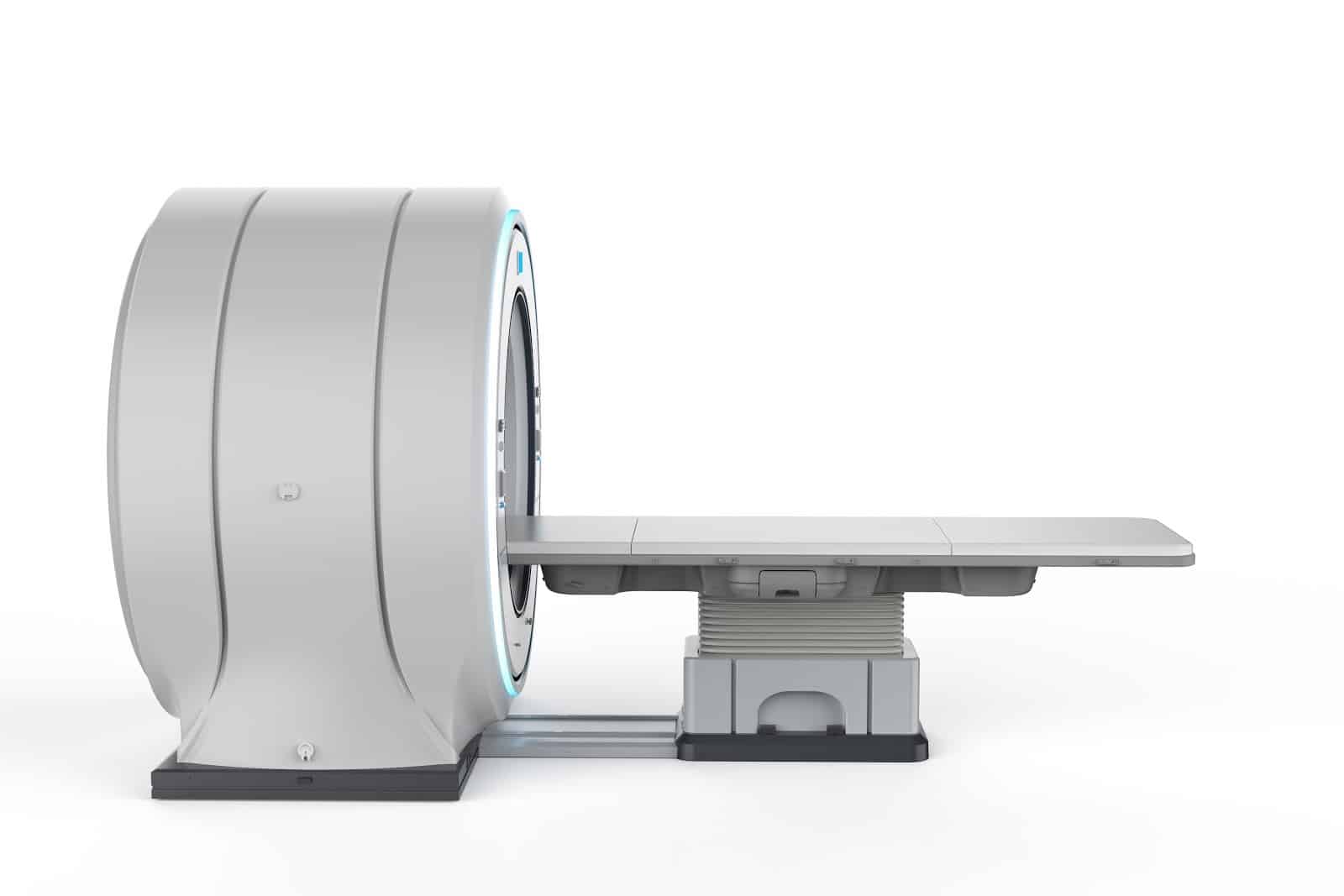
Magnetic Resonance Imaging (MRI) is a non-invasive imaging technique that produces detailed three-dimensional anatomical images, often used for detecting diseases, diagnosing them, and monitoring a patient’s treatment journey. It relies on an advanced technique that excites and detects changes in the orientation of the proton spins found in water, which forms the tissues of the body.

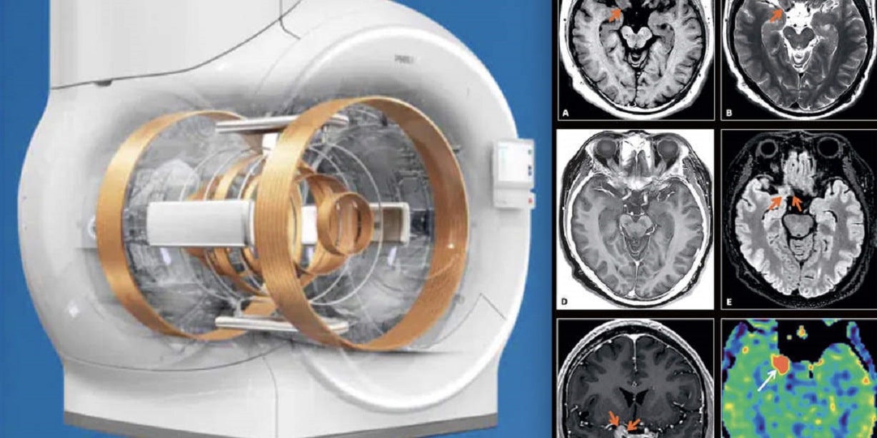
MRI machines use powerful magnets to create a magnetic field that reorients the protons in the body to align with it. When radiofrequency pulses are applied, the protons are stimulated, causing them to fall out of alignment. When the pulses are stopped, the machine measures the energy emitted as the protons realign with the magnetic field. The time it takes for the protons to realign, and the amount of energy emitted depend on the tissue characteristics and the chemical nature of the compounds. These differences help doctors distinguish between different tissues and diagnose medical conditions with greater accuracy.
The History of MRI and Its Development Over Time
The history of MRI technology dates back to the study of magnetic resonance and how the nuclei of electrons and atoms respond to magnetism. In the 1930s, physicist I.I. Rabi developed a technique for measuring the magnetic properties and movement of atoms, which laid the foundation for what is now known as Nuclear Magnetic Resonance (NMR), which became the basis for medical MRI.
In the 1940s, physicists Felix Bloch and Edward Purcell studied the magnetic resonance properties of materials, paving the way for the use of water content in the body to develop MRI images. In 1952, Purcell and Bloch were awarded the Nobel Prize in Physics for their work.
In 1969, Dr. Raymond Damadian proposed the possibility of using MRI to distinguish cancer cells from healthy cells, and he successfully demonstrated his hypothesis in mice. Damadian discovered that MRI could differentiate between tissues based on differences in relaxation times, leading him to develop a full-body MRI scanner. In 1972, Damadian filed the first patent for this technology, and in 1977, he captured the first MRI image of the human body, a cross-sectional image of his assistant’s chest, marking the beginning of MRI imaging as we know it today.
Applications of MRI Technology
The development of MRI technology has been a significant achievement in the medical field, allowing doctors and scientists to examine the inside of the human body in fine detail using a non-invasive tool. MRI is used to diagnose a wide range of conditions, including:
- Brain and spinal cord abnormalities
- Tumors, cysts, and lesions in different parts of the body
- Early screening for breast cancer in women at high risk
- Injuries or deformities of joints such as the back and knee
- Certain types of heart problems
- Liver diseases and other abdominal organ conditions
- Assessment of pelvic pain in women, such as fibroids and endometriosis
- Suspected uterine abnormalities in women undergoing infertility evaluation
The Importance of MRI in Medicine
MRI stands out for its ability to capture detailed images of any part of the body from any imaging direction, making it an essential tool for providing high contrast of soft tissues compared to other imaging techniques like CT scans. This ability to distinguish between fat, water, muscles, and soft tissues helps doctors diagnose a variety of medical conditions more effectively.

Among the key benefits of MRI is its ability to provide detailed three-dimensional images of the targeted area, which enhances doctors’ ability to diagnose diseases accurately and efficiently. It also does not use radiation, as in X-rays or CT scans, making it a safer option for radiation-sensitive individuals such as pregnant women and children.
In addition, MRI scans have become faster and more comfortable thanks to recent technological advancements. Scans that typically take between 15 to 30 minutes provide accurate results quickly. Moreover, modern MRI machines with wide openings help reduce patient anxiety, making the scan more comfortable and secure.
Functional MRI (fMRI) also measures what the tissues are doing rather than just their appearance, helping doctors assess brain activity. By identifying regions involved in vital functions like speaking, movement, sensation, or planning, fMRI can be used to accurately assess the risks of brain surgery.
Uses of Functional MRI
- Determining the impact of tumors on the brain
- Assessing the effects of strokes
- Studying head and brain injuries
- Monitoring neurodegenerative diseases like Alzheimer’s
Technological Advancements in MRI
The field of MRI has seen remarkable progress with the emergence of new techniques that improve image accuracy and scan speed. High-field MRI systems such as 7T and 10T have been developed, offering higher image resolution and more detailed visuals than traditional systems with 1.5T or 3T. Additionally, fast imaging techniques have been developed that reduce scan time without compromising image quality. Functional MRI (fMRI) now enables real-time brain activity monitoring. There has also been significant advancement in imaging techniques for white matter in the brain, as well as advanced methods for examining brain tumors and other brain conditions. These developments help improve medical diagnoses and provide more accurate and faster results.

Recent Innovations in MRI Technology
- Advancements in both device technologies and image sequencing have led to faster scan times. Techniques such as parallel imaging, simultaneous multi-slice imaging, and compressed sensing have contributed to reducing scan times while improving image quality.
- Functional Magnetic Resonance Imaging (fMRI) allows for direct monitoring of brain activity, providing new insights into blood flow and oxygen levels in the brain. This enhances our understanding of brain functions and disorders.
- Diffusion Tensor Imaging (DTI) visualizes the movement of water molecules within tissues, offering a clear view of connectivity within the brain’s white matter. This is particularly useful for detecting changes in conditions like multiple sclerosis and brain injuries.
- Magnetic Resonance Spectroscopy (MRS) is an advanced technique used to study brain tumors, strokes, neurological disorders, and other diseases affecting the brain.
- Advanced Coil Technology has led to improved designs such as array and surface coils, enhancing image quality and sensitivity. This, in turn, reduces scan times and increases diagnostic accuracy.
- Intraoperative MRI (iMRI) integrates MRI imaging with surgical procedures, providing real-time, live images during surgery to guide surgeons and ensure precision.
- Weight-Bearing MRI is performed when the patient is in a weight-bearing position, such as standing or sitting. This type of MRI is helpful for diagnosing disorders resulting from changes in posture or load bearing, such as spinal and joint diseases.
How Artificial Intelligence Impacts the Future of MRI Imaging
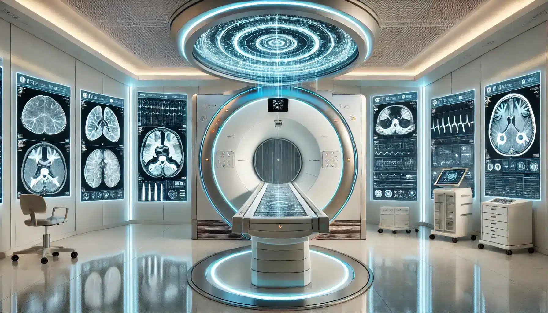
The integration of Artificial Intelligence (AI) into MRI technology represents a significant turning point in medical diagnostics. AI provides substantial improvements in image accuracy and quality, enhancing early and precise diagnosis. Algorithms such as machine learning and deep learning improve scan speed and efficiency, while also automating image analysis and reducing human errors. These advancements open up new possibilities for applying MRI in areas like oncology, neurology, and cardiology, allowing for more personalized and effective medical care.

Key Contributions of Artificial Intelligence in MRI
- Improved Diagnostic Accuracy: AI enhances the precision of MRI image analysis, allowing for more accurate diagnoses.
- Increased Efficiency: Through the automation of image analysis, AI reduces the time required for interpretation, enabling faster decision-making.
- Personalized Medicine: AI offers tailored insights into patient health, aiding in the development of customized treatment plans.
- In Oncology: AI assists in detecting tumors and distinguishing between benign and malignant lesions, improving cancer diagnosis and treatment.
- In Neurology: AI helps in identifying early signs of neurological diseases such as Alzheimer’s and Parkinson’s, facilitating earlier intervention.
- In Cardiology: AI is used to analyze heart images and guide treatment decisions regarding heart function and blood flow.
- Patient Comfort: By reducing scan times and enhancing image quality without compromising accuracy, AI contributes to a more comfortable MRI experience for patients.
Future Directions in MRI Technology
Diagnostic imaging technologies are advancing rapidly, offering more precise, non-invasive methods for early disease detection. Notable developments in this field include artificial intelligence and machine learning, where algorithms are used to analyze medical images with a speed and accuracy that sometimes surpasses that of radiologists. These advancements allow for earlier detection of tumors and other diseases and help in tailoring treatments for individual patients based on their specific needs.
On the other hand, 3D and 4D Imaging provides more detailed views of the body compared to traditional 2D images, facilitating the diagnosis of complex conditions such as bone diseases and heart diseases. Additionally, Molecular Imaging allows physicians to visualize biological processes at the cellular level, aiding in the early detection of cancer.
Portable Point-of-Care Devices are also an emerging technology, enabling physicians to perform real-time scans close to patients, whether in hospitals or remote areas. In conclusion, the future of diagnostic imaging holds great promise for improving diagnostic accuracy, personalizing patient care, reducing costs, and expanding access to healthcare.
In conclusion, with the rapid advancements in the field of Magnetic Resonance Imaging (MRI), it has become essential to keep up with the latest technologies and modern practices to ensure accurate diagnoses and advanced healthcare. Therefore, HSI offers specialized courses in MRI techniques aimed at equipping medical professionals with the practical and scientific knowledge necessary to utilize this technology in the best possible way.
Source: The Future of Magnetic Resonance Imaging (MRI)